Diagnosis of primary ciliary dyskinesia*
Diagnóstico de discinesia ciliar primária
Mary Anne Kowal Olm1, Elia Garcia Caldini2, Thais Mauad3
Abstract
Primary ciliary dyskinesia (PCD) is a genetic disorder of ciliary structure or function. It results in mucus accumulation and bacterial colonization of the respiratory tract which leads to chronic upper and lower airway infections, organ laterality defects, and fertility problems. We review the respiratory signs and symptoms of PCD, as well as the screening tests for and diagnostic investigation of the disease, together with details related to ciliary function, ciliary ultrastructure, and genetic studies. In addition, we describe the difficulties in diagnosing PCD by means of transmission electron microscopy, as well as describing patient follow-up procedures.
Keywords: Kartagener Syndrome; Cilia; Mucociliary clearance; Ciliary motility disorders; Diagnosis.
“Ciliary beating was one of the first characteristics—the Alpha—of animal life on Earth, and it will be the last—the Omega.”
– J. C. de Man, Dutch physician and historian
1. Postdoctoral Student. Department of Pathology, University of São Paulo School of Medicine, São Paulo, Brazil. 2. Associate Professor. Department of Pathology, University of São Paulo School of Medicine, São Paulo, Brazil. 3. Tenured Professor. Department of Pathology, University of São Paulo School of Medicine, São Paulo, Brazil. *Study carried out in the Department of Pathology, University of São Paulo School of Medicine, São Paulo, Brazil. Correspondence to: Mary Anne Kowal Olm. Avenida Dr. Arnaldo, 455, sala 1150, CEP 01246-903, São Paulo, SP, Brasil. Tel. 55 11 5686-7301. E-mail: [email protected]
Financial support: None.
Submitted: 1 October 2014. Accepted, after review: 22 January 2015.
Introduction
Primary ciliary dyskinesia (PCD) is a disease characterized by a change in ciliary beat frequency, ciliary beat pattern, or both and has a heterogeneous genetic basis, typically being an autosomal recessive disorder; the main consequence of ciliary dysfunction is a reduced efficiency of mucociliary clearance of the upper and lower airways, leading to chronic infections and inflammation.(1,2)
Few countries have records of the prevalence, diagnosis, and prognosis of PCD, the data varying greatly across countries. The prevalence of PCD ranges from 1:2,200 to 1:40,000, PCD occurring most frequently in families with consanguineous marriages. Evidence suggests that the diagnosis of PCD is often delayed, which is mainly due to a failure to recognize the disease and the need for sophisticated technical resources for PCD screening.(1)
In Europe, there are reports of more than 1,000 PCD patients at 223 centers in 26 countries.(1) In
the United States, there are reports of fewer than 1,000 patients with an established diagnosis of PCD.(3) In Brazil, most pulmonology clinics lack the
resources required for the investigation of PCD; therefore, there are few reported cases,(4,5) which
are not representative of the prevalence of PCD in the country. According to a European consensus statement, the diagnosis of PCD should be based on the presence of a phenotype consistent with the disease and confirmed by diagnostic tests performed at specialized centers. (6) Screening
tests should precede the diagnostic investigation of PCD.(6) The objective of the present study was
to review the literature on the diagnosis of PCD.
Ciliary biology
bending (a forward power stroke and a backward recovery stroke).(9)
Motile ciliated airway epithelial cells are present in the nasal cavity, paranasal sinuses, middle ear, fallopian tube, cervix, vasa deferentia, and ependyma. In the airways, cilia can be found up to the 16th bronchial division. Ciliated respiratory epithelial cells are characterized by long cytoplasmic projections, with approximately 200 cilia per cell. Each cilium is 5-7 µm in length in the trachea and 2-3 µm in length in the seventh airway generation, being 0.25-0.33 µm in diameter.(10)
The main function of ciliated airway epithelial cells is to mediate the propulsion of the mucus gel layer toward the head through coordinated movements. In the lungs of healthy individuals, mucus is propelled at a ciliary beat frequency of 12-15 Hz, in the form of waves, at a speed of 4-20 mm/min.(11)
Clinical picture
The presence of general clinical indicators (for all age groups) and age-specific indicators should raise the clinical suspicion of PCD.(12)
General indicators include situs abnormalities (dextrocardia and isomerism) and a personal or family history of ciliopathies. Age-specific indicators include prenatal indicators (situs abnormalities on ultrasound), neonatal indicators (rhinorrhea at birth, neonatal respiratory distress with no apparent cause in full-term infants, abnormal situs, complex congenital heart disease—especially with laterality disorders—and a family history of PCD), childhood indicators (chronic productive cough, atypical asthma unresponsive to treatment, idiopathic bronchiectasis, rhinosinusitis—the presence of nasal polyposis is rare—agenesis of one or more sinuses, severe otitis media with effusion, prolonged otorrhea after ventilation tube insertion, and having a family member diagnosed with PCD), and adulthood indicators (childhood data plus male infertility due to immotile sperm, ectopic pregnancy, and subfertility due to static cilia in the fallopian tube).(12) In addition to the
aforementioned indicators, other authors consider the presence of pectus excavatum, which occurs in approximately 10% of patients with PCD and in 0.3% of the general population, and a high prevalence of scoliosis (5-10%).(13)
With regard to ear, nose, and throat problems, approximately 85% of all children with PCD with or without a central pair of microtubules,
the microtubular patterns being designated 9+2 and 9+0, respectively. Cilia are classified as motile or nonmotile.(7,8)
Motile cilia play a role in cell motility and extracellular fluid movement. Motile cilia with a 9+0 arrangement of microtubules are those in notochordal plate cells. During embryonic development (gastrulation), cells in the ventral node contain a single motile cilium per cell. This specialized motile cilium has nine pairs of peripheral microtubules with dynein arms but no central pair of microtubules; its rotational motion contributes to the development of organ laterality during embryogenesis. In the absence of normal nodal ciliary function, organ placement is random. Motile cilia with a 9+2 arrangement of microtubules are those in ciliated airway epithelial cells, fallopian tube epithelial cells, ependymal cells, and sperm tails.
Nonmotile cilia play a role in the perception of extracellular physical and biochemical signals. Nonmotile cilia with a 9+0 arrangement of microtubules are those in renal tubular epithelial cells (monocilia), bile duct epithelial cells, pancreatic duct epithelial cells, chondrocytes, and osteocytes, as well as the connecting cilia in retinal photoreceptor cells. Nonmotile cilia with a 9+2 arrangement of microtubules are those in the inner ear (i.e., kinocilia and stereocilia).
Normal ciliary ultrastructure in ciliated
airway epithelial cells
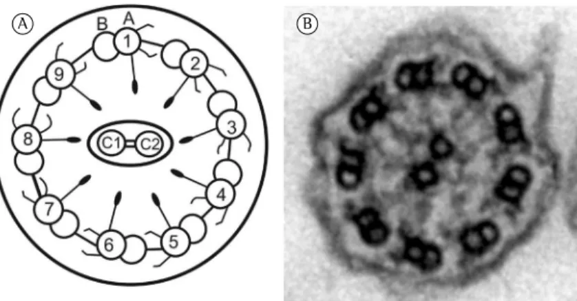
An axial view of a cilium (Figure 1) shows nine peripheral microtubule doublets. Each doublet consists of the A and B tubules. The uniform space between the microtubule doublets is maintained by nexin, which keeps the adjacent microtubules together. In addition, there are the outer and inner dynein arms throughout the A tubule, a central pair of isolated microtubules connected and surrounded by a discontinuous central sheath of protein, and radial spokes, which connect the central microtubules to the peripheral microtubules.
and genetic analysis. In cases of idiopathic bronchiectasis, PCD is a diagnosis of exclusion, given that other causes of bronchiectasis should be ruled out before screening for PCD.(14)
The diagnosis of PCD depends on appropriate training and resources. The consensus among American and British researchers is that the PCD phenotype and nasal NO measurements are important; ciliary motion has been studied in greater detail by European researchers,(15,16)
as has ciliated cell culture. However, American centers for the diagnosis of PCD have reported difficulties in standardizing the interpretation of ciliary motion and electron microscopy. Therefore, at those centers, the diagnosis of PCD is based on the presence of a phenotype consistent with the disease and abnormal nasal NO values, associated with genetic testing to identify the mutations.(15)
High-speed video imaging of the ciliary beat frequency and pattern contributes to the understanding of the effects of ciliary defects on mucus transport. It allows the visualization of the normal pattern of ciliary beating; that is, a forward power stroke followed by a slow, slightly sideways, backward recovery stroke. Changes in the normal pattern of ciliary beating can be associated with specific genetic defects.(16-18)
Chart 1 shows the correlations among reported present with severe otitis media with effusion,
which usually improves by the age of 13 years. Mucus accumulation in the Eustachian tube causes conductive hearing loss that varies with time. There is also an underdevelopment of the frontal and sphenoid sinuses in PCD patients, and nasal polyposis can occur in up to 18% of cases.(6) More than half of all PCD patients
usually have problems in the paranasal sinuses, with frequent radiological investigations and local surgical procedures.(13)
Diagnosis
According to a European consensus statement, diagnostic tests should be performed in the following groups: patients with situs inversus or heterotaxy; children with chronic productive cough, idiopathic bronchiectasis, or severe upper airway disease; children with cerebral ventriculomegaly; siblings of patients with PCD; infants with unexplained neonatal respiratory distress; males with immotile sperm; and females with recurrent ectopic pregnancy.(1)
Patients should be referred to a specialized center(6) for clinical history taking and screening
tests, as well as ciliary function tests (ciliary beat frequency and ciliary beat pattern),(2) ciliary
ultrastructural analysis, immunofluorescence,
Figure 1 - Schematic illustration and electron micrograph of a normal airway cilium. In A, schematic illustration of an axial section of a normal cilium in a ciliated airway epithelial cell, in which the peripheral microtubular doublets (comprising the A and B tubules) are numbered from 1 to 9; the central microtubules are designated C1 and C2. The A tubule contains the outer and inner dynein arms, which interact dynamically with the B tubule of the adjacent microtubule and produce the sliding of the peripheral microtubular doublets relative to one another. The illustration also shows nexin links, which connect the microtubular doublets (thus preventing structural disarray during their sliding motion), and radial spokes, which extend from the periphery to the center of the ciliary axis. In B, electron micrograph (original magnification, ×50,000) showing the ultrastructural appearance of an axial section of a normal airway cilium. Source: Department of Pathology, University of São Paulo School of Medicine, São Paulo, Brazil, 2010.
and flagellar axonemes(24,25) are controlled by
common genes and different genes.(26) Therefore,
patients with abnormal semen analysis results should be referred to tertiary care facilities for analysis of respiratory cilia, in order to establish a diagnosis.
Definitive diagnosis
In order to establish a definitive diagnosis of PCD, certain phenotypic characteristics (at least three characteristics, typically five or more characteristics) should be present: neonatal respiratory distress in full-term infants; laterality defects; chronic, year-round nasal congestion; chronic, year-round productive cough; recurrent lower respiratory tract infections; bronchiectasis; chronic otitis media with effusion for more than 6 months; chronic pansinusitis; male infertility; and a history of ciliary dyskinesia in a close relative.(27)
ciliary ultrastructural defects, gene mutations, and ciliary beat patterns.(16) The most common
genetic mutations include the DNAH5 gene mutation (in 15-28% of cases) and the DNAI1 gene mutation (in 2-10% of cases).(19)
The study of ciliary beat frequency should be accompanied by ciliary beat patterns analysis, given that approximately 10% of individuals with normal ciliary beat frequency can present with an abnormal ciliary beat pattern.(20,21)
Immunofluorescence assays of cilia collected by nasal brushing, performed with specific antibodies and based on established mutations, can aid in the genetic diagnosis of PCD.(22,23)
Semen analysis is used in some centers in Brazil as an indirect indicator of PCD, given that sperm cells behave like modified cilia, with reduced motility. However, in patients with Kartagener syndrome, sperm flagella and respiratory cilia vary across individuals and might not be equal in the same patient; this suggests that ciliary
Chart 1 - Ciliary ultrastructural defects, genetic mutations, and ciliary beat pattern in patients with primary ciliary dyskinesia.
Ultrastructural changes, genetic mutations, and associated chromosomes
Ciliary beat pattern
Outer dynein arm defects
DNAI1 (9p21-p13); DNAI2 (17q25); DNAH5
(5p15.2); DNAL1 (14q24.3); NME8/TXNDC3
(7p14-p13); CCDC114 (19q13.32); and
ARMC4 (10p12.1-p11.23)(3,19,25)
DNAH5, DNAI1, DNAI2, ARMC4, and
CCDC103: residual, disorganized ciliary beat with uncoordinated neighboring cilia(6,16)
Proteins involved in the formation of outer and inner
dynein arms
DNAAF1 (LRRC50): 16q24.1; DNAAF2
(KTU): 14q21.3; DNAAF3 (C19orf51): 19q13.42; CCDC103 (17q21.31); C21orf59
(21q.22.1); DYX1C1 (15q21.3); LRRC6
(8q24); HEATR2 (7p22.3); SPAG1 (8q22); and ZMYND10 (3p21.31)(3,19,25)
KTU/DNAAF2, LRRC50/DNAAF1, LRRC6, and ZMYND10: complete ciliary immotility;
DYX1C1: complete ciliary immotility or reduced ciliary beat amplitude with a few
static cilia(16)
Inner dynein arm defects and
microtubular disorganization
CCDC39 (3q26.33) and CCDC40 (17q25.3)
(3,19,25)
Stiff cilia with reduced ciliary beat amplitude(16)
Nexin links
CCDC164 (DRC1), 2p23.3: nexin link missing, axonemal disorganization in a
small proportion of cilia(23,25); CCDC65
(DRC2):12q13.12(23,25)
CCDC164: reduced ciliary beat amplitude(16)
Changes in the central pair of microtubules
HYDIN (16q22.2): mostly normal; RSPH9
(6p21.1), RSPH4 (6q22.1), and RSPH1
(21q22.3): central pair defects in a small proportion of cilia(3,19,25)
HYDIN: reduced ciliary bending; some cilia showing circular motion and others being immotile; RSPH4A: stiff cilia; RSPH1: stiff cilia and cilia showing circular motion(16);
RSPH9: circular motion(6)
Normal ultrastructure DNAH11 (7p21): changes in outer dynein arm proteins(3,19,25)
Reduced ciliary bending(16); stiff,
hyperkinetic, or static cilia(6,15)
Reduced generation
of motile cilia CCNO/MCIDAS (5q11)(60) Reduced number of motile cilia(60)
processes of nasal NO production and metabolism in PCD patients have yet to be fully elucidated.(29)
The leading hypotheses to explain the reduced nasal NO levels in over 95% of patients with PCD are related to ciliated epithelial cells per se and anatomical obstructions. At the epithelial level, it has been suggested that there is reduced NO biosynthesis or increased NO metabolism caused by the accumulation of thick mucus or the presence of bacteria. At the anatomical level, it has been suggested that NO is sequestered in blocked nasal sinuses or, alternatively, nasal NO biosynthesis or NO storage capacity is limited because of agenesis of the paranasal sinuses.(29)
Nasal NO levels are extremely low in PCD patients, for whom nasal NO measurement is strongly recommended.(30-32) In the airways, NO
plays many roles—it mediates inflammation and stimulates ciliary motility, for example—and NO concentrations are much higher in the upper airways than in the lower airways (200-2,000 ppb vs. 4-160 ppb). Nasal NO production (nL/min) is calculated by multiplying nasal NO concentration by the sampling flow rate. Values of less than 100 nL/min indicate the possibility of PCD.(27)
However, values of less than 77 nL/min have a sensitivity of 0.98 and a specificity of more than 0.999 for PCD (in patients over 5 years of age, with the use of palate closure maneuvers and nonmanual chemiluminescence analyzers). (32)
Further studies are needed in order to define cut-off points for tidal volume measurements in uncooperative young children.(28,31) Of all
PCD-associated gene mutations described to date, the RSPH1 gene mutation is an exception in that patients with that mutation can have normal nasal NO values.(15)
The saccharin test
The saccharin test is a good test to assess nasal mucociliary transport, which is usually prolonged in individuals with PCD. It consists of placing a particle of saccharin of 1 mm in diameter on the floor of the nasal cavity, approximately 1 cm into the inferior turbinate. The patient sits quietly with the head bent forward and must not sniff, sneeze, cough, eat, or drink for the duration of the test. The time (in min) to tasting saccharin in the pharynx is measured. The result of the test is considered abnormal when the time to tasting saccharin in the pharynx is greater than 60 min. (14) However, a study conducted
According to BESTCILIA, a European Commission-funded consortium dedicated to improve PCD care and knowledge,(23) a diagnosis of
PCD requires a) a clinical presentation consistent with the disease and b) confirmation by at least two of the following methods: unequivocally abnormal high-speed video microscopy finding; unequivocally abnormal transmission electron microscopy finding; unequivocally abnormal immunofluorescence microscopy finding; abnormally low nasal NO concentration/production; and demonstration of unequivocal biallelic disease-causing mutations by genotyping. In cases in which only high-speed video microscopy and nasal NO concentration/ production are abnormal, high-speed video microscopy should be repeated at least three times and show the same abnormal results each time.(23) Patients with typical clinical symptoms and
only one abnormal diagnostic test are considered to have a possible diagnosis of PCD, exceptions being made on an individual basis.
At several centers worldwide, the use of nasal NO testing is recommended in order to confirm the diagnosis,(15) and the diagnostic tests should
be repeated when the phenotype and low levels of nasal NO do not correlate with ciliary ultrastructure and beat frequency. Secondary defects should be excluded when the results of nasal NO testing are normal and accompanied by ciliary motility defects or ciliary ultrastructural defects.
A genetic test result is considered positive for PCD when there are two genes with trans mutations—in which the wild-type allele (A) and mutant allele (b) of one gene are located on one chromosome and the mutant allele (a) and wild-type allele (B) of another gene are located on the homologous chromosome—and no correcting mutations.(12)
Screening tests
Screening tests are important in order to select which of the patients with signs and symptoms suggestive of PCD should undergo analysis of ciliary function and ultrastructure. Nasal NO testing and the saccharin test are used as screening tests in 46% and 36%, respectively, of all centers in 26 European countries.(2)
Exhaled nasal NO measurement
fibrocystic liver disease, retinitis pigmentosa, and hydrocephalus.
Ciliopathies constitute a group of diseases associated(12) with genetic mutations that result
in changes in ciliary formation or function. Given that cilia are components of many cell types, ciliary dysfunction can manifest as a constellation of clinical features such as retinal degeneration, kidney disease, and cerebral abnormalities. Molecular genetic studies conducted in recent years suggest a clear relationship between primary cilium development and function and various clinical conditions.(35)
Syndromic manifestations of ciliopathies are found in Joubert syndrome, Meckel-Gruber syndrome, Senior-Loken syndrome, oral-facial-digital syndrome, Leber congenital amaurosis, Bardet-Biedl syndrome, Alström syndrome, asphyxiating thoracic dystrophy (Jeune syndrome), Ellis-van Creveld syndrome, and Sensenbrenner syndrome.(12)
Kartagener syndrome is a rare congenital malformation consisting of the triad of situs inversus, bronchiectasis, and sinusitis.(37) The
association between immotile cilia and situs inversus was based on the hypothesis that, in the early stages of normal embryogenesis, the position of nodal and notochordal cilia and their ciliary beat orientation are predetermined, and that their ciliary beat frequency determines organ laterality through a cascade of molecular signaling. When the aforementioned cilia are immotile, organ placement is random, resulting in many cases of situs inversus, which usually occurs in 50% of PCD patients,(36,37) some of whom are
diagnosed with Kartagener syndrome.(38)
Standardization of electron microscopy
analysis of ciliary ultrastructure
Although several transmission electron microscopy facilities have been working on the standardization of diagnostic criteria to be used in ciliary ultrastructural analysis, no proposal has been universally accepted. The variety of PCD-associated defects and the rarity of the disease make it difficult to standardize the interpretation of electron microscopy.(39)
There are multiple factors that limit the use of electron microscopy as a diagnostic test for PCD: secondary ciliary changes caused by infection or inflammation; difficulties in the fixation and processing of ciliated cells; the need for ultrathin sections; the technical complexity of in Brazil(33) and involving 238 children (in the
10-16 year age bracket) established a different cut-off point (of 30 min), showing that the test should not be performed during an acute viral infection or in the subsequent month. In patients with abnormal ciliary ultrastructure, the saccharin test has good sensitivity (95% of cases for values greater than 30 min and 75% of cases for values greater than 60 min). However, false positives can occur in 0.4-15% of cases in healthy adults. Nasal mucociliary transport can be slower in patients with septal deviation or rhinoscleroma. In a recent review,(6) the saccharin test was reported to be
difficult to perform correctly and unreliable in children under 12 years of age. In addition, cases with extremely uncoordinated ciliary beating might be missed by the saccharin test.
Pulmonary radioaerosol mucociliary
clearance testing
Current clinical experience is insufficient to recommend the use of pulmonary radioaerosol mucociliary clearance tests in clinical practice.(2)
Genetics
Genetic studies have identified mutations in several genes encoding ciliary structure and functional proteins; however, genetic tests are not readily available in clinical practice.(8) The
finding of a PCD-associated gene mutation constitutes laboratory evidence for a definitive diagnosis of PCD.(27) Chart 1 presents the most
important PCD-associated genes described to date. Mutations in genes such as RSPH1, RSPH4A, RSPH9, HYDIN, MCIDAS, and CCNO, which cause no laterality defects, allow clinical correlations to be established.(3,23)
Associated clinical conditions
There is evidence that ciliary disorders are related to various developmental problems and clinical conditions, which are known as ciliopathies. A family history of ciliopathy should raise the suspicion of PCD in patients or their relatives with characteristics suggestive of PCD. (34)
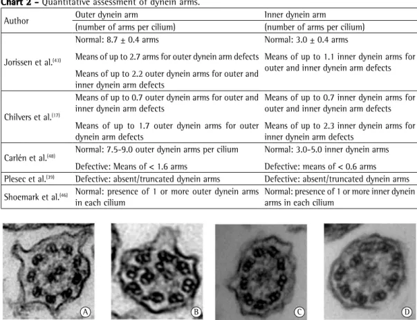
across centers.(17,39,43,46,48) Chart 2 summarizes
the results of studies involving quantitative assessment of dynein arms.
With regard to dynein arm defects, absence of dynein arms can be found in more than 90% of cilia. (17) Isolated inner dynein arm defects
constitute only a small fraction of confirmed PCD cases and require confirmation by repetition of the test with a healthy epithelium sample.(3)
Partial absence of dynein as a primary defect is considered controversial and requires further studies for confirmation.(43) Figure 2 shows the
major ultrastructural defects described to date.
Normal ciliary ultrastructure and PCD
Extremely reduced nasal NO levels and abnormal ciliary function (ciliary beat frequency, ciliary beat pattern, or both) with normal ciliary ultrastructure require genetic testing for a mutation consistent with the disease (i.e., the DNAH11 gene mutation).(8,27) Patients with a clinical history
consistent with PCD, low levels of exhaled nasal NO, and abnormal ciliary motility have been diagnosed with PCD, despite the recommendation that genetic testing be performed in order to confirm the diagnosis.(8,27)
It has been reported that 3-30% of patients with PCD have uncertain or normal ciliary ultrastructure, the diagnosis of PCD in such patients not being based on ciliary ultrastructure alone.(44) Advances in the molecular genetics of
PCD have allowed the identification of mutations in approximately 70% of patients.(15,25)
Dynein arm defects are the most common defects in patients with PCD: outer dynein arm defects, in 30-43% of cases; outer and inner dynein arm defects, in 9-36%; inner dynein arm defects, in 11-30%; normal ultrastructure, in 25%; transposition, in 14%; central pair defects, in 9%; radial spoke defects, in 7%; and ciliary aplasia, in 6%.(34)
Rare defects
In patients with PCD, ultrastructural changes include nexin link defects,(49) absence of the
central pair of microtubules, and absence of the basal bodies and sheath (alternatively, the basal bodies and sheath are present but have a reduced number of cilia).(47,48)
transmission electron microscopy; the need for an adequate number of interpretable images; and the fact that interpretation requires recognition of normal variability and nonspecific changes.(40)
In addition, samples from children are usually less suitable for ciliary ultrastructural analysis than are those from adults (60% vs. 87%).(40,41)
Collection of ciliated cells
For ciliated cell collection, nasal brushing has lower morbidity than does nasal biopsy, as well as being less costly and easier to perform. (14,42)
Bronchoscopy brushes are used in order to collect ciliated cells from the inferior nasal turbinate, near the transverse septum. Some of the material is separated for ciliary beat frequency and ciliary beat pattern analysis, and the remainder is sent for electron microscopy analysis.(12,14)
For ciliated cell collection, patients are required to be free of acute respiratory infection for 4-6 weeks in order to minimize the presence of changes caused by secondary dyskinesia.
Parameters for evaluating ciliary
ultrastructure
Ciliary orientation
Ciliary disorientation is associated with PCD. In cases of ciliary disorientation, ciliary ultrastructure is normal and ciliary beat frequency is normal or near normal, but ciliary motion is inefficient because of ciliary beat disorientation; that is, it does not correctly propel the mucus.(42)
Microtubules
The peripheral microtubules can show disorganization and inner dynein arm defects,(3)
as well as transposition defects (the central pair being replaced by a peripheral microtubule).(43,44)
Transposition defects(44,45) or translocation
defects(3) are characterized by the absence of
the central pair of microtubules in certain cross sections (9+0 arrangement), some sections showing the central pair replaced by an outer microtubule doublet (8+2 arrangement).(43,45) The absence of
the central pair alone has been reported as a primary defect.(47)
Quantitative assessment of dynein arms
ciliary defect elsewhere in the airways. Patients with normal ciliary ultrastructure and abnormal ciliary function require ciliary orientation studies.(14)
Diagnosis confirmed by ciliated cell culture
Ciliated cell cultures are performed only at specialized centers and are followed by transmission electron microscopy analysis, being recommended to differentiate between primary and secondary defects. Culture duration is approximately 6 weeks. The success rate of ciliated cell cultures is 75%, cultures being conclusive in 85% of cases.(42,43)
The absence of secondary defects after growth in culture media (ciliogenesis) contributes to the diagnosis of PCD.
Ciliary ultrastructure report and final
results
At internationally renowned PCD research centers, the diagnosis (report) of ciliary
Secondary ciliary dyskinesia
Secondary or acquired ciliary dyskinesia can be caused by injury to ciliated airway epithelial cells by physical and chemical agents.
Respiratory infections and the inflammatory immune response to the infections can affect ciliary function, inducing secondary ciliary dyskinesia. Secondary lesions include compound cilia (fused membranes or multiple axonemes within a single membrane), peripheral and central microtubular abnormalities, swelling of the membranes, shortened dynein arms, ciliary membrane blebs, and absence of the ciliary membrane.(50,51)
In healthy individuals and in patients without ciliary dyskinesia, there are variations (of 4 ± 3%) in normality for secondary ciliary changes in healthy individuals, and the presence of up to 10% of altered cilia can be considered normal.(50,52) In
cases of ciliary aplasia, bronchial mucosal biopsy is required in order to confirm the presence of a Chart 2 - Quantitative assessment of dynein arms.
Author Outer dynein arm Inner dynein arm
(number of arms per cilium) (number of arms per cilium)
Jorissen et al.(43)
Normal: 8.7 ± 0.4 arms
Means of up to 2.7 arms for outer dynein arm defects
Means of up to 2.2 outer dynein arms for outer and inner dynein arm defects
Normal: 3.0 ± 0.4 arms
Means of up to 1.1 inner dynein arms for outer and inner dynein arm defects
Chilvers et al.(17)
Means of up to 0.7 outer dynein arms for outer and inner dynein arm defects
Means of up to 1.7 outer dynein arms for outer dynein arm defects
Means of up to 0.7 inner dynein arms for outer and inner dynein arm defects
Means of up to 2.3 inner dynein arms for inner dynein arm defects
Carlén et al.(48) Normal: 7.5-9.0 outer dynein arms per cilium
Defective: Means of < 1.6 arms
Normal: 3.0-5.0 inner dynein arms
Defective: means of < 0.6 arms Plesec et al.(39) Defective: absent/truncated dynein arms Defective: absent/truncated dynein arms
Shoemark et al.(46) Normal: presence of 1 or more outer dynein arms
in each cilium
Normal: presence of 1 or more inner dynein arms in each cilium
A B C D
structural defects—has been reported.(53) However,
in a recent longitudinal study of 74 patients, lung function varied widely over the course of 10 years, up to 34% of the patients having experienced a significant decline in lung function despite treatment; this shows that PCD poses a serious threat to lung function.(55,56)
Treatment
Ciliary defects cannot be treated with the conventional pharmacological armamentarium, and there is no specific treatment for ciliary dysfunction. Therapy is aimed at improving mucociliary clearance, treating infections, and improving or stabilizing lung function, preventing chronic lung injury. The recommendations are based on expert opinion, being inferred from the available evidence for cystic fibrosis, although there are differences between the two diseases in terms of their pathophysiology.(6) Education
and information are considered important tools for patients and their families.(14)
Outpatient treatment is multidisciplinary, involving pulmonologists, otolaryngologists, nurses, physiotherapists, psychologists, and social workers.(19)
Patients should be advised to avoid environmental allergens and smoking, and physical exercise can be a better bronchodilator than beta-agonists. Regular visits to referral centers are to take place every 2-3 months in children and every 6-12 months in adults, as needed.(14)
In addition to basic immunization, patients should receive annual influenza vaccination and pneumococcal vaccination.
Respiratory monitoring
The two pillars upon which respiratory treatment stands are antibiotic therapy and chest physiotherapy. Chest physiotherapy should be performed twice a day for 20 min, increasing during exacerbations. (14)
Sputum culture and serial spirometry should be performed in the follow-up of patients with PCD. Antibiotic therapy should be initiated at the first sign of any increase in respiratory symptoms or deterioration of lung function, lasting two weeks in general. Antibiotics should be given on the basis of culture sensitivity testing. Intravenous treatment should be used if symptoms do not respond to oral antibiotics. In adults, colonization with Pseudomonas aeruginosa is not rare and might ultrastructural changes is based on the observation
of 200-300 cilia,(46) a minimum of 100 cilia(44) being
evaluated in cross sections (and approximately 30% of dynein arms being visualized). Abnormalities found in less than 10% of cilia are considered to be within the normal range.(50) PCD-related defects
include absence of outer dynein arms, absence of outer and inner dynein arms, microtubular disorganization,(43) and changes in the central
pair of microtubules (transposition defect).(44)
The presence of inner dynein arm defects(3)
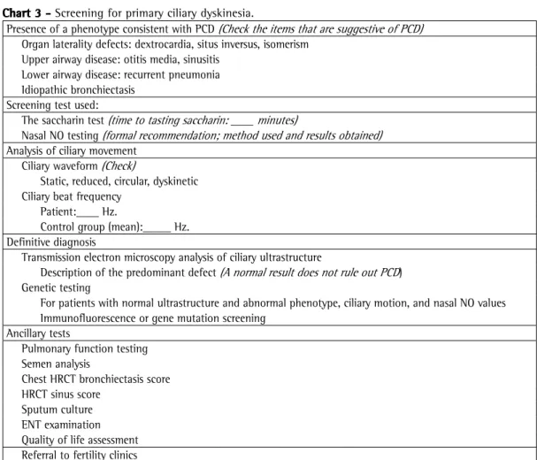
or ciliary disorientation alone requires new samples in order to confirm the diagnosis. The ciliary ultrastructure report should be conclusive regarding the presence or absence of PCD-related defects. The results of all investigations should be expressed as a definitive diagnosis (Chart 3).
Radiology
In patients with PCD, a HRCT scan of the chest (Figure 3) shows middle and lower lobe involvement—the middle and lower lobes being more affected than the upper lobes in PCD patients when compared with cystic fibrosis patients (in whom the upper lobes are more affected than the middle and lower lobes)—with subsegmental atelectasis, peribronchial thickening, mucus plugging, evidence of air trapping, ground-glass opacities,(25) areas of consolidation, and
well-defined bronchiectasis.(19)
The presence of bronchiectasis is related to age. In a study of 72 patients, 98% of those over 18 years of age (in the 19-73 year age bracket) had signs and symptoms of bronchiectasis, as did 61% of those under 18 years of age.(53) Adult
patients present with advanced lung disease.(54)
Pathophysiology of the disease in
the airways and lung function
Genetic defects in respiratory epithelial cilia cause a significant reduction in mucociliary transport, with retention of secretions, recurrent infections, and, consequently, bronchiectasis. Impaired alveolar gas exchange can occur in the long term, causing respiratory failure, pulmonary hypertension, and right heart failure.(52)
require more aggressive intravenous therapy and long-term use of inhaled antibiotics.(2,3,6,23)
Chart 3 - Screening for primary ciliary dyskinesia.
Presence of a phenotype consistent with PCD (Check the items that are suggestive of PCD)
Organ laterality defects: dextrocardia, situs inversus, isomerism Upper airway disease: otitis media, sinusitis
Lower airway disease: recurrent pneumonia Idiopathic bronchiectasis
Screening test used:
The saccharin test (time to tasting saccharin: ____ minutes)
Nasal NO testing (formal recommendation; method used and results obtained)
Analysis of ciliary movement Ciliary waveform (Check)
Static, reduced, circular, dyskinetic Ciliary beat frequency
Patient:____ Hz.
Control group (mean):_____ Hz. Definitive diagnosis
Transmission electron microscopy analysis of ciliary ultrastructure
Description of the predominant defect (A normal result does not rule out PCD) Genetic testing
For patients with normal ultrastructure and abnormal phenotype, ciliary motion, and nasal NO values Immunofluorescence or gene mutation screening
Ancillary tests
Pulmonary function testing Semen analysis
Chest HRCT bronchiectasis score HRCT sinus score
Sputum culture ENT examination Quality of life assessment Referral to fertility clinics
PCD: primary ciliary dyskinesia; ENT: ear, nose, and throat. Source: Department of Pathology, University of São Paulo School of Medicine, São Paulo, Brazil, 2014.
Figure 3 - Axial HRCT scan of the chest of a 30-year-old patient with primary ciliary dyskinesia (absence of outer and inner dynein arms) and advanced lung disease. Note significant involvement of the lung bases, with bronchial wall thickening, the signet ring sign, areas of consolidation, and attenuation differences. Source: Department of Clinical Pulmonology, Heart Institute, University of São Paulo School of Medicine - Hospital das Clínicas, São Paulo, Brazil, 2014.
The use of recombinant human DNase, which reduces the viscoelasticity of respiratory mucus, shows conflicting results and requires further investigation before it can be recommended for PCD patients. Hypertonic saline can be effective in improving mucociliary clearance; however, to date, there have been no controlled clinical trials to support its use.(6,23)
Auditory monitoring
At otolaryngology clinics, patients should be monitored for hearing loss, which requires specific procedures.(13) The results of studies on
the treatment of otitis media with effusion are contradictory regarding the use of ventilation tubes, larger samples being required in order to draw conclusions.(57) Endoscopic sinus surgery
appears to improve local symptoms.(13)
Other referrals
require psychosocial support in order to cope with the stigmata attached to the disease, including infertility and possible school problems.(14)
Surgical resection for localized bronchiectasis can be beneficial in some cases.(58) Although lung
transplantation is a possibility, only a few cases have been reported in the literature.(59)
Prognosis
To date, there have been no large-scale, long-term studies for a more detailed prognosis of PCD. The high genetic variability of the disease determines differences in progression among patients. Pulmonary impairment is more severe in patients diagnosed in adulthood than in those diagnosed in adolescence. A minority of patients might progress to severe lung disease with respiratory failure requiring lung transplantation.(25)
Final considerations
The diagnostic report of PCD should include the results of all investigations that led to the diagnosis of the disease, including phenotyping, screening tests, analysis of ciliary function (ciliary beat frequency, ciliary waveform, or both), qualitative and quantitative assessment of ciliary ultrastructure, immunofluorescence, and gene mutation screening. Ciliated cell cultures can aid in the diagnosis of PCD. Normal ciliary ultrastructure does not rule out PCD. The results of all investigations should be expressed as a definitive diagnosis.
Acknowledgments
We would like to thank Dr. Samia Rached for the CT image.
References
1. Kuehni CE, Frischer T, Strippoli MP, Maurer E, Bush A, Nielsen KG, et al. Factors influencing age at diagnosis of primary ciliary dyskinesia in European children. Eur Respir J. 2010;36(6):1248-58. http://dx.doi. org/10.1183/09031936.00001010
2. Strippoli MP, Frischer T, Barbato A, Snijders D, Maurer E, Lucas JS, et al. Management of primary ciliary dyskinesia in European children: recommendations and clinical practice. Eur Respir J. 2012;39(6):1482-91. http://dx.doi. org/10.1183/09031936.00073911
3. Knowles MR, Daniels LA, Davis SD, Zariwala MA, Leigh MW. Primary ciliary dyskinesia. Recent advances in diagnostics, genetics, and characterization of clinical disease. Am J Respir Crit Care Med. 2013;188(8):913-22. http:// dx.doi.org/10.1164/rccm.201301-0059CI
4. Olm MA, Kögler JE Jr, Macchione M, Shoemark A, Saldiva PH, Rodrigues JC. Primary ciliary dyskinesia: evaluation using cilia beat frequency assessment via spectral analysis of digital microscopy images. J Appl Physiol (1985). 2011;111(1):295-302. http://dx.doi. org/10.1152/japplphysiol.00629.2010
5. Santos JW, Waldow A, Figueiredo CW, Kleinubing DR, Barros SS. Discinesia ciliar primária. J Pneumol. 2001;27(5):262-68. http://dx.doi.org/10.1590/ S0102-35862001000500006
6. Barbato A, Frischer T, Kuehni CE, Snijders D, Azevedo I, Baktai G, et al. Primary ciliary dyskinesia: a consensus statement on diagnostic and treatment approaches in children. Eur Respir J. 2009;34(6):1264-76. http:// dx.doi.org/10.1183/09031936.00176608
7. Fliegauf M, Benzing T, Omran H. When cilia go bad: cilia defects and ciliopathies. Nat Rev Mol Cell Biol. 2007;8(11):880-93. Erratum in: Nat Rev Mol Cell Biol. 2008;9(1):88. http://dx.doi.org/10.1038/nrm2278 8. Leigh MW, Pittman JE, Carson JL, Ferkol TW, Dell
SD, Davis SD, et al. Clinical and genetic aspects of primary ciliary dyskinesia/Kartagener syndrome. Genet Med. 2009;11(7):473-87. http://dx.doi.org/10.1097/ GIM.0b013e3181a53562
9. Cowan MJ, Gladwin MT, Shelhamer JH. Disorders of ciliary motility. Am J Med Sci. 2001;321(1):3-10. http:// dx.doi.org/10.1097/00000441-200101000-00002 10. Houtmeyers E, Gosselink R, Gayan-Ramirez G, Decramer
M. Regulation of mucociliary clearance in health and disease. Eur Respir J. 1999;13(5):1177-88. http://dx.doi. org/10.1034/j.1399-3003.1999.13e39.x
11. Tilley AE, Walters MS, Shaykhiev R, Crystal RG. Cilia Dysfunction in Lung Disease. Annu Rev Physiol. 2014 Oct 29. [Epub ahead of print]
12. Bush A, Hogg C. Primary ciliary dyskinesia: recent advances in epidemiology, diagnosis, management and relationship with the expanding spectrum of ciliopathy. Expert Rev Respir Med. 2012;6(6):663-82. http://dx.doi. org/10.1586/ers.12.60
13. Sommer JU, Schäfer K, Omran H, Olbrich H, Wallmeier J, Blum A, et al. ENT manifestations in patients with primary ciliary dyskinesia: prevalence and significance of otorhinolaryngologic co-morbidities. Eur Arch Otorhinolaryngol. 2011;268(3):383-8. http://dx.doi. org/10.1007/s00405-010-1341-9
14. Bush A, Cole P, Hariri M, Mackay I, Phillips G, O’Callaghan C, et al. Primary ciliary dyskinesia: diagnosis and standards of care. Eur Respir J. 1998;12(4):982-8. http://dx.doi. org/10.1183/09031936.98.12040982
15. Lucas JS, Leigh MW. Diagnosis of primary ciliary dyskinesia: searching for a gold standard. Eur Respir J. 2014;44(6):1418-22. http://dx.doi. org/10.1183/09031936.00175614
16. Raidt J, Wallmeier J, Hjeij R, Onnebrink JG, Pennekamp P, Loges NT, et al. Ciliary beat pattern and frequency in genetic variants of primary ciliary dyskinesia. Eur Respir J. 2014;44(6):1579-88. http://dx.doi. org/10.1183/09031936.00052014
17. Chilvers MA, Rutman A, O’Callaghan C. Ciliary beat pattern is associated with specific ultrastructural defects in primary ciliary dyskinesia. J Allergy Clin Immunol. 2003;112(3):518-24. http://dx.doi.org/10.1016/S0091-6749(03)01799-8 18. Chilvers MA, Rutman A, O’Callaghan C. Functional analysis
Rev. 2009;10(2):44-50. http://dx.doi.org/10.1016/j. prrv.2008.10.001
35. Waters AM, Beales PL. Ciliopathies: an expanding disease spectrum. Pediatr Nephrol. 2011;26(7):1039-56. http:// dx.doi.org/10.1007/s00467-010-1731-7
36. Afzelius BA. Asymmetry of cilia and of mice and men. Int J Dev Biol. 1999;43(4):283-6.
37. Brueckner M. Cilia propel the embryo in the right direction. Am J Med Genet. 2001;101(4):339-44. http://dx.doi. org/10.1002/1096-8628(20010715)101:4<339::AID-AJMG1442>3.0.CO;2-P
38. Sutherland MJ, Ware SM. Disorders of left-right asymmetry: heterotaxy and situs inversus. Am J Med Genet C Semin Med Genet. 2009;151C(4):307-17. http:// dx.doi.org/10.1002/ajmg.c.30228
39. Plesec TP, Ruiz A, McMahon JT, Prayson RA. Ultrastructural abnormalities of respiratory cilia: a 25-year experience. Arch Pathol Lab Med. 2008;132(11):1786-91. 40. Leigh MW, O’Callaghan C, Knowles MR. The challenges
of diagnosing primary ciliary dyskinesia. Proc Am Thorac Soc. 2011;8(5):434-7. http://dx.doi.org/10.1513/ pats.201103-028SD
41. Papon JF, Coste A, Roudot-Thoraval F, Boucherat M, Roger G, Tamalet A, et al. A 20-year experience of electron microscopy in the diagnosis of primary ciliary dyskinesia. Eur Respir J. 2010;35(5):1057-63. http:// dx.doi.org/10.1183/09031936.00046209
42. Jorissen M, Willems T. Success rates of respiratory epithelial cell culture techniques with ciliogenesis for diagnosing primary ciliary dyskinesia. Acta Otorhinolaryngol Belg. 2000;54(3):357-65.
43. Jorissen M, Willems T, Van der Schueren B, Verbeken E, De Boeck K. Ultrastructural expression of primary ciliary dyskinesia after ciliogenesis in culture. Acta Otorhinolaryngol Belg. 2000;54(3):343-56.
44. Lucas JS, Burgess A, Mitchison HM, Moya E, Williamson M, Hogg C, et al. Diagnosis and management of primary ciliary dyskinesia. Arch Dis Child. 2014;99(9):850-6. http://dx.doi.org/10.1136/archdischild-2013-304831 45. Sturgess JM, Chao J, Turner JA. Transposition of ciliary
microtubules: another cause of impaired ciliary motility. N Engl J Med. 1980;303(6):318-22. http://dx.doi. org/10.1056/NEJM198008073030606
46. Shoemark A, Dixon M, Corrin B, Dewar A. Twenty-year review of quantitative transmission electron microscopy for the diagnosis of primary ciliary dyskinesia. J Clin Pathol. 2012;65(3):267-71. http://dx.doi.org/10.1136/ jclinpath-2011-200415
47. Stannard W, Rutman A, Wallis C, O’Callaghan C. Central microtubular agenesis causing primary ciliary dyskinesia. Am J Respir Crit Care Med. 2004;169(5):634-7. http:// dx.doi.org/10.1164/rccm.200306-782OC
48. Carlén B, Stenram U. Primary ciliary dyskinesia: a review. Ultrastruct Pathol. 2005;29(3-4):217-20. http://dx.doi. org/10.1080/01913120590951220
49. Carlén B, Lindberg S, Stenram U. Absence of nexin links as a possible cause of primary ciliary dyskinesia. Ultrastruct Pathol. 2003;27(2):123-6. http://dx.doi. org/10.1080/01913120309930
50. Bush A. Primary ciliary dyskinesia. Acta Otorhinolaryngol Belg. 2000;54(3):317-24.
51. Bertrand B, Collet S, Eloy P, Rombaux P. Secondary ciliary dyskinesia in upper respiratory tract. Acta Otorhinolaryngol Belg. 2000;54(3):309-16.
children and young adults. Thorax. 2003;58(4):333-8. http://dx.doi.org/10.1136/thorax.58.4.333
19. Boon M, Jorissen M, Proesmans M, De Boeck K. Primary ciliary dyskinesia, an orphan disease. Eur J Pediatr. 2013;172(2):151-62. http://dx.doi.org/10.1007/ s00431-012-1785-6
20. Bush A, O’Callaghan C. Primary ciliary dyskinesia. Arch Dis Child. 2002;87(5):363-5; discussion 363-5. http:// dx.doi.org/10.1136/adc.87.5.363
21. Stannard WA, Chilvers MA, Rutman AR, Williams CD, O’Callaghan C. Diagnostic testing of patients suspected of primary ciliary dyskinesia. Am J Respir Crit Care Med. 2010;181(4):307-14. http://dx.doi.org/10.1164/ rccm.200903-0459OC
22. Djakow J, O’Callaghan C. Primary ciliary dyskinesia. Breathe [serial on the Internet]. 2014 Jun [cited 2014 Oct 1];10(2):122-33. Available from: http://breathe. ersjournals.com/content/10/2/122.full
23. Werner C, Onnebrink JG, Omran H. Diagnosis and management of primary ciliary dyskinesia. Cilia. 2015;4(1):2. http://dx.doi.org/10.1186/s13630-014-0011-8 24. Munro NC, Currie DC, Lindsay KS, Ryder TA, Rutman
A, Dewar A, et al. Fertility in men with primary ciliary dyskinesia presenting with respiratory infection. Thorax. 1994;49(7):684-7. http://dx.doi.org/10.1136/thx.49.7.684 25. Lobo LJ, Zariwala MA, Noone PG. Primary ciliary dyskinesia.
QJM. 2014;107(9):691-9. http://dx.doi.org/10.1093/ qjmed/hcu063
26. Escudier E, Escalier D, Pinchon MC, Boucherat M, Bernaudin JF, Fleury-Feith J. Dissimilar expression of axonemal anomalies in respiratory cilia and sperm flagella in infertile men. Am Rev Respir Dis. 1990;142(3):674-9. http://dx.doi.org/10.1164/ajrccm/142.3.674
27. Leigh MW, Zariwala MA, Knowles MR. Primary ciliary dyskinesia: improving the diagnostic approach. Curr Opin Pediatr. 2009;21(3):320-5. http://dx.doi.org/10.1097/ MOP.0b013e328329cddb
28. Marthin JK, Nielsen KG. Choice of nasal nitric oxide technique as first-line test for primary ciliary dyskinesia. Eur Respir J. 2011;37(3):559-65. http://dx.doi. org/10.1183/09031936.00032610
29. Walker WT, Jackson CL, Lackie PM, Hogg C, Lucas JS. Nitric oxide in primary ciliary dyskinesia. Eur Respir J. 2012;40(4):1024-32. http://dx.doi. org/10.1183/09031936.00176111
30. American Thoracic Society; European Respiratory Society. ATS/ERS recommendations for standardized procedures for the online and offline measurement of exhaled lower respiratory nitric oxide and nasal nitric oxide, 2005. Am J Respir Crit Care Med. 2005;171(8):912-30. http:// dx.doi.org/10.1164/rccm.200406-710ST
31. Collins SA, Gove K, Walker W, Lucas JS. Nasal nitric oxide screening for primary ciliary dyskinesia: systematic review and meta-analysis. Eur Respir J. 2014;44(6):1589-99. http://dx.doi.org/10.1183/09031936.00088614 32. Leigh MW, Hazucha MJ, Chawla KK, Baker BR, Shapiro
AJ, Brown DE, et al. Standardizing nasal nitric oxide measurement as a test for primary ciliary dyskinesia. Ann Am Thorac Soc. 2013;10(6):574-81. http://dx.doi. org/10.1513/AnnalsATS.201305-110OC
33. Adde FV, Rozov T. Teste da sacarina em crianças. J Pneumol. 1997;23(2):66-70.
Respir J. 1997;10(10):2376-9. http://dx.doi.org/10.11 83/09031936.97.10102376
57. Campbell RG, Birman CS, Morgan L. Management of otitis media with effusion in children with primary ciliary dyskinesia: a literature review. Int J Pediatr Otorhinolaryngol. 2009;73(12):1630-8. http://dx.doi. org/10.1016/j.ijporl.2009.08.024
58. Smit HJ, Schreurs AJ, Van den Bosch JM, Westermann CJ. Is resection of bronchiectasis beneficial in patients with primary ciliary dyskinesia? Chest. 1996;109(6):1541-4. http://dx.doi.org/10.1378/chest.109.6.1541
59. Date H, Yamashita M, Nagahiro I, Aoe M, Andou A, Shimizu N. Living-donor lobar lung transplantation for primary ciliary dyskinesia. Ann Thorac Surg. 2001;71(6):2008-9. http://dx.doi.org/10.1016/S0003-4975(00)02276-1 60. Boon M, Wallmeier J, Ma L, Loges NT, Jaspers M, Olbrich
H, et al. MCIDAS mutations result in a mucociliary clearance disorder with reduced generation of multiple motile cilia. Nat Commun. 2014;5:4418. http://dx.doi. org/10.1038/ncomms5418
52. Rossman CM, Newhouse MT. Primary ciliary dyskinesia: evaluation and management. Pediatr Pulmonol. 1988;5(1):36-50. http://dx.doi.org/10.1002/ ppul.1950050109
53. Noone PG, Leigh MW, Sannuti A, Minnix SL, Carson JL, Hazucha M, et al. Primary ciliary dyskinesia: diagnostic and phenotypic features. Am J Respir Crit Care Med. 2004;169(4):459-67. http://dx.doi.org/10.1164/ rccm.200303-365OC
54. Kennedy MP, Noone PG, Leigh MW, Zariwala MA, Minnix SL, Knowles MR, et al. High-resolution CT of patients with primary ciliary dyskinesia. AJR Am J Roentgenol. 2007;188(5):1232-8. http://dx.doi.org/10.2214/ AJR.06.0965
55. Marthin JK, Petersen N, Skovgaard LT, Nielsen KG. Lung function in patients with primary ciliary dyskinesia: a cross-sectional and 3-decade longitudinal study. Am J Respir Crit Care Med. 2010;181(11):1262-8. http:// dx.doi.org/10.1164/rccm.200811-1731OC