881 881 881 881 881 Mem Inst Oswaldo Cruz, Rio de Janeiro, Vol. 97(6): 881-885, Septem ber 2002
The Influence of H ydrocortisone on Cellular D efence M echanisms
of
Biomphalaria glabrata
D eborah Regina Serrano/+, Eliana M aria Zanotti-M agalhães, Luiz Augusto M agalhães, José Ferreira de Carvalho*
Departamento de Parasitologia, Instituto de Biologia, Universidade Estadual de Campinas, Caixa Postal 6109, Cidade Universitária, 13083-970 Campinas, SP, Brasil *Statistika Consultoria, Campinas, SP, Brasil
Since the internal defense system of mollusks consists of cellular and humoral mechanisms, we examined the role of hydrocortisone in mollusks defense cells and the influence of this steroid on the development of Schistosoma mansoni in its intermediary host. Hydrocortisone had an immunosuppressive action in Biomphalaria glabrata, as reflected in the reduced number of defense cells and the altered cell physiology. Histopathological analysis showed that hydrocortisone facilitated the intramolluscan development of S. mansoni, by reducing the extent of the inflam-matory response, seen as a greater number of viable sporocysts with no surrounding hemocytes.
Key words:Schistosoma mansoni - Biomphalaria glabrata - hydrocortisone - sporocyst - hemocyte reaction
Hydrocortisone, like other corticosteroids, has immu-nosuppressive and anti-inflammatory actions, and is widely used to control inflammatory responses in infections, al-lergies and anaphylaxis (Stites et al. 1997). At high con-centrations, hydrocortisone attenuates cellular defense reactions and delays the migration of phagocytic cells to the traumatized area by reducing vasodilatation and the subsequent vascular permeability. Corticosteroids inhibit the late manifestations of the inflammatory process, such as capillary and fibroblast proliferation, collagen deposi-tion and wound healing (Ferri et al. 1977, Grodsky 1977). The internal defense system of snails consists of cel-lular and humoral defense mechanisms (Ratcliffe 1985, Van der Knaap & Loker 1990). The hemocytes are freely circu-lating cells found in the hemolymph and represent the principal means of internal defense in mollusks and other invertebrates (Sima & Vetvicka 1990, Shiff 1994). These cells are brought about “Amebocytes Producing Organ” – APO (Amebocytes = Hemocytes). This organ is located at renopericardic region (Lie at al. 1975, Jeon et al. 1983, Sima & Vetvicka 1990). Hemocytes can move freely to and within tissues since mollusks have an open vascular sys-tem (Lie et al. 1987, Loker & Bayne 1988, Van der Knaap & Loker 1990).
Two main types of hemocytes (granulocytes and hyalinocytes) occur in the hemolymph of the mollusk
Biomphalaria glabrata. The granulocytes, which are rec-ognized by these many pseudopodia and are similar in appearance to mammal nerve cells, are responsible for phagocytosis and the immobilization of parasites by en-capsulation (Muller 1985, Ratcliffe 1985). The cytoplas-mic granules of these cells, which are enzymes producer,
are know as real lysosomes (Cheng & Garrabant 1977, Cheng & Butler 1979, Lie et al. 1987, Ottaviani & Franchini 1988). The hyalinocytes, which are smaller than granulo-cytes, are spherical and have no pseudopodia. These cells have a poorly defined role in defense and there is evi-dence that they react to soluble antigen (Cheng & Garrabant 1977). The granulocytes and hyalinocytes have been suggested to be two different cell types (Sminia & Van der Knaap 1987, Lie et al. 1987), although others (Seta et al. 1996) believe that these cells represent different stages of development of the same cell type. Pan (1996) suggested that, under appropriate conditions, B. glabrata
hyalinocytes can become granulocytes.
Reis et al. (1995) noted a direct correlation between the resistance to infection by Schistosoma mansoni and sporocyst death; this observation suggested that he-mocytes were an important factor in fighting infections. Kassim and Richards (1979), Sullivan and Richards (1981), and Guaraldo et al. (1981) confirmed that sporocyst de-velopment in susceptible mollusks was slow, whereas in non-susceptible mollusks the sporocysts were surrounded by defense cells and were quickly killed. The granuloma-tous reaction, which provokes phagocytosis and larval killing soon after parasite penetration, is mediated by granulocytes which have a large phagocytic capacity (Pan 1965, Bayne et al. 1980, Lie et al. 1980, Guaraldo et al. 1981).
In previous work, verifying the influence of hydrocor-tisone on S. mansoni development in B. glabrata, we observed larger infection rate in the mollusks treated with hydrocortisone. In addition, mollusks treated with hydro-cortisone released a greater number of cercariae and the time to the start of larvae elimination was shorter than that observed in untreated snails (Serrano et al. 2002). These results, as well as hydrocortisone effect in mammal defence mechanisms, principally in phagocytic cells, led us to examine whether hydrocortisone could exert an im-munosuppressive action on the hemocytes of B. glabrata
and prevent the hemocyte reaction around trematode lar-vae, hereby facilitating the development of the parasite. This work was supported by Capes.
+Corresponding author. Fax: + 55-19-3788.6282. E-mail:
882 882 882 882
882 Hydrocortisone Action in Snails • D eborah Regina Serrano et al.
MATERIALS AND METHODS
Albino B. glabrata, 7-8 mm in diameter, from Belo Horizonte (BH). The snails were housed in the Depart-ment of Parasitology, Institute of Biology, Unicamp. The
S. mansoni strain used was from BH (Paraense & Correa 1963).
The experimental groups used were: group I - non-infected mollusks which were not treated with hydrocor-tisone; group II - non-infected mollusks treated with hy-drocortisone; group III - infected mollusks not treated with hydrocortisone; group IV - infected mollusks treated with hydrocortisone.
Eighty mollusks (20 per group) were used.
For the treatment with hydrocortisone, the mollusks were placed in chlorine-free water (4 mollusks per 100 ml) containing 0.3 ml of hydrocortisone solution (Solu-Cortef, 125 mg/ml). The water was changed every 24 h for four days, with new hydrocortisone added each time. On the fifth day the mollusks were placed in chlorine-free water. The treated (group IV) and not treated (group III) mol-lusks were infected on the second day of hydrocortisone treatment in group IV by exposing them individually to 10 miracidia at 28°C.
For a differential counts of circulating hemocytes, hemolymph was collected from all groups at 0.5, 1, 3, 5, 7, 9, 11, 24, 48 and 72 h after the end of treatment in groups II and IV. Two mollusks from each group were used at each time interval. Hemolymph was collected via the cephalopodal region with a Pasteur pipette (Michelson 1966) and the samples immediately placed in a Neubauer chamber for cell counting using phase contrast micros-copy. Differential counting allowed the identification of granulocytes and hyalinocytes.
Histopathological analyses were done on mollusks from groups III and IV (n = 24 each). The snails were infected individually by exposing them to 100 miracidia of
S. mansoni. Three mollusks from each group were then fixed in Bouin’s fixative at 12, 24, 48, 72 h and 7, 14, 21 and
28 days after ending the treatment with hydrocortisone in group IV. After a 48 h fixation, the snails were removed from their shells and the body embedded in paraffin. Body sections 5 µm thick were stained with Gomori’s trichromic (Guaraldo et al. 1981). The sections were examined by light microscopy and the primary sporocysts were scored for viability, for the presence of hemocyte reactions around the larvae and for the type of cells involved in the reac-tions. The location, viability and maturity of secondary sporocysts were also recorded.
RESULTS
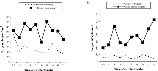
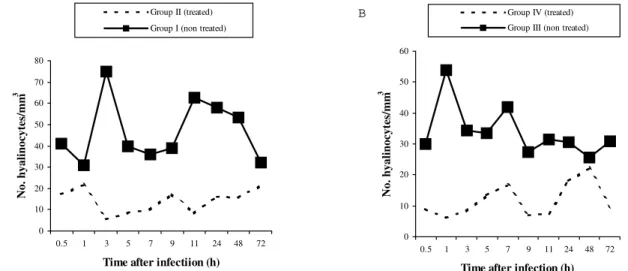
The number of hemocytes in treated mollusks was al-ways smaller than in untreated mollusks. In all groups and at all times, the number of granulocytes was greater then that of hyalinocytes (Figs 1, 2). The hemocytes in the hemolymph were differentiated into granulocytes and hyalinocytes. The granulocytes are star-shaped cells with cytoplasmic granules and are able to form pseudopodia which allow them to adhere to surfaces. In contrast, hyalinocytes are round cells which do not emit pseudopo-dia and are not adhesive.
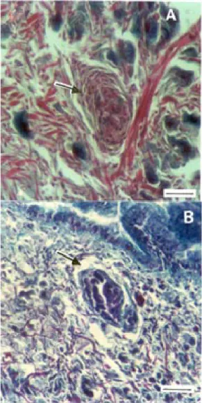
There was a predominance of degenerated primary sporocysts in untreated mollusks (group III), whereas in treated mollusks (group IV) the number of viable primary sporocysts was greater (Fig. 3). Whereas 86.2% of the primary sporocysts in the tissues of group III snails were degenerated (13.8% viable), in group IV snails, 74.2% of the primary sporocysts were viable (25.8% degenerated). The presence of a pyknotic nucleus and an eosinophilic cytoplasm characterized the degenerated sporocysts, most of which were surrounded by hemocytes that iso-lated the larva of the surrounding circumjacent tissue. The hemocytes in this case appeared extended, with char-acteristics similar to fibroblasts (Fig. 5A)
Fig. 4 shows the number of hemocyte reactions around primary sporocysts in mollusks of groups III and IV. In untreated B. glabrata, most of the primary sporocysts
A B
Fig. 1: average number of circulating granulocytes in A: uninfected Biomphalaria glabrata; B: B. glabrata infected with Schistosoma mansoni.
0 20 40 60 80 100 120 140 160 180
0.5 1 3 5 7 9 11 24 48 72
Time after infection (h)
No. granulocytes/mm
3
Group II (treated) Group I (non treated)
0
50
100
150
200
250
300
350
0.5 1 3 5 7 9 11 24 48 72
Time after infection (h)
No. granulocytes/mm
3
883 883883 883883 Mem Inst Oswaldo Cruz, Rio de Janeiro, Vol. 97(6), Septem ber 2002
(75.4%) were surrounded by an amebocyte reaction (Fig. 5A), whereas in treated mollusks most of the primary spo-rocysts (74.1%) were not involved in such a reaction. With few exceptions, the hemocytes in reactions around spo-rocysts were elongated, as in mammalian fibroblasts.
The first secondary sporocysts were seen in treated mollusks (group IV), and most were viable; degenerated secondary sporocysts predominated in untreated mol-lusks (group III). The tissue distribution of these sporo-cysts was similar in both groups but, quantitatively, there were more of these cells in treated mollusks.
Cercarial shedding was not examined in this experi-ment but, in a previous study (Serrano et al. 2002), we observed more cercarial shedding in mollusks treated with hydrocortisone. Thus in snails treated with hydrocorti-sone, a total of 69,519 cercariaes was obtained, whereas in untreated mollusks, only 2,514 larva were obtained. This data were significantly different (p = 0.0001).
A B
Fig. 2: average number of circulating hyalinocytes in A: uninfected Biomphalaria glabrata; B: B. glabrata infected with Schistosoma mansoni.
A B
Fig. 3: average number of primary sporocysts in Biomphalaria glabrata exposed to 100 Schistosoma mansoni miracidia. A: degenerated sporocysts; B: viable sporocysts.
Fig. 4: average number of primary sporocysts with a surrounding reaction in Biomphalaria glabrata exposed to 100 Schistosoma mansoni miracidia.
0 20 40 60 80 100 120
3.5 4 5 6 10 17 24 31
Time afte r infe ction (days)
Espor. with hemoc. reaction
G roup III (infected, non treated)
G roup IV (infected, treated)
0 10 20 30 40 50 60 70 80
0.5 1 3 5 7 9 11 24 48 72
Time after infectiion (h)
No. hyalinocytes/mm
3
Group II (treated) Group I (non treated)
0 10 20 30 40 50 60
0.5 1 3 5 7 9 11 24 48 72
Time after infection (h)
No. hyalinocytes/mm
3
Group IV (treated) Group III (non treated)
0 20 40 60 80 100 120
3.5 4 5 6 10 17 24 31
Time afte r infe ction (days)
Degenerated (sporocysts)
G roup III (infected, non treated)
G roup IV (infected, treated)
0 10 20 30 40 50 60 70 80 90 100
3.5 4 5 6 10 17 24 31
Time after infection (days)
Viable sporocysts
884 884 884 884
884 Hydrocortisone Action in Snails • D eborah Regina Serrano et al.
DISCUSSION
Our results confirmed the data of Cheng (1975), Cheng and Auld (1977), and Jeong and Heyneman (1976) re-garding the existence of two types of defense cells in molluscan hemolymph with granulocytes being more nu-merous than hyalinocytes. Hydrocortisone had an immu-nosuppressive effect on both cell types in S. mansoni
infected (group IV) and non-infected (group II) mollusks. Granulocytes were the most affected by hydrocortisone and were fewer in treated mollusks (Figs 1, 2).
Joky et al. (1985) and Seta et al. (1996) observed an increase in the number of hemocytes in the first 72 h after infection with S. mansoni. Since in our study hydrocorti-sone affected the number of hemocytes, it is probable that the steroid may act on the APO
(amebocyte-produc-ing organ) of the mollusks (Jeong et al. 1983) to reduce the number of defense cells produced and/or retard their maturation. The reduction in hemocytes after treatment with hydrocortisone could also result from a direct action of the drug on these cells.
There was a significant interaction between treatment and infection in altering the number of granulocytes since group IV mollusks (infected-treated) showed the greatest reduction in the number of defense cells. Hyalinocytes were less affected by this treatment. Time had no effect on the number of cells.
Ferri et al. (1977) reported that hydrocortisone inhib-ited macrophage mobilization in humans. Granulocytes, like macrophages, have pseudopodia and phagocytic ac-tivity. In addition to affecting the number of defense cells, hydrocortisone also reduced the number of sporocysts with a surrounding hemocyte reaction (Fig. 4). This ac-tion may be directly on the hemocytes to affect their mo-bilization and the production of pro-phagocytic sub-stances. This response could help to prevent hemocyte reactions around the trematode larvae, thereby allowing development of the parasite.
Histopathological analysis revealed similar numbers of viable and degenerated primary sporocysts. The no-table characteristic of viable sporocysts is the absence of an hemocyte reaction surrounding the larva. These spo-rocysts contain somatic and germ cells (Cheng & Bier 1972). The somatic cells are round and of variable size, with a modestly basophilic cytoplasm and slender baso-philic granulations that extend to the external membrane. The single nucleus is generally central and round and occupies about ¼ of the cell volume. The nucleolus is sometimes prominent. The nucleus of germ cells is ve-sicular and the cytoplasm is very rich in granulations. Both cells are surrounded by a polysaccharide membrana (Guaraldo et al. 1981).
The degenerated sporocysts (Fig. 5A) generally have a pyknotic nucleus; the cytoplasm is eosinophilic and the cytoplasmic granules tend to group together. The hemocytes concentrate in large numbers around the larva in degeneration isolating the adjacent tissue. The nucleus and cytoplasm of the hemocytes are similar to those of a fibroblast. The cytoplasm contain eosinophilic granula-tions that perhaps stores of secretion produts. During degeneration, the sporocysts loose their usual appear-ance to become eosinophilic and granulous, and form a mass surrounded by hemocytes. When degeneration is advanced, only hemocytes remain surrounding a region that gradually filled up with conjunctive tissue. However, some spaces were occupied by larvae (Guaraldo et al. 1981).
The treated group (group IV) showed a greater num-ber of viable sporocysts with no hemocyte reaction (Fig. 3); but when this reaction occurred, it was more discrete than in group III (Fig. 5B). This attenuated response prob-ably reflected the reduced number of defence cells and the inhibition of their ability to penetrate the traumatized site and to immobilize the larvae by encapsulation (Lie et al. 1987).
Hydrocortisone did not affect the distribution of sec-ondary sporocysts since in both groups of mollusks these Fig. 5: cephalopodal region of Biomphalaria glabrata infected
885 885885 885885 Mem Inst Oswaldo Cruz, Rio de Janeiro, Vol. 97(6), Septem ber 2002
cells occurred principally in the cephalopodal region, mantle border, ureter, hepatopancreas, ovotestis and in-testine, with no significant difference between the groups.
S. mansoni developed more rapidly in treated mollusks, with mature secondary sporocysts appearing 10 days af-ter exposure to miracidia. In the untreated group, mature secondary sporocysts were observed only 24 days after infection.
In conclusion, hydrocortisone exerts immunosuppres-sive and anti-inflammatory effects in B. glabrata. The re-duction in the activity and quantity of defense cells led to a more discrete inflammatory process, with a reduced or no hemocyte reaction around the sporocysts and in-creased the production of viable sporocysts with no hemocyte reaction.
These data are in agreement with observation of Serrano et al. (2002) who described an increased number of cercariae released by mollusks treated with hydrocorti-sone.
REFERENCES
Bayne CJ, Buckley PM, de Wan PC 1980. Macrophage-like hemocytes of resistant Biomphalaria glabrata are cyto-toxic for sporocysts of Schistosoma mansoni in vitro. J Parasitol 66:413-419.
Cheng TC 1975. Functional morphology and biochemistry of molluscan phagocytes. Ann N Y Acad Sci 266: 343-379. Cheng TC, Auld KR 1977. Hemocytes of the pulmonate
gas-tropod Biomphalaria glabrata. J Invertebr Pathol 30: 119-122.
Cheng TC, Bier JW 1972. Studies on molluscan schistosomia-sis: an analysis of the development of the cercariae of Schis-tosoma mansoni. Parasitology 64: 129-141.
Cheng TC, Butler MS 1979. Experimentally induced elevations in acid phosphatase acitivity in the hemolymph of Biomphalaria glabrata (Mollusca). J Invertebr Pathol 34: 119-129.
Cheng TC, Garrabant TA 1977. Acid phosphatase in granulo-cytic capsules formed in strains of Biomphalaria glabrata totally and partially resistant to Schistosoma mansoni. Int JParasitol 77:467-472.
Ferri RG, Calich VLG, Vaz CAC 1977. Imunologia, Edgard Blucher Ltda, São Paulo, 317 pp.
Grodsky GM 1977. Química e função dos hormônios. In AH Harper, Manual de Química Fisiológica, 4th ed., Atheneu, São Paulo, p. 447-508.
Guaraldo AMA, Magalhães LA, Rangel HA, Pareja G 1981.
Evolução dos esporocistos de Schistosoma mansoni
(Sambon, 1907) em Biomphalaria glabrata (Say, 1818) e Biomphalaria tenagophila (D’Orbigny, 1835). Rev Saúde Pública 15:436-448.
Jeong KH, Heyneman D 1976. Leukocytes of Biomphalaria
glabrata – Morphology and behavior of granulocytic cells in vitro. J Invertebr Pathol 28:357-362.
Jeong KH, Lei KJ, Heyneman D 1983. The ultrastructure of the amoebocyte-producing organ in Biomphalaria glabrata. Dev Comp Immunol 7:217-228.
Joky A, Matricon-Gondran M, Benex J 1985. Response to the amoebocyte-production organ of sensitized Biomphalaria glabrata after exposure to Echinostoma caproni miracidia. J Invertebr Pathol 45:28-33.
Kassim OO, Richards CS 1979. Host reactions in Biomphalaria glabrata to Schistosoma mansoni miracidia involving varia-tions in parasite strains, numbers and sequence of expo-sure. J Parasitol 9:565-570.
Lie KL, Heyneman D, Yan P 1975. The origin of amoebocytes in Biomphalaria glabrata. J Parasitol 63: 574-576. Lie KL, Jeong KH, Heyneman D 1980. Tissue reactions
in-duced by Schistosoma mansoni in Biomphalaria glabrata. Ann Trop Med Parasitol 74:157-166.
Lie KL, Jeong KH, Heyneman D 1987. Molluscan host reac-tion to helminthic infecreac-tion. In EJL Soulsby, Immune Re-sponses in Parasitic Infections: Immunology, Immunopa-thology and Immunoprophilaxis. Vol. IV: Protozoa, Arthropodes and Invertebrates, Chapter 7, CRC Press, Florida, p. 211-270.
Loker ES, Bayne CJ 1988. Immune mechanisms in
Trematode-snail interactions. In A Lackie, Immune Mechanisms in Invertebrate Vectors, Clarendon Press, Oxford, p. 199-220. Michelson EH 1966. Specificity of hemolymph antigens in taxo-nomic discrimination of medically important snail. J Parasitol 52:466-472.
Muller WEG 1985. Invertebrate immunity: basic concepts and recent advances. Int Rev Cytol 97: 183-351.
Ottaviani E, Franchini A 1988. Ultrastructural study of haemocytes of the freshwater snail Planorbarius corneus (L) (Gastropoda, Pulmonata). Acta Zool 69:157-162. Pan CT 1965. Studies on the host-parasite relationship
be-tween Schistosoma mansoni and the snail Australorbis glabratus. Am J Trop Med Hyg 14:931-976.
Pan CT 1996. Schistosoma mansoni: the ultrastructure of lar-val morphogenesis in Biomphalaria glabrata and of associ-ated host-parasite interactions. J Med SciBiol 49:129-149. Paraense WL, Correa LR 1963. Variation in susceptibility of populations of Australorbis glabratus to a strain of Schis-tosoma mansoni. Rev Inst Med Trop São Paulo 5:15-22. Ratcliffe NA 1985. Invertebrate immunity – A primer for the
now specialist (review). Immunol Lett 10: 253-270. Reis SMPM, Magalhães LA, Carvalho JF 1995. Ação da
inoculação de hemolinfa no mecanismo de defesa de Biomphalaria tenagophila (Orbigny, 1835). Rev Saúde Pública29:259-264.
Serrano DR, Zanotti-Magalhães EM, Magalhães LA, Carvalho JF 2002. Influência da hidrocortisona no desenvolvimento do Schistosoma mansoni em Biomphalaria glabrata. Rev Soc Bras Med Trop 35: 149-153.
Seta L, Magalhães LA, Carvalho JF 1996. Comportamento dos amebócitos circulantes de moluscos planorbídeos frente ao parasitismo por larvas de Schistosoma mansoni, inoculação de tinta nanquim e fratura da concha. Rev Saúde Pública 3: 332-340.
Shiff CJ 1994. Molluscan defence mechanisms: immunity or population biology? Parasitol Today 10: 188-190. Sima P, Vetvicka V 1990. Evolution of Immune Reaction, CRC
Press, Boca Raton, 247 pp.
Sminia T, Van der Knaap WPW 1987. Cells and molecules in molluscan immunology. Dev Comp Immunol 11: 17-28. Stites DP, Terr AI, Parslow TG 1997. Medical Immunology,
9th ed., Appleton & Lange, São Francisco, 900 pp. Sullivan JT, Richards CS 1981. Schistosoma mansoni
NIH-SM-PR-2 strain in susceptible stocks of Biomphalaria glabrata: comparative histology. J Parasitol 67: 702-708. Van der Knaap WPW, Loker ES 1990. Immune mechanism in
886 886 886 886