Treatment of osteochondral injuries with platelet gel
Marcus Vinicius Danieli,I,IIHamilton da Rosa Pereira,IICarlos Augusto de Sa´ Carneiro,I,II Se´rgio Luiz Felisbino,III Elenice DeffuneII
IUniort.e Ortopedia Especializada, Private Clinic and Hospital, Londrina/PR, Brazil.IIUniversidade Estadual Paulista ‘‘Ju´lio de Mesquita Filho’’ (UNESP), Faculty of Medicine of Botucatu, Botucatu/SP, Brazil.IIIUniversidade Estadual Paulista ‘‘Ju´lio de Mesquita Filho’’ (UNESP), Institute of Biosciences, Botucatu/SP, Brazil.
OBJECTIVES: Treatments for injured articular cartilage have not advanced to the point that efficient regeneration is possible. However, there has been an increase in the use of platelet-rich plasma for the treatment of several orthopedic disorders, including chondral injuries. Our hypothesis is that the treatment of chondral injuries with platelet gel results in higher-quality repair tissue after 180 days compared with chondral injuries not treated with gel.
METHODS: A controlled experimental laboratory study was performed on 30 male rabbits to evaluate
osteochondral injury repair after treatment with or without platelet gel. Osteochondral injuries were surgically induced in both knees of each rabbit at the medial femoral condyle. The left knee injury was filled with the platelet gel, and the right knee was not treated. Microscopic analysis of both knee samples was performed after 180 days using a histological grading scale.
RESULTS:The only histological evaluation criterion that was not significantly different between treatments was metachromasia. The group that was treated with platelet gel exhibited superior results in all other criteria (cell morphology, surface regularity, chondral thickness and repair tissue integration) and in the total score. CONCLUSION: The repair tissue was histologically superior after 180 days in the study group treated with platelet gel compared with the group of untreated injuries.
KEYWORDS: Platelet-Rich Plasma; Articular Cartilage; Injuries; Regenerative Medicine.
Danieli MV, Rosa Pereira H, Sa´ Carneiro CA, Felisbino SL, Deffune E. Treatment of osteochondral injuries with platelet gel. Clinics. 2014;69(10):694-698.
Received for publication onApril 6, 2014;First review completed onMay 15, 2014;Accepted for publication onJune 9, 2014 E-mail: [email protected]
Tel.: 55 43 9146-4799
& INTRODUCTION
Treatments for injured articular cartilage have not advanced to the point that efficient regeneration is possible because chondral tissue consists of few cells and a rich, complex extracellular matrix. Chondral tissue also has no nerves, lymphatic drainage or blood vessels, which are essential to the processes of clot formation and repair (1-4). Moreover, scar tissue from cartilage injury does not support loads in the same manner as normal tissue and a long-term evolution to osteoarthritis often results (4). Osteoarthritis is an incurable, disabling disease. The 50 million cases of the disease in the USA (5) are associated with estimated treatment costs of approximately 65 billion dollars per year (6). Surgical options may be considered in the treatment of chondral injuries to prevent or slow the onset of osteoarthritis.
Most of the surgical techniques are based on the stimulation of mesenchymal cell migration, which leads to the formation of scar tissue (5,7). More recently, the use of autologous chondrocyte transplantation has increased, but the cost of this technique remains high and the reported results are variable and not guaranteed to prevent the onset of symptomatic degenerative joint disease (2,7,8,9-11).
The use of platelet-rich plasma (PRP) for the treatment of several types of orthopedic disorders, including chondral injuries, has increased recently (12).
PRP is a plasma fraction that contains a high concentra-tion of platelets and is rich in many growth factors. These growth factors are part of the natural process of tissue healing and homeostasis and they have the capacity to stimulate cell proliferation, mesenchymal stem cell chemo-taxis and cell differentiation. PRP also has the capacity to stimulate debris scavenging, angiogenesis, extracellular matrix synthesis (13), bone synthesis and the production of intra-articular hyaluronic acid (12,14,15) and to reduce pain (14,15). A literature review by Kon et al. (16) also cites the potential of PRPs to decrease hemorrhage, cartilage fibrillation and hypertrophy of synovial tissue. PRP is obtained from the patient using a minimally invasive technique that is relatively easy and inexpensive (5,12).
Copyrightß2014CLINICS– This is an Open Access article distributed under the terms of the Creative Commons Attribution Non-Commercial License (http:// creativecommons.org/licenses/by-nc/3.0/) which permits unrestricted non-commercial use, distribution, and reproduction in any medium, provided the original work is properly cited.
No potential conflict of interest was reported.
However, the literature has few well-designed studies on the use of PRP in chondral injuries, with only controversial pre-clinical and clinical results (16).
Therefore, this study treated osteochondral injuries of the articular cartilage of the knee with PRP gel, using rabbits as experimental models. The hypothesis was that treated injuries would have higher-quality repair tissue than non-treated injuries.
& MATERIALS AND METHODS
The Ethics Committee for Animal Research of the associated institution approved this study under protocol 717.
Thirty New Zealand rabbits, minimum age of 3 months (average: 4.6 m) and weighing over 2 kg (average: 3.1 kg) were obtained from the Orthopedics and Experimental Surgery Laboratory. The animals were confined to indivi-dual metal cages before and after the surgical procedure and fed standardized rations and waterad libitum.
The sample size was based on previous studies per-formed using the same animal model and methodology (5,17-19).
First, the blood of several rabbits was collected to prepare the PRP with the addition of 3.8% sodium citrate to avoid coagulation. This procedure was performed in the blood bank of our hospital. The blood was centrifuged at 1000 RPM for 10 minutes, which resulted in a separation into three layers: a lower layer of concentrated red blood cells, a middle layer of white blood cells and an upper layer of plasma. The upper layer was separated and centrifuged for another 10 minutes at 3000 RPM, which yielded two fractions: platelet-poor plasma (the upper fraction) and PRP (the lower fraction). This method was validated at the blood bank to produce a PRP of at least 1 million platelets per microliter of plasma (5). The PRP was activated 1 day before surgery via the addition of calcium and human thrombin.
Our PRP was P3-x-Bb according to the classification proposed by Delong et al. (20).
Surgical Technique
Animals were anesthetized intramuscularly with tileta-mine and zolazepam in combination with 2% xylazine hydrochloride at doses of 15.0-30.0 mg/kg and 4 mg/kg of body weight, respectively. Local anesthesia was applied
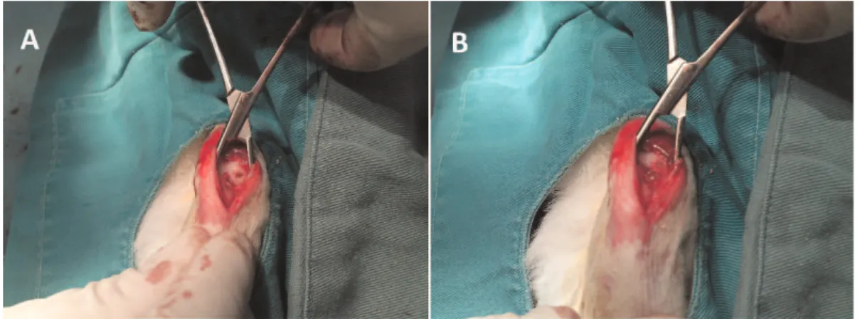
(Xylocaine 2%) at the point of incision. A medial para-patellar incision was made and the patella was dislocated to the side. The medial femoral condyles were exposed and perforated with a trephine with the knees bent to cause lesions 3.5 mm in diameter and 4.0 mm deep on both knees. The lesions were washed with sterile saline solution (0.9% NaCl) and the injury to the left knee was filled with PRP gel (Figures 1 and 2). The right knee was left untreated. The injuries were sutured in layers using 4-0 nylon monofilament.
Animals received 0.1 ml/kg of Enrofloxacin (antibiotic) subcutaneously for 5 days after surgery and 0.2 mg/kg of Meloxicam 0.2% (anti-inflammatory) subcutaneously for 2 days after surgery.
The animals were euthanized 180 days post-surgery with an overdose of sodium pentobarbital. The medial femoral condyles were removed, preserved in a 10% formalin solution and sent to the university’s Department of Morphology for histological analyses. Samples were dec-alcified, cut and stained with toluidine blue. A blinded morphologist evaluated and classified each sample accord-ing to the histological gradaccord-ing scale described by Wakitani et al. (21) (Table 1).
A Leica Qwin ColourH(RGB) image analyzer coupled to a LeicaH DM 2500 microscope was used for microscopic analyses.
Mann-Whitney and Wilcoxon’s non-parametric tests were used for statistical analyses in SPSS Inc. Version 15.0 (Chicago, IL, USA), with significance set atp,0.05.
& RESULTS
Between May 2009 and January 2010, 30 rabbits under-went surgery. There were 15 deaths: 13 from respiratory infection and 2 during the surgical procedure. These 15 animals were excluded from statistical analyses. A micro-scopic evaluation could not be performed on the right knee sample of rabbit number 10 due to decalcification problems. No surgical site infection was encountered in any case. Synovitis and arthrofibrosis were not evaluated.
Histological analyses, with separate consideration given to each criterion score, revealed significant differences in cell morphology (p= 0.002), surface regularity (p,0.001), chondral thickness (p,0.02) and lateral integration (p,0.02), with the best results observed in the treated group. There was no difference in metachromasia (p= 0.063). The
difference in final scores was statistically significant (p,0.001), with superior results obtained in the treated group (Table 2).
Examples of microscopic findings are shown in Figures 3 and 4.
A large amount of scar tissue was present bilaterally for the injuries of rabbit 21, but the scar was much better organized on the treated side (Figure 4).
& DISCUSSION
Rabbits were chosen for this study due to their wide-spread use as a model for chondral injury repair. The use of very young rabbits was avoided due to their greater repair potential (1,22,23). Euthanasia was delayed until the 180th day after injury to evaluate injury repair over a longer time period. Buckwalter (2) reported that chondral and osteo-chondral repair is fully completed within 6 weeks, but
remodeling continues for months or even years. The minimum time until analysis of repair should range from 4 to 6 weeks, but even the most promising methods require evaluation after 6 months. Bittencourt (5) observed good evolution of chondral repair with platelet gel at 90 days. Therefore, we selected the same treatment method, but we employed a larger injury and a single 180-day evaluation to assess the durability of the tissue.
A 3.5-mm-diameter injury was chosen because of its repair difficulty and to demonstrate the heightened poten-tial of PRP gel. Lesions larger than 3.0 mm in diameter rarely heal in rabbits without some form of treatment (17). A depth of 4 mm was chosen because this depth reaches the subchondral bone without causing bone marrow destruction (3,24).
Several other studies using similar models of injury in rabbits obtained superior results in treated specimens, but the treatment methods were different and different materi-als were used (3,10,19,25). Nonetheless, hyaline cartilage in its classic form did not appear in the repair tissues, which is consistent with our results.
Wei & Messner (22) demonstrated that the biomechanical behavior of repair tissue, even with 80% hyaline-like cartilage, is inferior to normal cartilage and it tends to deteriorate more slowly over time. Ozsoy et al. (25) made an interesting comment: even untreated control injuries present macroscopically adequate repair, but with a catabolic activity that is higher than the treated group. Therefore, treatment of chondral injuries could potentially increase the duration and function of the repair tissue, despite the fibrocartilaginous of the tissue. PRP is capable of reducing chondral catabolism (26) and inhibiting the apoptosis of chondrocytes subjected to trauma (27), which also plays an important role in the longevity of repair tissue. This result was also confirmed by Milano et al. (28), who showed that PRP gel associated with microfractures in chondral injuries in sheep yielded the best macroscopic, histological and biomechanical results.
The poor metachromasia results may demonstrate a more deficient matrix with a lower concentration of glycosami-noglycans, although this deficiency could improve over time with matrix production by chondrocytes. Drengk et al. (29) observed that cell proliferation of sheep chondrocytes and mesenchymal stem cells cultivated in vitro with the addition of PRP was up to 67% greater compared with cells cultivated without PRP, although type-II collagen mRNA expression was reduced. The suggested explanation was that increased proliferation negatively influenced cell
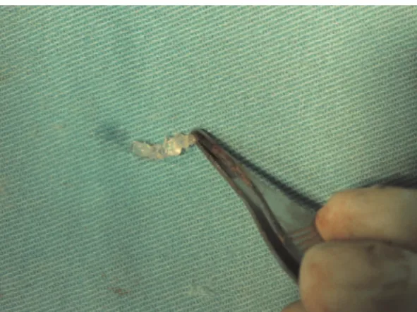
Figure 2 -Platelet gel before application to the chondral injury (source: personal file).
Table 1 -Histological Grading Scale (Wakitani et al. (27)).
Category Score
Cell Morphology
Hyaline cartilage 0
Mostly hyaline cartilage 1
Mostly fibrocartilage 2
Mostly non-cartilage 3
Non-cartilage only 4
Metachromasy (Matrix Staining)
Normal 0
Slightly reduced 1
Markedly reduced 2
Without metachromasy 3
Surface regularity
Smooth 0
Moderate 1
Irregular 2
Severely irregular 3
Cartilage thickness*
.2/3 0
1/3-2/3 1
,1/3 2
Integration with adjacent cartilage
Both sides integrated 0
One side integrated 1
No side integrated 2
*Average thickness of repair tissue compared with normal surrounding
cartilage.
Table 2 -Means and standard deviations of histological scores.
Right Left
Mean SD Mean SD p-value
CM 2.43 0.94 1.27 0.7 p= 0.002
M 1.71 0.82 1.07 0.6 p= 0.063
SR 1.86 0.66 0.6 0.63 p,0.001
CT 1 0.88 0.2 0.41 p,0.02
LI 0.64 0.63 0.07 0.26 p,0.02
Total 7.64 2.87 3.2 1.78 p,0.001
CM: Cell Morphology, M: Metachromasy, SR: Surface Regularity, CT: Cartilage Thickness,
differentiation, but this phenotype might be altered in anin vivoenvironment after cell proliferation reached its limit.
Several articles commonly used scaffolds, such as collagen matrix (17), polylactic-glycolic acid matrix (18) and polylactic acid matrix (19). However, scaffolding might not be necessary when using PRP in a gel form because we did not use any scaffolding and our results were similar to the literature.
This study has several limitations that should be considered. The multiple deaths may have modified results, but we chose
not to recruit more animals because our results demonstrated statistically significant differences. Nevertheless, the standard deviation may be considered high, even with the significant intergroup differences. Perhaps a larger sample of cases might address or compensate for this finding. Histological scoring may be subject to bias because it is examiner-dependent. We attempted to reduce this bias by having an experienced, blinded morphologist perform the microscopic analyses. The absence of clinical assessment parameters was also a limitation in our study. Synovitis or arthrofibrosis could have occurred
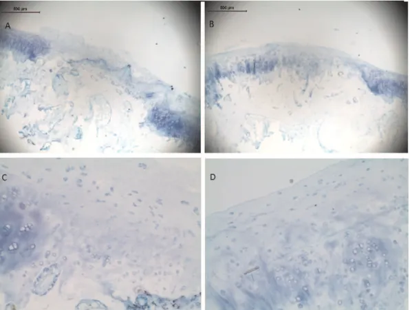
Figure 3 -Histological view of rabbit 8 samples, stained with toluidine blue. A) Right knee (46zoom): note the fibrocartilaginous tissue with an irregular surface; B) Left knee (46zoom): more cartilaginous tissue, better organized and smoother surface; C) Right knee (206 zoom): the presence of more elongated cells (fibroblasts); D) Left knee (206zoom): the presence of organized chondrocytes and a smooth surface (source: personal file).
more in one group and changed our results. We could have also performed immunohistochemistry for type II collagen data, but this technique was not in the original study project. We did not pursue this option because the results were already significantly different.
From these data, we concluded that chondral injuries in rabbit knees treated with platelet gel presented histologi-cally superior repair results following a 180-day period compared with identical untreated injuries.
& AUTHOR CONTRIBUTIONS
Danieli MV wrote the manuscript and performed the surgeries. Deffune E prepared PRP and revised the manuscript. Pereira HR revised the manuscript. Sa´ Carneiro CA assisted in surgeries and took care of rabbits. Felisbino SL was responsible for microscopic analyses.
& REFERENCES
1. Bhosale AM, Richardson JB. Articular cartilage: structure, injuries and review of management. Br Med Bull. 2008;87(1):77-95, http://dx.doi. org/10.1093/bmb/ldn025.
2. Buckwalter JA. Articular cartilage injuries. Clin Orthop Relat Res. 2002;402:21-37, http://dx.doi.org/10.1097/00003086-200209000-00004. 3. Ribeiro JL, Camanho GL, Takita LC. Estudo macrosco´pico e histolo´gico
de reparos osteocondrais biologicamente aceita´veis. Acta Ortop Bras. 2004;12(1):16-21, http://dx.doi.org/10.1590/S1413-78522004000100003. 4. Souza TD, Del Carlo RJ, Viloria MIV. Avaliac¸a˜o histolo´gica do processo
de reparac¸a˜o da superfı´cie articular de coelhos. Cienc Rural. 2000;30(3):439-44, http://dx.doi.org/10.1590/S0103-84782000000300011. 5. Bittencourt RAC. Cultura de condro´citos para uso terapeˆutico: recon-stituic¸a˜o de cartilagem [Tese]. Botucatu: Faculdade de Medicina, Universidade Estadual Paulista; 2008. 129f. Available online: http:// www.athena.biblioteca.unesp.br/exlibris/bd/bbo/33004064006P8/2008/ bittencourt_rac_dr_botfm.pdf.
6. D’Lima DC, Colwell Jr CW. Clinical objectives for cartilage repair. Clin Orthop Relat R. 2001;391:S402-5, http://dx.doi.org/10.1097/00003086-200110001-00037.
7. Alford JW, Cole BJ. Cartilage restoration, part 2. Techniques, outcomes, and future directions. Am J Sports Med. 2005;33(3):443-60.
8. Sgaglione NA. Biologic approaches to articular cartilage surgery: future trends. Orthop Clin North Am. 2005;36(4):485-95, http://dx.doi.org/10. 1016/j.ocl.2005.05.006.
9. Haleem AM, El Singergy AA, Sabry D, Atta HM, Rashed LA, Chu CR, et al. The Clinical Use of human culture-expanded autologous bone marrow mesenchymal stem cells transplanted on platelet-rich fibrin glue in the treatment of articular cartilage defects: a pilot study and preliminary results. Cartilage. 2010;1(4):253-61, http://dx.doi.org/10. 1177/1947603510366027.
10. Costa AJF, Oliveira CRGCM, Leopizzi N, Amatuzzi MM. O uso da matriz o´ssea desmineralizada na reparac¸a˜o de leso˜es osteocondrais. Estudo experimental em coelhos. Acta Ortop Bras. 2001;9(4):27-38. 11. Wasiak J, Clar C, Villanueva E. Autologous cartilage implantation for full
thickness articular cartilage defects of the knee. Cochrane Database of Systematic Reviews, In The Cochrane Library, Issue 3, 2009. Art N˚CD003323.DOI:10.1002/14651858.CD003323.pub4.
12. Foster TE, Puskas BL, Mandelbaum BR, Gerhardt MB, Rodeo AS. Platelet-rich plasma - from basic science to clinical applications. Am J Sports Med. 2009;37(11):2259-72, http://dx.doi.org/10.1177/ 0363546509349921.
13. Mehta S, Watson JT. Platelet rich concentrate: Basic science and current clinical applications. J Orthop Trauma. 2008;22(6):433-8.
14. Lopez-Vidriero E, Goulding KA, Simon DA, Sanchez M, Johnson DH. The use of platelet-rich plasma in arthroscopy and sports medicine: optimizing the healing environment. Arthroscopy. 2010;26(2):269-78, http://dx.doi.org/10.1016/j.arthro.2009.11.015.
15. Sanchez M, Anitua E, Orive G, Mujika I, Andia I. Platelet-rich therapies in the treatment of orthopaedic sport injuries. Sports Med. 2009;39(5):345-54, http://dx.doi.org/10.2165/00007256-200939050-00002. 16. Kon E, Filardo G, Di Martino A, Marcacci M. Platelet-rich plasma (PRP) to treat sports injuries: evidence to support its use. Knee Surg Sports Traumatol Arthrosc. 2011;19(4):516-27, http://dx.doi.org/10. 1007/s00167-010-1306-y.
17. Qi YY, Chen X, Jiang YZ, Cai HX, Wang LL, Song XH, et al. Local delivery of autologous platelet in collagen matrix simulated in situ articular repair. Cell Transplant. 2009;18(10):1161-9, http://dx.doi.org/ 10.3727/096368909X12483162197169.
18. Sun Y, Feng Y, Zhang CQ, Chen SB, Cheng XG. The regenerative effect of platelet-rich plasma on healing in large osteochondral defects. Int Orthop. 2010;34(4):589-97, http://dx.doi.org/10.1007/s00264-009-0793-2. 19. Yan H, Yu C. Repair of full-thickness cartilage defects with cells of different origin in a rabbit model. Arthroscopy. 2007;23(2):178-87, http://dx.doi.org/10.1016/j.arthro.2006.09.005.
20. Delong JM, Russel RP, Mazzoca AD. Platelet-rich plasma: the PAW classification system. Arthroscopy. 2012;28(7):998-1009, http://dx.doi. org/10.1016/j.arthro.2012.04.148.
21. Wakitani S, Goto T, Pineda SJ, Young RG, Mansour JM, Caplan AI, et al. Mesenchymal cell-based repair of large, full-thickness defects of articular cartilage. J Bone joint Surg Am. 1994;76(4):579-92.
22. Wei X, Messner K. Maturation-dependent durability of spontaneous cartilage repair in rabbit knee joint. J Biomed Mater Res. 1999;46(4):539-48, http://dx.doi.org/10.1002/(SICI)1097-4636(19990915)46:4, 539::AID-JBM12.3.0.CO;2-S.
23. Fuller JA, Ghadially FN. Ultrastructural observations on surgically produced partial-thickness defects in articular cartilage. Clin Orthop Relat R. 1972; 86:193-205, http://dx.doi.org/10.1097/00003086-197207000-00031.
24. Martin-Hernandes C, Cebamanos-Celma J, Molina-Ros A, Ballester-Jimenez J, Ballester-Soleda J. Regenerated cartilage produced by autogenous periosteal grafts: a histologic and mechanical study in rabbits under the influence of continuous passive motion. Arthroscopy. 2010;26(1):76-83, http://dx.doi.org/10.1016/j.arthro.2009.07.005. 25. Ozsoy MH, Aydogdu S, Taskiran D, Sezak M, Hayran M, Oztop F, et al.
The effects of early or late treatment of osteochondral defects on joint homoeostasis: an experimental study in rabbits. Knee Surg Sports Traumatol Arthrosc. 2009;17(6):578-89, http://dx.doi.org/10.1007/s00167-008-0675-y.
26. Kon E, Buda R, Filardo G, Di Martino A, Timoncini A, Cenacchi A, et al. Platelet-rich plasma: intra-articular knee injections produced favorable results on degenerative cartilage lesions. Knee Surg Sports Traumatol Arthrosc. 2010;18(4):472-9, http://dx.doi.org/10.1007/ s00167-009-0940-8.
27. Rezende MU, Silva RBB, Bassit ACF, Tatsui NH, Sadigursky D, Neto RB. Efeito do plasma rico em plaquetas na apoptose po´s-trauma´tica de condro´citos. Acta Ortop Bras. 2011;19(2):102-5.
28. Milano G, Passino ES, Deriu L, Careddu G, Manunta L, Manunta A, et al. The effect of platelet rich plasma combined with microfractures on the treatment of chondral defects: an experimental study in a sheep model. Osteoarthritis Cartilage. 2010;18(7):971-80, http://dx.doi.org/10.1016/j. joca.2010.03.013.