517 517 517 517 517 Mem Inst Oswaldo Cruz, Rio de Janeiro, Vol. 92(4): 517-522, Jul./Aug. 1997
Comparative Histopathology of
Biomphalaria glabrata, B.
tenagophila
and
B. straminea
with Variable Degrees of
Resistance to
Schistosoma mansoni
Miracidia
Cecilia Pereira de Souza, Claudia Cunha Borges/
+, Andréa Guimarães
Santana*, Zilton A Andrade*/
++Laboratório de Malacologia, Centro de Pesquisas René Rachou-FIOCRUZ, Av. Augusto de Lima 1715, 30190-002 Belo Horizonte, MG, Brasil *Laboratório de Patologia Experimental, Centro de Pesquisas Gonçalo
Moniz-FIOCRUZ, Rua Valdemar Falcão 121, 40295-001 Salvador, BA, Brasil
A comparative histopathological study of three snails species - Biomphalaria glabrata, B. tenagophila and B. straminea - which had been infected with Schistosoma mansoni miracidia revealed similar qualitative features, consisting of areas of sporocyst proliferation and differentiation associated with reactive host reaction, at the time they were actively eliminating great number of cercariae. However, in specimens that were exposed to miracidia but failed to eliminate cercariae later on, different histo-pathological pictures were observed in different snail species.
While B. glabrata exhibited frequent focal (granulomatous) proliferation of amebocytes in several organs, B. tenagophila and B. straminea only rarely showed such reactive changes, suggesting that the mechanism of resistance to miracidial infection probably follows different pathways in the snail species studied.
Key words: Biomphalaria histopathology - Schistosoma mansoni - differential susceptibility
Biomphalaria glabrata is highly susceptible to infection with Schistosoma mansoni miracidia and represents the main intermediate host for that trematode in the large endemic area of northeast-ern and southeastnortheast-ern Brazil (Paraense 1970). How-ever, susceptibility to infection is variable among different specimens, a high degree of resistance be-ing sometimes exhibited by some of them (Pan 1965). Recently, Godoy et al. (1997) reported that
B. glabrata, which completely failed to emit cer-cariae after a relatively prolonged time following exposition to S. mansoni miracidia, exhibited fo-cal inflammatory and granulomatous lesions in several organs in the absence of demonstrable para-sites, histopathologically.
Such lesions were interpreted as the aftermath of a successful clearance reaction against invad-ing sporocysts, suggestive of high degree of resis-tance.
There are two other important S. mansoni in-termediate host snails in Brazil: B. tenagophila and
B. straminea. Several studies have attempted to comparatively evaluate the behavior of those sus-ceptible snails regarding their interaction with S.
+ CNPq Research Fellow
++ Corresponding author. Fax: +55-71-356.4292
Received 19 February 1997 Accepted 12 May 1997
mansoni (Barbosa 1975, Paraense & Correa 1978, Souza et al. 1995a, b) but whether the peculiar tis-sue reactivity described by Godoy et al. (1997) in resistant B. glabrata does ever occur in these other two species, and to which extent and frequency, has hitherto not been reported.
Present investigation is concerned with a com-parative study of the tissue reactions in three S. mansoni intermediate hosts: B. glabrata, B. tenagophila and B. straminea. The purpose is to comparatively describe the histopathologic find-ings and attempt to correlate them with previous malacological data indicative of different degrees of susceptibility among the three snail species.
MATERIALS AND METHODS
Laboratory bred B. glabrata, B. tenagophila
exami-518 518 518 518
518 Comparative Histopathology of Biomphalaria Species • CP de Souza et al.
nation. Since B. glabrata presented high mortality rate, the cases for histological study was comple-mented with previously obtained material. As con-trols, non-exposed snails belonging to the same three species were also used. The snails were re-moved from the shell under menthol anesthesia, fixed in totum in Bouin’s fluid for 6 hr and then transferred to 70% alcohol. Further procedures in-cluded dehydration in 100% alcohol, clearing in xylol and paraffin embedding. Five µm paraffin-embedded sections were stained with hematoxy-lin and eosin, and eventually with the sirius-red method for collagen and the Periodic-acid Schiff (PAS) with and without previous diastase treat-ment. Slides stained with sirius-red were also mi-croscopically examined under polarized light.
RESULTS
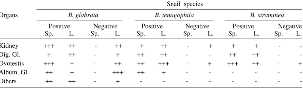
Table I shows the general comparative mala-cological results obtained with the infection of the three snail species. B. glabrata was the most sus-ceptible, followed by B. tenagophila and B. straminea in that order. Table II summarizes the main histological results. Quantitative histological findings were different for each snail species. De-scription will consider the qualitative changes which were common to all snails belonging to each group, according to cercarial elimination.
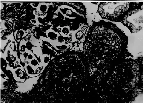
Snails shedding cercariae - Developing sporo-cysts were found in large numbers in all snails which were eliminating cercariae, but not in those that did not. The secondary sporocysts found ap-peared larger and more numerous in B. glabrata, and slightly more frequent in B. tenagophila than in B. straminea, but the general microscopical ap-pearance was similar regardless snail species. Spo-rocysts were usually formed as a space-occupying lesion, without any visible changes in the neigh-boring snail tissues. However, sporocysts were also found in several locations, side by side with vari-able degree of tissue reactions (Fig. 1). These lat-ter consisted of either focal and diffuse prolifera-tion of amebocytes, frequently forming encapsu-lating or granulomatous structures centered by spo-rocyst and cercaria remnants, and limited periph-erally by concentric laminations of thin fibers.
Sometimes an encapsulating, ring-like, cellular reaction was formed around a collection of viable cercariae, which exhibited different degrees of maturation (Fig. 2). Cells involved in these reac-tions were mainly macrophage-like amebocytes. They varied in shape from round to fusiform, with little variation in size. The stroma of the inflamma-tory foci was represented by an increased amount of the amorphous and fibrillar components of the extracellular matrix, sometimes mimicking the
pro-TABLE I
Comparative results on the infection of three Biomphalaria species by 50 miracidia of Schistosoma mansoni
Species Exposed No. of positivesa Infection Mortality
snails ratea ratea
B. glabrata 50 26 96.3% 46%
B. tenagophila 50 17 68 % 50%
B. straminea 50 6 18.7% 36%
a: 37 days after exposition
TABLE II
Comparative histopathological study of Biomphalaria glabrata, B. tenagophila and B. straminea exposed to
Schistosoma mansoni miracidia, emitting (Positives) or not cercariae (Negatives) after three months from exposure, regarding the presence of sporocysts (Sp.) and inflammatory lesions (L.)
Snail species
Organs B. glabrata B. tenagophila B. straminea
Positive Negative Positive Negative Positive Negative
Sp. L. Sp. L. Sp. L. Sp. L. Sp. L. Sp. L.
Kidney +++ ++ - ++ + ++ - + + + -
-Dig. Gl. + ++ - + ++ ++ - - ++ ++ -
-Ovotestis +++ + - ++ ++ +++ - + +++ ++ - +
Album. Gl. ++ + - +++ ++ + - - -
-Others ++ ++ - + - - -
519 519519 519519 Mem Inst Oswaldo Cruz, Rio de Janeiro, Vol. 92(4), Jul./Aug. 1997
cess of fibrosis seen in vertebrates. However, the newly formed “fibrous” tissue, both within and at the periphery of the inflammatory lesions, did not stain with sirius-red as does vertebrate collagen. In a few instances it did stain, but without the
charac-teristic polarization-light refringence. It should be mentioned here that some normal fibers of the ex-tracellular matrix of the snail (especially in the foot) do stain like vertebrate collagen and exhibit bire-fringence under polarized light.
Fig. 1: aspect of Biomphalaria glabrata digestive glands showing both parasites and tissue reaction. Parasites are seen above left, with sporocysts and cercariae at several stages of development. At lower right there is dense amebocyte proliferation forming three nodular (granulomatous) reactions. Structures of the digestive glands can be seen at upper right. Hematoxylin & eosin, 200X.
520 520 520 520
520 Comparative Histopathology of Biomphalaria Species • CP de Souza et al.
Exposed snails that failed to eliminate cercariae
- No viable sporocysts were found in histological sections taken from any of the three snail species. However, focal inflammatory tissue reactions were found in several organs in B. glabrata and only
rarely in the two other species. In the ovotestis and renal tubular region in the case of B. tenagophila
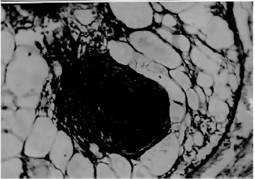
(Fig. 3) and only once, in the region of an atrophic ovo-testis of B. straminea (Fig. 4). These reactions were entirely similar to those seen in snails
shed-Fig. 3: region of the ovotestis of Biomphalaria straminea which had been infected, but did not eliminate cercariae. It shows infiltration and proliferation of amebocytes and the formation of three nodular lesions, some of them containing dark irregular non-identified material. The possibility of this latter being sporocyst remnants has not been excluded. Hematoxylin & eosiin, 200X.
521 521521 521521 Mem Inst Oswaldo Cruz, Rio de Janeiro, Vol. 92(4), Jul./Aug. 1997
ding many cercariae, and which were found encir-cling or in close vicinity of proliferating sporocysts. However, no evident sporocystic structures were identified in any of the foci or in any other part of the snail tissues.
Normal non-infected snails- They showed es-sentially normal histology. No focal or diffuse in-flammatory reaction was ever observed.
DISCUSSION
A tentative histological grading of susceptibil-ity for the three snail species was attempted by considering sporocyst distribution in different or-gans, their size and number, as well as the tissue reaction elicited by them. In the present investiga-tion this would place B. glabrata, B.tenagophila
and B. straminea, in that order, as the most sus-ceptible S. mansoni intermediate hosts. These re-sults are in keeping with previous ones obtained by a combination of malacological techniques (Souza et al. 1995b).
Susceptible snails give rise to variable num-bers of cercariae. Those which are very suscep-tible can shed numerous cercariae, with no overt reactions, their tissues appearing tolerant to the presence of the multiplying and growing sporo-cysts (Newton 1952). Others may eliminate only a few cercariae and in them variable degrees of tis-sue reactions take place (Pan 1965). These reac-tions usually consist of massive proliferation of amebocytes, with encapsulation and destruction of sporocysts, the cells probably acting in concert with soluble factors in the molluscan hemolymph (Bayne et al. 1980). Recently, some specimens taken from a resistant F15 generation of B. glabrata
completely failed to emit cercariae several months after being exposed to numerous miracidia. When they were histologically examined, no sporocysts were found, but focal and diffuse inflammatory reactions, similar to those seen in infected and re-sistant snails, appeared at several locations. Such peculiar reactions were not found in normal, non-exposed control snails. They were thought to rep-resent cicatrices left by destroyed sporocysts (Godoy et al. 1997). This kind of delayed-devel-oped resistance probably represents an alternative and novel kind of host defense mechanism against
S. mansoni miracidia, in spite of evidences sug-gesting that S.mansoni sporocysts can sometimes better develop their ability to interfere with the defense mechanism of the snail as they grow older (Lie et al. 1980). Thus, the amebocytic reaction that is already known to be partially inhibitory of sporocyst development, appears sometimes to be totally effective.
Therefore, protection against miracidial inva-sion could be provided by at least two mechanisms.
One being represented by the already known ex-ample of direct miracidium destruction which occurs soon after penetration. Locker et al. (1986) found miracidium-amebocyte contact within 3 hr and phagocytosis of sporocysts microvilli and pieces of tegument within 7.5 hr, while extensive pathology was demonstrated within 24 hr and by 48 hr only scattered remnants of sporocysts re-mained. The other mechanism would be repre-sented by the one here postulated, that is, a suc-cessful mounting of a destructive reaction when several sporocysts have already disseminated throughout the snail tissues.
This second mechanism appeared frequently and widespread in B. glabrata, but it was only rarely seen in the other two species. Strongly re-sistant B. tenagophila and B. straminea rather uti-lize the second mechanism, the killing of invading miracidia probably occurring soon after their pen-etration into the snail tegument, which would leave no residual changes.
REFERENCES
Barbosa FS 1975. Survival and cercaria production of Brazilian Biomphalaria glabrata and B. straminea
infected with Schistosoma mansoni. J Parasitol 61: 151-152.
Bayne CJ, Buckley PM, Dewan PC 1980. Macrophagelike hemocytes of resistant
Biomphalaria glabrata are cytotoxic for sporocysts of Schistosoma mansoni in vitro. J Parasitol 66:
413-419.
Godoy A, Souza CP, Guimarães CT, Andrade ZA 1997. Unusual histological findings in Biomphalaria glabrata with high degree of resistance to Schisto-soma mansoni miracidia. Mem Inst Oswaldo Cruz 92: 121-122.
Lie KJ, Jeong KH, Heyneman D 1980. Tissue reaction induced by Schistosoma mansoni in Biomphalaria glabrata. Ann Trop Med Parasitol 78: 157-166. Loker ES, Bayne CJ, Yui MA 1986. Echinostoma
paraensei: hemocytes of Biomphalaria glabrata as targets of Echinostoma mediate interference with host snails resistance to Schistosoma mansoni. Exper Parasitol 62: 149-154.
Newton WL 1952. The comparative tissue reaction of two strains of Australorbis glabratus to infection with Schistosoma mansoni. J Parasitol 38: 362-366.
Pan C-T 1965. Studies on the host-parasite relationship between Schistosoma mansoni and the snail
Australorbis glabratus. Am J Trop Med Hyg 14: 931-975.
Paraense WL 1970. Planorbídeos hospedeiros intermediários do Schistosomamansoni, p. 13-20. In AS Cunha, Esquistossomose mansoni. Sarvier & USP, São Paulo.
522 522 522 522
522 Comparative Histopathology of Biomphalaria Species • CP de Souza et al.
Paraense WL, Correa LR 1981. Observations of two biological races of Schistosoma mansoni. Mem Inst Oswaldo Cruz 76: 287-291.
Souza CP, Janotti-Passos LK, Freitas JR 1995a. Degree of host-parasite compatibility between Schistosoma mansoni and their intermediate molluscan hosts in
Brazil. Mem Inst Oswaldo Cruz 90: 5-10.
Souza CP, Cunha RCP, Andrade ZA 1995b. Develop-ment of Schistosoma mansoni in Biomphalaria tenagophila, Biomphalaria straminea and