Studies on
resistance and
response to
vancomycin in
Enterococcus
faecalis:
a last resort
antibiotic
Tânia Catarino Ribeiro
Oeiras, January 2011
S
tu
d
ies
o
n
r
e
s
is
ta
nc
e
a
n
d
r
e
s
p
o
n
s
e
t
o
v
a
ncom
yc
in
in
En
te
r
oc
oc
c
u
s f
a
e
c
a
lis
:
a l
a
s
t r
e
s
o
rt a
n
tib
io
tic
Enterococci are members of the gastrointestinal tract microbial con-sortium of humans and other animals but the genus has proven adept at causing opportunistic infections in immunocompromised patients.
Enterococci were the first clinically relevant etiological agents acquir
-ing vancomycin resistance. The appearance of vancomycin resist -ance in enterococci and its successful spread within the genus and
to bacteria from other genus directly implies less therapeutic options for critically ill patients infected with these opportunistic pathogens. Moreover, when antimicrobial therapy is prescribed to treat infection,
both the bacteria which are the target of antibiotic treatment and the
others are exposed to a range of subinhibitory concentrations in the
host, during and after treatment with the therapeutic dose.
In the first part of this thesis we focused on molecular and microbio
-logical approaches to characterize the dissemination of vancomycin
resistance determinants among enterococcal strains, contributing to draw a bigger picture of the scenario in Portugal. On the second part
we carried out a more extensive study on the extent of the effects of vancomycin exposure and how metabolically E. faecalis cope with this exposure. This work contributes to the knowledge of the transcription
events that E. faecalis undergoes under stress, and hopefully could be
used in future to elucidate the biology of this bacterium.
Oei
Enterococci are members of the gastrointestinal tract microbial con-sortium of humans and other animals but the genus has proven adept at causing opportunistic infections in immunocompromised patients. Enterococci
were
the first clinically relevant etiological agents acquir-ing vancomycin resistance. The appearanceof
vancomycin resist-ance in enterococci and its successful spread within the genus and to bacteria from other genus directly implies less therapeutic options for critically ill patients infected with these opportunistic pathogens. Moreover, when antimicrobial therapy is prescribed to treat infection, both the bacteria which are the target of antibiotic treatment and the others are exposed to a range of subinhibitory concentrations in the host, during and after treatment with the therapeutic dose.In the first part of this thesis we focused on molecular and microbio-logical approaches to characterize the dissemination
of vancomycin
resistance determinants among enterococcal strains, contributing to draw a bigger picture of the scenario in Portugal. On the second part we carried out a more extensive study on the extent of the effects of vancomycin exposure and how metabolicallyE. faecsJis
cope with this exposure. This work contributes to the knowledge of the transcriptionevents
that E.faecsJis
undergoes under stress, and hopefully could be used in future to elucidate the biology of this bacterium.Studies on
resistance and
response
10 .
vancomycin In
Enterococcus
faeca/is:
Studies on resistance and response
to vancomycin in
Enterococcus faecalis
:
a last resort antibiotic
Tânia Catarino Ribeiro
Dissertation presented to obtain a Ph.D. degree in Biochemistry by
Instituto de Tecnologia Química e Biológica
Universidade Nova de Lisboa.
Instituto de Tecnologia Química e Biológica
Universidade Nova de Lisboa
grant - SFRH/BD/21535/2005.
Cover conception and design by
André Loureiro
João Brilhante (www.joaobrilhante.com)
Professor Doutora Maria de Fátima Silva Lopes – Investigadora Auxiliar do Laboratório Associado de Oeiras, Instituto de Tecnologia Química e Biológica, Universidade Nova de Lisboa.
Examiners:
Professor Doutor Michael S. Gilmore – Research Scientist of the Massachussetts Eye and Ear Infirmary, Sir William Osler Professor Department of Ophthalmology, Department of Microbiology and Molecular Genetics, Biological and Biomedical Sciences Program of the Harvard Medical School, Boston, USA (Principal Examiner).
Professor Doutor Axel Hartke – Senior Scientist (PR1 of Molecular Microbiology), Leader of the group Stress and Virulence of Enterococci of Caen University, France (Principal Examiner).
Professor Doutor Francisco Dionísio – Professor Auxiliar do Departamento de
inteiramente boa, muitas vezes não é inteiramente má -, sim, fazer uma coisa
completa causa-me, talvez, mais inveja do que outro qualquer sentimento. É
como um filho: é imperfeita como todo o ente humano, mas é nossa como os
nossos filhos são.”
Bernardo Soares, Livro do Desassossego, 1982
“Do something complete, whole, good or bad – and, if is never entirely good, most
of the times it is not entirely bad -, yes, make a complete thing causes me,
perhaps, more envy than any other feeling. It's like a child: it is imperfect like any
human being, but it is ours like our children are.”
Bernardo Soares, Book of Disquiet, 1982
From what I have heard the acknowledgment section is usually the last one
to be written, for me it worked exactly on the opposite way. Even before I started
this thesis I already knew most of the people I would be grateful for supporting me
because are the same group of people that would support me in any decision and
adventure I would decide to embrace. When I actually started, a couple of more
people were immediately added to list of people that would have a crucial role in
this thesis and in my growth process as a scientist and as a person. The rest was
easy, just add here and there the good friends, colleagues and roomies that
crossed my path and inevitably remove a few disappointments along the way.
I also heard that in general people start acknowledging the Institutions (fair
enough) but institutions are made of people and I would prefer to start there….
with people!
To my supervisor Dr. Fátima Lopes, for accepting me in her laboratory. For
her supervision and for provide the conditions without which the work presented
here would not have been possible.
To Dr. Axel Hartke and Dr. Yannick Auffray for receiving me on their lab and
sharing with me their expertise.
To Dr. Michael S. Gilmore - I want to thank Mike for welcoming me in his
laboratory. For his support, expertise and guidance. For sharing with me his
inspiring enthusiasm for science, his remarkable knowledge, his sense of humour.
Thank you also for all the wise advices and for allowing me to live in Boston, the
most wonderful town where I feel at home.
To ITQB for taking me as a student and for providing all the conditions
To the Schepens Eye Research Institute - Harvard Medical School, for
accepting me as a visiting student. For all the facilities they offer and for allow me
to come in contact with an excellent scientific environment.
To Laboratoire de Microbiologie D’Environment - Caen University for
receiving me and give me the chance to have my first experience in a foreign lab.
I greatly acknowledge the financial support of Fundação para a Ciência e
Tecnologia.
To all the colleagues from the SAVE lab, past and present. To all my
colleagues in all the labs I have visited during this thesis, in Boston, Caen and
Lausanne. To all the colleagues from neighbour labs. Every one of you taught me
something. I kept everything and learned and grew from there. Thank you!
I have to start with Doudou. Since the first day he got to the lab, laughs
were guaranteed. Then we got on a plane together, Boston was the destination
and I could have not been in better company. Overseas, we became roommates,
cookmates, gymmates, laundrymates, gossipmates, travelmates and other mates
I can´t recall. Well, everything that is good ends fast and we were back 3 months
later. Good thing we are now even closer. I really admire you and thank you for
being you!
Marta A. is the girl next desk! Always there to help, for support, for coffee, for
late lunches, gossip and chitchatting. Thank you for all the cathartic moments
cursing.
Kelli is a strength of Nature. And, as Nature, she is of a bewildering
Marcus is the quiet low profile, tough when I realised he was under my skin.
Thank you for the morning coffees, for showing me the benefits of taking the
stairs, the walks along the Charles River and for all the priceless moments. I am
glad we share the sense of humour and an understanding that I am slightly
funnier.
Emmy is the force. Gave me the privilege to get into her life and opened the
door of her house, family and heart for me and I am so grateful!
Tom, the awesomium boy! I am so happy that I had the chance to meet you.
You impressed me with your enormous sensitivity and maturity.
Stephen, thank you for all the hilarious moments, for making me laugh until
my belly hurts, for listening to me, for the wise advices and being present when I
most need. You are the example that friends are not measured in time!
Sarah, the unstoppable. Thank you for being such a great friend and even
with a zillion things going on you were always aware of me and took care of me
very well!
Chris is my big boy! Thanks for always eat my food without complaining, for
watching TV with me in the early Saturday mornings but mostly for barbecuing!
I also want to thank to the North End Penthouse for being cozy, comfy, fun
and big enough to receive all the friends and the best parties.
Sandra is the pacifying. It was/is on her door that I knock when I need
someone to see problems and difficulties with other eyes. She always finds the
best perspective of everything. This is how it works: I get in her office, pull a chair,
I do some non-sense talking, she calms me down, I get up and leave. Best of all
is that I know I can come back for another round next week, next month or within
Ana B. is the optimism. Thank you for all the fun lunches and breaks and for
showing me the best bakeries in town. But, most of all, thank you for kick my butt
on the right moments, always with that huge smile of you!
Zé is a well kept secret. I can´t really figure out how we manage to be friends
because we are so damn alike! Well, not everything needs a scientific
explanation. Thank you.
Patrícia A. is the old friend. It has been a long time and a long journey
together. Thank you for always being so generous, supportive and optimistic.
Carlos, thank you for your so honest friendship, for the long talks, for the
long night in the couch watching bad TV and for the hilarious dinners!
Pedro Nina… listen carefully, I shall say this only once “I am so proud to
have you as a friend”!
Diogo, for believing me more than I do!
Miguel, thank you for all the help and support, for the discrete indeed
meaningful compliments, and for being part of the family.
To all of you (Zé, Marta, Doudou, Ana B., Sandra, Mónica, Miguel, Sara,
João, Patrícia) for this spontaneous mobilization to help me in this stressful
(though clarifying) moment and did not hesitate in picking your red pens to review
this thesis, made me company and kept me awake on the long nights, feed me,
and made me laugh in the breaks. To you all I owe this thesis being ready on time
but mostly I owe you my mental health.
To my family, that always motivated me and supported me in my decisions.
To Inês, for having helped me to put things into perspective, making me
realize that the most important things are not always cherished enough and for
having helped me grow stronger. I hope I can do the same for you throughout
your life.
To Mónica and Arne for being yourselves and for help me become who I am
today. For loving me no matter what and for being supportive without questioning
my choices. One for all and all for one!
I never wondered how it would be to have the best parents in the world.
Probably because I am not the best daughter. My parents, unlike the perfect
parents, are loaded with defects and faults that I only can forgive and apologize
because they have even more virtues. My parents sometimes have bad temper
and are stubborn. My parents, told me "no" several times and to several things
but they never let me miss anything. My parents, those that are not the best in the
world, compelled me to read, books and travel. My parents let me alone taking
risks and making difficult decisions, but where there to support me even if I did
not made the right decision, just the best one I could at that time. My parents,
always had a shoulder available, and never asked me why. My parents always
told me that if we survived the May of 1969 we can survive anything… and I know
exactly what they mean. My parents are not the best parents in the world. But I
never thought I would have been happier with others. And that is just too good to
be true.
Thank you ALL for standing by over the years.
Abstract
Enterococci are part of the normal human and animal gut microbiota and
hardly cause infections in healthy individuals. In the last 20 years enterococci
have emerged as common causes of hospital-acquired infections. One of the
major reasons why these microorganisms easily survive in the hospital
environment is their intrinsic resistance to several commonly used antibiotics, and
more importantly, their ability to acquire resistance to many currently used
antibiotics, including glycopeptides.
Development of resistance to the glycopeptide vancomycin in the
Enterococcus genus presents a worldwide major problem. Infections with
vancomycin resistant enterococci are not only difficult to treat but the organisms
show a strong propensity to disseminate and spread from patient to patient in the
hospital setting. Accurate knowledge of the real scenario of vancomycin
resistance is essential to design national and global strategies and prevent
community and nosocomial transmission of vancomycin resistant organisms.
Vancomycin binds with high affinity to the D-Ala C-terminus of the
peptidoglycan pentapeptide, thus blocking the addition of late precursors by
transglycosylation to the nascent peptidoglycan chain and preventing subsequent
cross-linking by transpeptidation. In enterococci, glycopeptide resistance is
conferred by the presence of operons that encode enzymes for the synthesis of
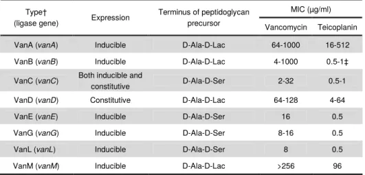
low-affinity peptidoglycan precursors to the glycopeptides. Eight types of
glycopeptide resistance have been characterized on both phenotypic and a
genotypic basis in enterococci. The classification of glycopeptide resistance is
currently based on the primary sequence of the resistance ligases (vanA, vanB,
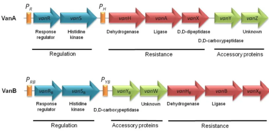
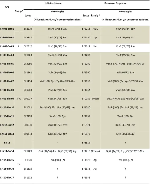
vanC, vanD, vanE, vanG, vanL, vanM). The most frequently detected operons are
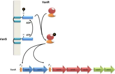
vanA and vanB operons and both harbor genes with regulatory functions (VanRS,
vanW). Their transcription is regulated by the VanRS and VanRBSB
two-component systems.
In the first part of this thesis we screened phenotypically susceptible strains,
from different origins in Portugal, for the presence of vancomycin resistance
genes. vanA and vanB genes were both found in dairy food isolates and in clinical
human and veterinary isolates. vanA gene was found to be more disseminated in
the clinical setting and vanB gene was only detected in a restricted number of
clinical isolates. However, none of the isolates carrying vanA and vanB genes had
complete vancomycin resistance operons. This finding suggests that the studied
strains were, at some point in time, in contact with vancomycin resistance
determinants, and probably due to absence of selective pressure, allowed genetic
events (deletions or insertions) ultimately leading to vanA and vanB operons
incomplete or corrupted, respectively. However, the presence of some of the
vancomycin resistance determinants in susceptible strains may suggest that
those genes could play some other biological role in those strains as if they could
confer any selective advantage in the presence of vancomycin.
Furthermore, we characterized a set of human clinical enterococcal strains
including three enterococcal vancomycin resistant Enterococcus faecium
(VREfm) strains carrying the insertion sequence ISEf1 and two Enterococcus
hirae strains responsible for nosocomial infection in two different patients. To our
knowledge this is the first report of E. hirae strains as etiological agents of
nosocomial infections in a portuguese hospital. We also point out to the
vulnerability of VITEK system alone in providing a correct assessment of
vancomycin resistant enterococci (VRE).
Following findings from the first part of this work, namely the hypothesis that
and recent evidences of involvement of two-component systems (other than
VanRS) in response to vancomycin; in the second part of this thesis we aimed
identifying complementary and/or alternative mechanisms that may be involved in
response to vancomycin.
During antimicrobial therapeutics the concentration of antibiotics in circulation
and in the different tissues reach frequently doses that are lower than the
therapeutic dose administered. Thus, we found important to study the
transcriptional response of E. faecalis to different vancomycin doses, thus
response to a subinhibitory and to a therapeutic dose of vancomycin was studied
in E. faecalis. The subinhibitory dose strictly and specifically induced the
vancomycin resistance genes. However, the therapeutic dose had a major effect
on the overall expression profile, as 14% of the genes were differentially
transcribed. These results demonstrate that E. faecalis responds globally to a
therapeutic vancomycin dose, suggesting that survival/resistance to vancomycin
may involve other genes and pathways.
We observed that after vancomycin exposure one ORF presented the same
level of transcription than the van genes. Our work led to the identification of a
new gene belonging to the vanB operon of E. faecalis (vanV). We provide
evidence of co-transcription of vanV together with vanYBWHBBXB and demonstrate that its expression is under the control of vanRBSB. However, vanV was not found to be required for vancomycin resistance and likely codes for an
accessory protein.
Taking together the transcriptomic results we obtained under vancomycin
exposure and the available literature we suspect that other enterococcal
two-component systems might be involved in sensing and responding to vancomycin.
found that CroRS is needed for VanRS induction and for TCS06 induction by
vancomycin.
To investigate the possibility of vanB operon genes influencing the
expression of other genes in E. faecalis, we used a genome-wide transcriptomic
approach to evaluate the differential expression of E. faecalis genes in vanB and
vanR independent mutants. In vanB mutant 205 and 309 of the V583 E. faecalis
genes showed differential expression, depending on the growth phase sampled.
For the vanR mutant 157 and 147 of the genes were differentially expressed.
Moreover, both mutants showed a very high number of genes located on mobile
genetic elements had their expression levels affected. These results suggest that
the vanB system may play a wider role in the cell.
In conclusion, the main findings presented in this thesis are that van
-resistance genes are spread among enterococcal strains from different origins.
Although these strains may keep a susceptible background in the presence of
vancomycin selective pressure, resistance may arise, suggesting that other
alternative mechanisms exist and may develop, by which enterococci might
survive in the presence of vancomycin. We also provide evidence that
vancomycin resistance is not strictly a product of vanB operon fully expression
and other two-component systems are involved and contribute to the high-level
resistance to the antibiotic. Furthermore, vanB operon is likely to have a broader
Os Enterococcus fazem parte da microbiota normal do tracto gastrointestinal
de humanos e animais e raramente causam infecções em indivíduos saudáveis.
Nos últimos 20 anos os enterococos emergiram como uma das causa mais
comuns de infecções adquiridas em ambiente hospitalar. Uma das principais
razões pela qual estes microrganimos sobrevivem com sucesso em ambiente
hospitalar deve-se à sua resistência intrínseca a vários antibióticos e, ainda mais
relevante, à sua capacidade para adquirir resistência a muitos dos antibióticos
frequentemente utilizados em terapêutica, incluindo os glicopéptidos. No género
Enterococcus o desenvolvimento de resistência ao glicopéptido vancomicina
constitui um sério problema a nível global. Infecções provocadas por enterococos
resistentes à vancomicina são, não apenas difíceis de tratar, como estas
bactérias também apresentam uma marcada propensão para se disseminarem
de doente para doente em ambiente hospitalar.
O conhecimento do cenário real do estado da disseminação da resistência à
vancomicina é essencial para poderem ser planeadas e adoptadas estratégias
globais de forma a prevenir a transmissão nosocomial de bactérias resistentes à
vancomicina, bem como a sua transmissão na comunidade.
A vancomicina liga-se com elevada afinidade ao D-Ala C-terminal do
pentapéptido do peptidoglicano, bloqueando a adição de mais precursores à
cadeia nascente do peptidoglicano e prevenindo o estabelecimento subsequente
das ligações cruzadas por transpeptidação. Em enterococos a resistência a
glicopéptidos é conferida pela presença de operões que codificam enzimas para
a síntese de precursores do peptidoglicano com reduzida afinidade para estes
antibióticos. Em enterococos, oito tipos de resistência a glicopéptidos já foram
caracterizados aos níveis fenotípico e genotípico. A classificação dos tipos de
codificados nos diferentes operões (vanA, vanB, vanC, vanD, vanE, vanG, vanL,
vanM). Os operões mais frequentemente detectados são os vanA e vanB que
possuem genes com funções reguladoras (VanRS, VanRBSB), de resistência
(VanHAXY, VanYBHBBXB) e proteínas acessórias (VanZ, vanW). A sua
transcrição é regulada pelos sistemas de dois componentes VanRS e VanRBSB,
respectivamente
Na primeira parte desta tese pesquisámos a presença de genes de
resistência à vancomicina em estirpes de enterococos fenotipicamente
susceptíveis ao antibiótico, de diferentes origens. Os genes vanA e vanB foram
detectados em isolados alimentares provenientes de alimentos lácteos, bem
como em isolados clínicos humanos e veterinários. O gene vanA está mais
disseminado no ambiente clínico e o gene vanB foi apenas detectado num
número reduzido de isolados clínicos. No entanto, nenhum dos isolados que
possuem os genes vanA e vanB apresentam operões completos de resistência à
vancomicina. Estas observações sugerem que as estirpes estudadas estiveram,
nalgum momento, em contacto com genes de resistência à vancomicina e,
provavelmente devido à ausência de pressão selectiva, ocorreram eventos
genéticos (delecções ou inserções) que, em última instância, levaram a operões
vanA e vanB incompletos ou corrompidos, respectivamente. No entanto, a
presença de alguns genes de resistência à vancomicina em estirpes susceptíveis
pode sugerir que estes genes desempenham outros papéis biológicos nestas
estirpes, como se constituíssem alguma vantagem selectiva em presença de
vancomicina. Além disso, caracterizámos um conjunto de estirpes clínicas
humanas, incluindo três estirpes de Enterococcus faecium vancomicina
resistentes (VREfm) que têm a sequência de inserção ISEf1 e duas estirpes de
diferentes. Esta é a primeira descrição de estirpes de E. hirae enquanto agentes
etiológicos de infecção nosocomial em hospitais Portugueses. Também
salientámos a vulnerabilidade do sistema VITEK, por si só, em identificar
correctamente enterococos resistentes à vancomicina (VRE).
Seguindo os resultados da primeira parte deste trabalho, nomeadamente a
hipótese de que operões vanA e vanB incompletos continuam a ter um papel na
célula, e as recentes evidências do envolvimento de outros sistemas de dois
componentes (que não o VanRS) na resposta à vancomicina, a segunda parte
desta tese teve por objectivo identificar mecanismos alternativos e/ou
complementares que estejam envolvidos na resistência à vancomicina.
Durante o processo terapêutico com um agente antimicrobiano a
concentração do antibiótico em circulação em nos diferentes tecidos atinge
frequentemente doses inferiores à dose terapêutica administrada Por este
motivo, considerámos importante estudar as respostas transcricionais de E.
faecalis a diferentes doses de vancomicina. Assim, a resposta a uma dose
sub-inibitória e a uma dose terapêutica de vancomicina foi estudada em E. faecalis. A
dose sub-inibitória induz estrita e especificamente os genes de resistência à
vancomicina. No entanto, a dose terapêutica tem um efeito muito significativo no
perfil geral de transcrição, com 14% dos genes a serem transcritos
diferencialmente. Estes resultados demonstram que E. faecalis responde
globalmente a uma dose terapêutica de vancomicina, sugerindo que a
sobrevivência/resistência à vancomicina pode envolver outros genes e vias.
Observámos que após exposição à vancomicina uma ORF apresentava o
mesmo nível de transcrição que os genes van. O nosso trabalho permitiu
identificar um novo gene pertencente ao operão vanB de E. faecalis (vanV).
vanYBWHBBXB e demonstramos que a sua expressão está sob o controlo de
vanRBSB. No entanto, o vanV não é necessário para a resistência à vancomicina
e provavelmente codifica para uma proteína acessória.
Considerando os resultados obtidos no estudo de transcriptómica sob
exposição à vancomicina e a literatura disponível fomos levados a suspeitar do
envolvimento de outros sistemas de dois componentes na detecção do sinal e
resposta à vancomicina. Assim, analisámos e descrevemos o envolvimento de
CroRS e TCS06 na resistência à vancomicina e percebemos que o CroRS é
necessário para a indução do VanRS e para a indução do TCS06 pela
vancomicina.
Para investigar a possibilidade dos genes do operão vanB influenciarem a
expressão de outros genes em E. faecalis, usámos uma abordagem
transcriptómica para avaliar a expressão diferencial dos genes de E. faecalis em
dois mutantes independentes, vanB e vanR. No mutante vanB, 205 e 309 genes
de E. faecalis V583 mostraram-se diferencialmente expressos, dependendo da
fase de crescimento. No mutante vanR, 157 e 147 genes foram diferencialmente
expressos. Nos dois mutantes, um elevado número de genes localizados em
elementos genéticos viram os seus níveis de expressão afectados. Estes
resultados sugerem que o sistema vanB poderá ter um papel muito mais
alargado na célula.
As principais conclusões apresentadas nesta tese são, portanto, que os
genes de resistência van estão disseminados entre estirpes de enterococos de
diferentes origens. No entanto estas estirpes mantêm um background susceptível
mas, em presença de pressão selectiva por vancomicina, a resistência pode
surgir, sugerindo que existem mecanismos alternativos que se podem
Fornecemos ainda evidências de que a resistência à vancomicina não é
estritamente produto de uma expressão plena do operão vanB e que outros
sistemas de dois componentes estão envolvidos e contribuem para a elevada
resistência ao antibiótico. É provável que o operão vanB tenha uma papel mais
Scientific Articles Peer Reviewed in International Journals:
Tânia Ribeiro, Axel Hartke, Michael S. Gilmore, Fátima Lopes. Transcriptomic
profile of E. faecalis V583 in response to vancomycin reveals bacterial support for full expression of high-level vancomycin resistance (in preparation).
Tânia Ribeiro, Fátima Lopes. CroRS integrity is essential for VanRS induction
and high-level vancomycin resistance phenotype (in preparation).
Tânia Ribeiro, Sofia Santos, Fátima Lopes. Identification of a new gene in the
vanB operon of E. faecalis (submitted for publication).
Tânia C Ribeiro, Vera Pinto, Frèdèric Gaspar, Maria FS Lopes (2008)
Enterococcus hirae causing wound infections in a hospital. Journal of Chinese Clinical Medicine 3:150-152.
Tânia Ribeiro, Marta Abrantes, Maria de Fátima Silva Lopes, Maria Teresa
Barreto Crespo (2007) Vancomycin-susceptible dairy and clinical enterococcal isolates carry vanA and vanB genes. International Journal of Food Microbiology 113:289-295.
Benoît Zuber, Marisa Haenni, Tânia Ribeiro, Fátima Lopes, Philippe Moreillon,
The objectives proposed for this dissertation were all focused on vancomycin resistance determinants dissemination and vancomycin response in enterococci. In particular we intended to learn more about the current real amplitude of vancomycin determinants dissemination in different environments and what are the mechanisms that mediate vancomycin response in enterococci.
The present Dissertation is divided in two main parts and into nine chapters. On the first part we present data related to the screening and detection of vancomycin resistance determinants in strains from different environmental sources. On the second part we focus in genes and pathways that might be involved in vancomycin resistance and response
Chapter 1 consists of a general introduction to contextualize the subject. A brief overview of vancomycin resistance in enterococci and its importance as a last resort antibiotic, to treat multidrug resistant infections caused by Gram-positive bacteria, and as stress agent is made. The results of this Doctoral work are presented in the following chapters. Each chapter has its own Introduction, Results and Discussion of the respective work.
In chapter 2 we describe the dissemination and prevalence of vancomycin resistance determinants (vanA and vanB) in a considerably large group of dairy
isolates and human and veterinary clinical isolates and this way study the dissemination of these determinants in different ecological niches. The arrangement of the prototype resistance element Tn1546 is determined and high
heterogeneity of this mobile element was observed.
Chapter 3 consists on the characterization of enterococcal strains in a Portuguese Hospital. We report the detection of E. faecium strains belonging to
cases of E. hirae as etiological agent of nosocomial infections in Portugal
associated with wound infections.
In chapter 4 we describe and discuss microarray data on how E. faecalis
V583 responds to a therapeutic dose of vancomycin.
In chapter 5 we demonstrate that vanB operon has one more gene on its
constitution than previously acknowledged.
Chapter 6 characterizes the involvement of two two-component systems in vancomycin sensing and potential induction of vancomycin resistance.
Chapter 7 describes the behavior of vancomycin susceptible strains when exposed to serial passages in presence of antibiotic and discuss the possible mechanims behind the observed adaptation.
Chapter 8 is centered on unraveling the role of vanB operon genes in
enterococci in absence of vancomycin induction. We present and discuss transcriptomic results on vanB and vanR mutants.
A adenine
AI after induction
Ala alanine
AMP ampicillin
APS antimicrobial peptide sensor
AS aggregation substance
Asp aspartate
ATCC american type culture collection
ATP adenosine triphosphate
B bacitracin
BC before Christ
BHI brain hearth infusion
BI before induction
bp base pair
°C degree Celsius
CA catalytic and ATP-binding domain
CAT chloramphenicol acetyltransferase
CC clonal complex
cDNA complementary desoxyribonucleic acid
CDP cytidine diphosphate
CECT colección española de cultivos tipo
CESR cell envelope stress response
CFP cefoperazone
CHEF clamped homogeneous electric fields
CIP ciprofloxacin
CLSI clinical and laboratory standards institute
CN gentamicin
CoNS coagulase-negative staphylococci
CS coefficient of similarity
CTP cytidine triphosphate
CWAP cell-wall anchored proteins
D aspartate
DA clindamycin
DHF dihydropholate
DHp dimerization and histidine-containing
phosphotransfer domain
DNA desoxiribonucleic acid
dNTP deoxynucleotide triphosphate
DSMZdeutsche sammlung von mikroorganismen
und zellkulturen
E erythromycin
EARSS european antimicrobial resistance
surveillance system
ECF extracytoplasmic function sigma factor
EDTA ethylenediaminetetraacetic acid
Ehk enterococcal histidine kinase
Err enterococcal response regulator
Ery erythromycin
Esp enterococcal surface protein
F nitrofurantoin
G guanine
GI gastrointestinal
GISA glycopeptide intermediate Staphylococcus
aureus
GlcN glucosamine
GlcNAc N-acetyl glucosamine
GRE glycopeptide resistant enterococci
H histidine
HiRCC high risk clonal complex
His histidine
HK histidine kinase
HLGR high-level gentamicin resistance
HLVR high-level vancomycin resistance
HPT histidine phosphotransferase
HSP heat shock protein
IE infectious endocarditis
IPN imipenem
IPP isopentenyl pyrophosphate
IR intergenic region
IS insertion sequence
K kanamycin
kb kilobase
Lac lactate
LMG Laboratorium voor Microbiologie
LTA lipoteichoic acid
µ µ µ
µg microgram
µ µ µ
µl microliter
µ µ µ
µM micromolar
MDR multi-drug resistance
MGE mobile genetic element
MIC minimum inhibitory concentration
min minute
ml milliliter
MLST multilocus sequence typing
mM milliMolar
MOPS 3-(N-morpholino)propanesulfonic acid
mRNA messenger ribonucleic acid
MRSA methicillin resistant Staphylococcus
aureus
MurNAc N-acetyl muramic acid
NA nicotinic acid
NAD nicotinamide adenine dinucleotide
Nam nicotinamide
NamND nicotinamide mononucleotide
NCCLS national committee on clinical laboratpry
standards
ng nanogram
nm nanometer
NMR nuclear magnetic resonance
NR nicotinamide riboside
OA oxaloacetate
ORF open reading frame
P penicillin B
PABA para-aminobenzoic acid
PAI pathogenicity island
PBP penicillin binding protein
PCR polymerase chain reaction
PFGE pulsed field gel electrophoresis
PG peptidoglycan
pH hydrogen potential
pmol picomol
ppGpp, pppGpp guanosine
3’,bispyrophosphate, guanosine 3’-diphosphate
5’-triphosphate, collectivelly ppGpp and pppGpp are
termed (p)ppGpp
PRPP phosphoribosyl pyrophosphate
PTS phosphoenolpyruvate phosphotransferase
system
RD rifampicin
RDO registered designation of origin
RNA ribonucleic acid
RNase ribonuclease
rpm rotations per minute
RR response regulator
RT-PCR reverse transcriptase PCR
S streptomycin
s second
SAM S-adenosylmethionine
SDS sodium dodecyl sulphate
Ser serine
SLR signal log ratio
SNP single nucleotide polymorphism
Spp. species
sRNA small non-coding RNA
SSC saline-sodium citrate buffer
ST sequence type
T thymine
TAE tris acetate EDTA buffer
TAIL-PCR thermal asymmetric interlaced-PCR
TBE tris borate EDTA buffer
TCS two-component system
TE/Tet tetracycline
THF trihydropholate
ThMP hydroxymethyl pyrimidine
Tn tranposon
TPP thiamine pyrophosphate
TRAP tripartite ATP-independent periplasmic
Tris trishydroxymethylaminomethane
(2-Amino-2-(hydroxymethyl)propane-1,3-diol)
U unit
UDP uridine diphosphate
UMP uridine monophosphate
UPGMA unweighted pair group method with
arithmetic mean
UPP undecaprenyl pyrophosphate
US United States
UTI urinary tract infections
UV ultraviolet radiation
V volt
Van vancomycin
VISA vancomycin-intermediate Staphylococcus
aureus
VRE vancomycin resistant enterococci
VREfm vancomycin resistant E. faecium
VREfs vancomycin resistant E. faecalis
VRSA vancomycin-resistant Staphlococcus
aureus
VSE vancomycin susceptible enterococci
VSEfm vancomycin susceptible Enterococcus
faecium
wt wild type
Abstract xv
Resumo xxi
List of Publications xxix
Dissertation Outline xxxiii
Abbreviations xxxv
Table of Contents xliii
Chapter 1
General Introduction
1
PART1:SPREAD OF VANCOMYCIN RESISTANCE DETERMINANTS AMONGST ISOLATES FROM DIFFERENT ENVIRONMENTS
Chapter 2
Vancomycin-Susceptible Dairy and Clinical Enterococcal Isolates carry vanA and vanB genes
55
Chapter 3
Molecular Characterization of Uncommonly found Enterococci in Portuguese Hospitals: VREfm from CC17 Carrying ISEf1 and
Enterococcus hirae Causing Infections
89
PART2:RESPONSE TO VANCOMYCIN:PLAYERS AND THEIR ROLES IN RESPONSE TO VANCOMYCIN
Chapter 4
Transcriptomic Profile of Enterococcus faecalis V583 in Response to Vancomycin Reveals Bacterial Support for Full Expression of High-Level Vancomycin Resistance
127
Chapter 5
Identification of a New Gene in vanB Operon of Enterococcus faecalis
195
Chapter 6
CroRS Integrity is Essencial for VanRS Induction and High-Level Vancomycin Resistance Phenotype
Induction of Resistance to Vancomycin in Susceptible Enterococcus faecalis strains
Chapter 8
Comparison of V583 with vanB and vanR Mutants by Transcriptional Aanalysis: vanB Operon Genes Are More Than Just Antibiotic Resistance Tools
265
Chapter 9
General Discussion and Future Perspectives
335
Chapter 1
CONTENTS
I. Vancomycin 5
Historical Introduction to Vancomycin Discovery 5
Vancomycin Mode of Action 9
II. Enterococcus 12
General Description of the Genus 12
Enterococci as Pathogens 13
III. Vancomycin Resistance 17
Mechanisms of Vancomycin Resistance in Enterococci 17
Vancomycin Resistance in Portugal 22
Vancomycin Resistance in other Bacteria 24
Regulation of Vancomycin Resistance 27
Enterococcal Two-Component Systems 32
Two-Component Systems and Cross-Regulation 36
Bacterial Response to Vancomycin and other Cell Wall-Active
Antibiotics
39
I.
VANCOMYCIN
HISTORICAL INTRODUCTION TO VANCOMYCIN DISCOVERY
The use of substances with antibacterial activity has been documented
throughout history. Over 50,000 years ago the Neanderthals used certain herbs
and natural substances to kill bacteria in the wounds and stop their propagation.
Human remains have been found with various herbs that are said to be used for
therapeutic purposes by the prehistoric man. There is evidence in the Ebers
papyrus (1550 BC) that the Egyptians have used beer yeast on wounds and
numerous other ancient civilizations used molds in one form or another for cures
and rituals. A chemical analysis of the bones of ancient Nubian mummies, shows
that they were regularly consuming tetracycline, most likely in their beer since the
grain used to make the fermented gruel contained the soil bacteria streptomyces,
which produces tetracycline (Levy, 2002). The ancient Chinese also used molds
to treat boils, carbuncles and other skin infections.
Nevertheless, it is only in the end of the 19th century that the first scientific
report of mold use with the specific aim of killing bacteria appeared. In 1897
Ernest Duchesne reported that certain penicillium molds could indeed destroy
Salmonella typhi and Escherichia coli in the test tube. He further showed that
inoculating intraperitoneally a guinea pig with a solution containing Penicillium
glaucum and E. coli, would protect the guinea pig against the lethal dose of E.
coli. He had shown that certain molds could kill bacteria – he had discovered
penicillin (reviewed in Duckett, 1999).
However, it is generally accepted that the discovery of penicillin dates from
1928 (reviewed in Podolsky, 1997) when the young bacteriologist Merlin Pryce
noticed that one of the petri dishes floating in the sink and ready to be discarded
lysed bacteria. Fleming, who was interested in the lysis phenomena, got intrigued
by this observation and decided to pursue it further. After several experiments he
showed that an active substance, produced by this mold, could kill bacteria such
as staphylococci, streptococci, gonococci but not others such as those
responsible for typhoid fever or cholera. Later he and his young assistant, Stuart
Craddock, proved that injection of the mold filtrate could not only save rabbits and
mice from lethal infections of staphylococci but it could also cure his assistant´s
sinus and eye infections.
Despite the successful experiments done by Fleming, the application of
penicillin to treat infections would have remained only an academic curiosity, if it
were not for the work of two other future Nobel Prize laureates. Fleming was
discouraged, with no research funds to continue the experiments and with a very
unstable compound of which only very small amounts could be purified. He was
further disappointed by his own unpublished observation that staphylococci
became resistant to penicillin very quickly (Podolsky, 1997). Fleming discretely
described his preliminary findings and then turned his back on penicillin. Later
Florey and Chain rescued penicillin and succeeded in purifying large amounts of
the compound, generating enough interest so that when World War II started
there was a bigger production of this antibiotic in order to decrease the fatalities in
Europe and Asia. By this time penicillin had been used in fewer than 100 people
in the United States of America.
On Sunday morning, November 29, 1942, the Boston Herald front page
announced the most devastating fire in the history of Boston, the Cocoanut Grove
Nightclub disaster. There were more than 450 victims and the physicians were
caring for the 200 survivors who resisted for the first 24 hours. The fire also
itself – the trial of a new, unique drug available only through government sources.
Limited amounts of penicillin were released to quell many of the infections
contracted by survivors of the fire. Actually, the Cocoanut Grove victims became
one of penicillin´s most important clinical trials and the success of skin grafting
and treating the severely burned patients was largely attributed to the action of
penicillin (Levy, 2002). Soon after, the use of antibiotics was widely spread and
penicillin was considered as a miraculous drug. Penicillin discovery marked the
beginning of the “antibiotic era”.
The success of penicillin encouraged scientists to search for and discover
new antibiotics that could treat other bacteria, including penicillin-resistant strains.
As more antibiotics were discovered, from the late 1940s into the 1970s, the
problem of resistance seemed little more than an annoyance.
By the 1950s, it was apparent that penicillin was not going to remain effective
against staphylococci. The pharmaceutical Eli Lilly and Company was soon
operating a large-scale screening program aimed solely at the isolation of
antibiotics with high level of specificity and activity against staphylococci. Soil
samples were obtained from all parts of the world and were subjected to the
screening procedures. An organic chemist at Lilly, Dr. E. C. Kornfeld, had a
missionary friend in Borneo who sent him dirt from the deep interior of the jungle.
In it, he found an organism that later was named Streptomyces orientalis.
Fermentation broths of the organism were highly active and bactericidal against
virtually all staphylococci tested. Studies in animals showed the level of toxicity to
be relatively low. The active compound in S. orientalis fermentation was labeled
compound 05865 (Levine, 2006).
The difficulty with which staphylococci would become resistant to the
Consequently, the earliest in vitro studies consisted on experiments on the
induction of resistance to compound 05865 by daily serial passage. The results
obtained lead to consider that the laboratory-induced level of staphylococcal
resistance to compound 05865 was negligible (Levine, 2006).
Since the impurities from the preparation and fermentation broth were linked
to ototoxicity and nephrotoxicity and due to its brownish color, compound 05865
earned the nickname "Mississippi mud". Soon, however, "Mississippi mud" was
given the generic name vancomycin, derived from the word vanquish, and efforts
were directed towards its purification (Levine, 2006).
Although the early clinical trials of vancomycin were modest in scope and
involved relatively few researchers, information concerning the efficacy of the
antibiotic in difficult staphylococcal infections quickly spread. With the increased
demand for vancomycin to be used in clinical studies and in emergency treatment
of serious staphylococcal infection, supplies were often inadequate. On various
occasions it was necessary for a representative of Eli Lilly and Company to locate
some vancomycin in one hospital and deliver it to another where it was more
urgently needed. The large volume of requests for vancomycin for emergency use
was followed by numerous reports of satisfactory therapeutic responses. Because
there was no other alternative in the treatment of serious infections caused by
staphylococci resistant to the then available antibiotics, the U.S. Food and Drug
Administration strongly urged Eli Lilly and Company to make vancomycin
generally available as soon as possible.
Early concerns regarding safety, which were based on results of studies in
animals, may have been responsible for the limited use of vancomycin for 20
years. It was reformulated and retested in animal models for toxicity in the 1970s.
vancomycin re-emerged as a key component in treating methicillin-resistant
Staphylococcus aureus (MRSA). The clinical need for an agent to fight MRSA
was apparent and vancomycin quickly became the drug of choice.
VANCOMYCIN MODE OF ACTION
The simultaneous worldwide emergence, first of methicillin-resistant S.
aureus, and then, the penicillin-resistant Streptococcus pneumonia, together with
the reported success on the treatment of pseudomembranous enterocolitis led to
the clinical re-introduction of vancomycin (Levine, 2006). In the 1980s
vancomycin was being widely used worldwide and its use increased specially in
the United States. In contrast, the rise of vancomycin use has been less
pronounced in Europe (Kirst et al., 1998).
Vancomycin is the most relevant member of the glycopeptide class of
antibiotics, which include also teicoplanin, telavancin and ramoplanin. The
chemical structure shared by the glycopeptide antibiotic family has been
determined by different methods as mass spectrometry, NMR and X-ray
crystallography (Barna & Williams, 1984), elucidating its mode of action.
Vancomycin, and other glycopeptide antibiotics, do not interact with the cell wall
biosynthetic enzymes but form complexes with the carboxy-terminal D-alanine
(D-Ala) residues of peptidoglycan precursors thus preventing their incorporation into
the cell wall, leading to cell wall synthesis inhibition (Reynolds, 1989).
The synthesis of peptidoglycan involves several steps (Figure 1). In the
cytoplasm, a racemase converts L-alanine to D-alanine (D-Ala), and then two
molecules of D-Ala are joined by a ligase, creating the dipeptide D-Ala-D-Ala,
which is then added to uracil diphosphate-N-acetylmuramyl-tripeptide to form
diphosphate-N-acetylmuramyl-pentapeptide is bound to the undecaprenol lipid carrier, which,
after the addition of N-acetyl glucosamine (GlcNAc) from uracil
diphosphate-GlcNAc, allows translocation of the precursors to the outer surface of the
cytoplasmic membrane. N-acetylmuramyl-pentapeptide is then incorporated into
nascent peptidoglycan by transglycosylation and allows the formation of
cross-bridges by transpeptidation (Reynolds, 1989).
Vancomycin binds with high affinity to the D-Ala-D-Ala C-terminus of the
pentapeptide (Figure 1A), therefore blocking the addition of late precursors by
transglycosylation to the nascent peptidoglycan chain and preventing subsequent
cross-linking by transpeptidation (Reynolds, 1989). As a secondary and less
relevant mechanism, glycopeptides inhibit the transglycosilation reaction of the
peptidoglycan unit into the growing polymer, presumably by steric hindrance from
the antibiotic-peptidoglycan precursor complex (Ge et al., 1999; Walsh, 1999).
At present, glycopeptides are used for the treatment of infections caused by
methicillin-resistant S. aureus (MRSA), coagulase-negative staphylococci (CoNS)
or by ampicillin-resistant enterococci. Furthermore, these drugs are indicated in
patients with serious β-lactam allergies (Levine, 2006). Glycopeptides are virtually
not active against Gram-negative bacteria which are protected by an outer
membrane.
Resistance to vancomycin in a clinical enterococcal isolate was first reported
in Europe in 1986 (Leclercq et al., 1988; Uttley et al., 1988) and in 1987 in the
United States (Sahm et al., 1989). Enterococci were the first clinically relevant
bacterial genus to acquire a resistance mechanism that specifically confers
Figure 1. Schematic representation of peptidoglycan biosynthesis in (A) vancomycin-susceptible and (B) vancomycin resistant bacteria. Binding of vancomycin to the C-terminal D-Ala-D-Ala of the late peptidoglycan precursors prevents the reactions catalyzed by transglycosylases, transpeptidases and D,D-carboxypeptidases. In vancomycin resistant strains there is still production of native peptidoglycan precursors ending in D-Ala-D-Ala resulting from the endogenous chromosomal pathway. Is on the native pathway that VanX and VanY act sequencing hydrolyzing the dipeptide D-Ala-D-Ala. Ddl, D-Ala-D-Ala ligase; MurF, a synthetase protein; UDP, uracil diphosphate. Adapted from (Courvalin, 2006).
racemase
Cytoplasm
Cell wall
L-Ala
D-Ala
D-Ala-D-Lac
UDP L-Ala-D-Glu-L-Lys VanA
ATP
MurF
UDP L-Ala-D-Glu-L-Lys-D-Ala-D-Lac
pentapeptide pyruvate D-Lac VanH NADH L-Ala-D-Glu-L-Lys-D-Ala-D-Lac L-Ala-D-Glu-L-Lys-D-Ala-D-Lac Carboxypeptidase (VanY) L-Ala-D-Glu-L-Lys-D-Ala-D-Lac transpeptidase L-Ala-D-Glu-L-Lys-D-Ala-D-Lac transglycosidase L-Ala-D-Glu-L-Lys-D-Ala-D-Lac
B
racemaseCytoplasm
Cell wall
L-Ala
D-Ala
D-Ala-D-Ala
UDP L-Ala-D-Glu-L-Lys Ddl ligase
MurF
UDP L-Ala-D-Glu-L-Lys-D-Ala-D-Ala
pentapeptide L-Ala-D-Glu-L-Lys-D-Ala-D-Ala
vancomycin L-Ala-D-Glu-L-Lys-D-Ala-D-Ala carboxypeptidase L-Ala-D-Glu-L-Lys-D-Ala-D-Ala transpeptidase L-Ala-D-Glu-L-Lys-D-Ala-D-Ala transglycosidase L-Ala-D-Glu-L-Lys-D-Ala-D-Ala N-acetylmuramic acid N-acetylglucosamine
Undecaprenyl lipid carrier
A
racemase
Cytoplasm
Cell wall
L-Ala
D-Ala
D-Ala-D-Lac
UDP L-Ala-D-Glu-L-Lys VanA
ATP
MurF
UDP L-Ala-D-Glu-L-Lys-D-Ala-D-Lac
pentapeptide pyruvate D-Lac VanH NADH L-Ala-D-Glu-L-Lys-D-Ala-D-Lac L-Ala-D-Glu-L-Lys-D-Ala-D-Lac Carboxypeptidase (VanY) L-Ala-D-Glu-L-Lys-D-Ala-D-Lac transpeptidase L-Ala-D-Glu-L-Lys-D-Ala-D-Lac transglycosidase L-Ala-D-Glu-L-Lys-D-Ala-D-Lac
B
racemaseCytoplasm
Cell wall
L-Ala
D-Ala
D-Ala-D-Lac
UDP L-Ala-D-Glu-L-Lys VanA
ATP
MurF
UDP L-Ala-D-Glu-L-Lys-D-Ala-D-Lac
pentapeptide pyruvate D-Lac VanH NADH pyruvate D-Lac VanH NADH L-Ala-D-Glu-L-Lys-D-Ala-D-Lac L-Ala-D-Glu-L-Lys-D-Ala-D-Lac Carboxypeptidase (VanY) L-Ala-D-Glu-L-Lys-D-Ala-D-Lac transpeptidase L-Ala-D-Glu-L-Lys-D-Ala-D-Lac transglycosidase L-Ala-D-Glu-L-Lys-D-Ala-D-Lac L-Ala-D-Glu-L-Lys-D-Ala-D-Lac L-Ala-D-Glu-L-Lys-D-Ala-D-Lac Carboxypeptidase (VanY) L-Ala-D-Glu-L-Lys-D-Ala-D-Lac Carboxypeptidase (VanY) L-Ala-D-Glu-L-Lys-D-Ala-D-Lac transpeptidase L-Ala-D-Glu-L-Lys-D-Ala-D-Lac transpeptidase L-Ala-D-Glu-L-Lys-D-Ala-D-Lac transglycosidase L-Ala-D-Glu-L-Lys-D-Ala-D-Lac transglycosidase L-Ala-D-Glu-L-Lys-D-Ala-D-Lac
B
racemaseCytoplasm
Cell wall
L-Ala
D-Ala
D-Ala-D-Ala
UDP L-Ala-D-Glu-L-Lys Ddl ligase
MurF
UDP L-Ala-D-Glu-L-Lys-D-Ala-D-Ala
pentapeptide L-Ala-D-Glu-L-Lys-D-Ala-D-Ala
vancomycin L-Ala-D-Glu-L-Lys-D-Ala-D-Ala carboxypeptidase L-Ala-D-Glu-L-Lys-D-Ala-D-Ala transpeptidase L-Ala-D-Glu-L-Lys-D-Ala-D-Ala transglycosidase L-Ala-D-Glu-L-Lys-D-Ala-D-Ala N-acetylmuramic acid N-acetylglucosamine
Undecaprenyl lipid carrier
A
racemase
Cytoplasm
Cell wall
L-Ala
D-Ala
D-Ala-D-Ala
UDP L-Ala-D-Glu-L-Lys Ddl ligase
MurF
UDP L-Ala-D-Glu-L-Lys-D-Ala-D-Ala
pentapeptide L-Ala-D-Glu-L-Lys-D-Ala-D-Ala
vancomycin L-Ala-D-Glu-L-Lys-D-Ala-D-Ala carboxypeptidase L-Ala-D-Glu-L-Lys-D-Ala-D-Ala transpeptidase L-Ala-D-Glu-L-Lys-D-Ala-D-Ala transglycosidase L-Ala-D-Glu-L-Lys-D-Ala-D-Ala L-Ala-D-Glu-L-Lys-D-Ala-D-Ala vancomycin L-Ala-D-Glu-L-Lys-D-Ala-D-Ala carboxypeptidase L-Ala-D-Glu-L-Lys-D-Ala-D-Ala carboxypeptidase L-Ala-D-Glu-L-Lys-D-Ala-D-Ala transpeptidase L-Ala-D-Glu-L-Lys-D-Ala-D-Ala transpeptidase L-Ala-D-Glu-L-Lys-D-Ala-D-Ala transglycosidase L-Ala-D-Glu-L-Lys-D-Ala-D-Ala transglycosidase L-Ala-D-Glu-L-Lys-D-Ala-D-Ala N-acetylmuramic acid N-acetylglucosamine
Undecaprenyl lipid carrier N-acetylmuramic acid N-acetylglucosamine
Undecaprenyl lipid carrier
During the first several decades of use, staphylococci did not mount a
resistance to vancomycin. It has only been within the last years that
vancomycin-intermediate Staphylococcus aureus (VISA) and vancomycin-resistant
Staphylococcus aureus (VRSA) have surfaced.
II.
ENTEROCOCCUS
GENERAL DESCRIPTION OF THE GENUS
In 1899, Thiercelin described Gram-positive coccoid-shaped bacteria from
the human intestine and used the name “entérocoque” to emphasize their
intestinal origin (Thiercelin, 1899). However, the term Streptococcus kept being
more commonly used. In 1937, Sherman developed a scheme classifying the
genus Streptococcus into four main groups: pyogenic, viridians, lactic and
enterococci (Sherman, 1937). Enterococci and streptococci were proven to be
different based on DNA hybridization experiments and subsequently the genus
Enterococcus was introduced in 1984 (Schleifer & Kilpper-Balz, 1984).
Enterococci are also separated from other Streptococcus species as they grow
between 10 and 45ºC, in 6.5% NaCl and at pH 9.6, survive heating at 60ºC for 30
min., hydrolyze esculine into esculitine (Devriese et al., 1991) and react with the
Lancefield group D antisera (Murray, 1990). Enterococci produce L(+)-lactic acid
homofermentatively from glucose.
Although about 40 Enterococcus species are now recognized
(http://www.dsmz.de/microorganisms/html/bacteria.genus/enterococcus.html), E.
faecalis and E. faecium are the two most common species found in the human
microbiota (Devriese et al., 1991). Enterococci are members of the
(Tannock, 2002). In humans enterococci also colonize the genito-urinary tract and
the oral and vaginal cavities (Murray, 1990). They are also capable of surviving in
many other environmental niches like soil, sand, water (usually as faecal
pollutants), food products and in plants (Mundt, 1986).
E. faecalis is currently the most intensively studied enterococcal specie due
to its prominence in the nosocomial setting. Until recently the only publically
sequenced genome available belonged to E. faecalis V583 (Paulsen et al., 2003),
which was the first reported vancomycin resistant clinical isolate in the United
States (Sahm & Olsen, 1990). In 2008 another E. faecalis strain, OG1RF, was
sequenced but the genome was not made publically available (Bourgogne et al.,
2008). Very recently the genome of other 28 enterococcal strains (including E.
faecalis, E. faecium, E. casseliflavus and E. gallinarum species) became available
(Palmer et al., 2010).
ENTEROCOCCI AS PATHOGENS
The staphylococci and the streptococci were the most prevalent hospital
pathogens in the 1960s and 1970s. Then, over a period of time, the distribution of
organisms involved in nosocomial infections shifted from positive to
Gram-negative in the 1970s and 1980s and since then Gram-positive organism have
re-emerged as major hospital pathogens (Levy, 1998). In large part this trend may
be attributed to the selection pressure created by antibiotics used in the hospital
setting. Although enterococci have been recognized as an important cause of
infectious endocarditis (IE) for more than a century (Singh et al., 2010), just in the
last two decades they were recognized as one of the leading causes of
nosocomial infections (Murray, 1990). The most prevalent infections caused by
infections and endocarditis (Moellering, 1992; Murray, 1998). Most of these
infections are caused by E. faecalis and only a small number are caused by E.
faecium (Witte et al., 1999). However, in recent years a progressive increase of
infections caused by E. faecium has been reported (Mundy et al., 2000; Treitman
et al., 2005). With exception of E. faecalis and E. faecium, other enterococcal
species are rarely associated to human pathogenesis and their clinical relevance
is often neglected, although there are reports of infections caused E. durans
(Stepanovic et al., 2004), E. hirae (Gilad et al., 1998; Poyart et al., 2002; Ribeiro
et al., 2008), E. raffinosus (Wilke et al., 1997; Sandoe et al., 2001, Savini et al.,
2008, Mastroiani, 2009), E. gallinarum (Reid et al., 2001; Takayama et al., 2003),
E. avium (Swaminathan & Ritter, 1999), E. casseliflavus (Reid et al., 2001;
Pappas et al., 2004; Iaria et al., 2005), E. cecorum (Hsueh et al., 2000) and E.
mundtii (Higashide et al., 2005). Enterococci are considered opportunistic
pathogens considering that they are generally harmless for healthy people but
able to cause serious infection in immunocompromised patients that are more
susceptible to infections.
Two main features contribute to enterococcal pathogenic aptitude: a wide
range of virulence factors, and the accumulation of different antimicrobial
resistance mechanisms.
E. faecalis possess a plethora of virulence factors designed to help
establishing infection and persist in the presence of the host immune response
(Shankar et al., 1999). There are four main stages in the pathogenesis of
enterococcal infections. The first is persistence in inanimate objects due to
intrinsic properties of bacteria, namely resistance to desiccation, heat and other