interactions and contribution to bacterial pathogenic traits
Renata Filipa Cruz de Matos
Dissertation presented to obtain the Ph.D degree in Biology
Instituto de Tecnologia Química e Biológica | Universidade Nova de Lisboa
Apoio financeiro da FCT e do FSE no âmbito do Quadro Comunitário de
apoio BD nº SFRH/BD/43461/2008
This thesis experimental work was performed at:
Supervisors
Doctor Maria de Fátima Gonçalves Ribeiro dos Santos Silva Lopes
Auxiliary investigator at Instituto de Tecnologia Química e Biológica,
Oeiras, Portugal.
Doctor Pascale Serror
Chargé de Recherche, MICALIS, Institut National de la Recherche
Agronomique, Jouy-en-Josas, France.
Examining Committee
Professor Doctor Carlos São-José
Auxiliary Professor at Dept. de Microbiologia e Imunologia,
Faculdade de Farmácia da Universidade de Lisboa, Portugal.
Professor Doctor Willem van Schaik
Assistant Professor at Dept. of Medical Microbiology, University
Medical Center Utrecht, The Netherlands.
Professor Doctor Bruno Gonzalez-Zorn
Full Professor at Universidad Complutense de Madrid, Spain.
Professor Doctor Mário Manuel Carmo de Almeida Santos
Full Professor at Faculdade de Ciências da Universidade de Lisboa,
Acknowledgments
I would like to start by thanking ITQB (Instituto de Tecnologia Química
e Biológica) and INRA (Institut National de la Recherche Agronomique) for
giving me the opportunity to do good research with the best conditions. In
addition I also thank the financial support from Fundação para a Ciência e
Tecnologia that allow me to carry on my research for 4 years and
participate in several international meetings.
Thanks to my supervisors, Fatima and Pascale, It was such a
pleasure to work with you both. Fatima, thanks for believing in me since
day 1 when I came to your lab to get interviewed for a Master. Thanks for
supporting my PhD application, but most of all, thanks for letting me
pursue my PhD abroad, which allowed me to grow up and meet
outstanding people. I was very happy to join your lab and to stay as your
student for almost seven years. I’ve learnt a lot from you, both at scientific
and personal levels. Pascale, my everyday supervisor. Thank you for
accepting me in your lab for a short stay of 1 year that became 4. Thanks
for sharing with me your enthusiasm for science, your organization and
scientific rigor (all those PCRs!). Thanks to have been there, sometimes
on the other side of the phone, on Saturdays, Sundays and holidays, for
all my nervous breakdowns and to cheer me up through the hard times.
Thanks to the members of my thesis commission: Marie-Agnès Petit,
Jan Kok and Bruno Gonzalez-Zorn. Thanks for your availability and
helpful suggestions. I wish to thank Bruno in particular. Thanks for being
there since the beginning and for making sure you would be present for
the end. The month I spent on your lab, some time ago, was THE very
nice experience that gave me the strength to go abroad for my thesis.
Because nice work is done together with nice people, I would like to
their availability, scientific discussions and priceless contributions.
Thanks to all past and present colleagues at Ex-UBLO and INRA
Jouy-en-Josas. It was very nice to share the common rooms and coffee
breaks with you for the last 4 years. Special thanks to the Phage Team.
Between Virus of Microbes, 1st and 2nd editions, scientific discussions and
the 16h chocolate breaks, we have shared a lot of time together! You
were always there for me with encouraging words and I will not forget your
support during the hard times. Marie-Agnes, thank you so much for
helping us finishing the paper and for the countless readings and
suggestions. Your contributions were priceless. Special thank to Colin for
the correction of my thesis summary.
A huge thanks to CPE members, past and present! Thanks to
Francis, Françoise, Stéphane, Naima, Violaine and Laureen. Thanks for
the nice scientific discussions and sometimes not that scientific. Thanks
for the coffee and tea breaks, the therapy, the jokes and the advices. Your
support was very important to me.
Thanks to my RNA colleagues back at ITQB: Inês, Sandra, Zé, Vânia
and Margarida. My weeks in Portugal during my PhD, were definitely more
pleasant because of you.
Thanks to my SAVE colleagues: Neuza, Marta and Tânia. Marta, I
return you acknowledgment in your thesis and confirm that our visits to
Amsterdam and Paris were great! And it will be a pleasure to receive you
in Lyon, who knows, for a Lyon x Sporting!
Elodie, thanks for showing me around, to have invited me for nice
barbecues in your house and, even from faraway, to have supported me
during my thesis with your friendship and encouraging words. Hope you’ll
visit us in Lyon!
Thanks to DA best office mates I could ask for! Albertas, a.k.a. Mr.
nobody wants to dance Beyoncé with me, no more colorful pictures with
the iPad…it was too sad! I really appreciate your time with us at the lab
(and not only for Beyoncé), the beers at Gif and the brunches at Paris.
Too bad you can’t come to my thesis defense!
Lionel, you were present every step of the way, in the worst and best
moments! You were in the P2 (upstairs and downstairs), in Lab 10, in
Paris, in Lyon, in Lisbon, for papers, for presentations, for job applications,
for thesis…well, everywhere and for everything! Thank you so much! Your
support was exceptional! And to pay all this we are waiting you for many
Bimby dinners in Lyon!!
A lot of thanks to ‘Família das Bolas’ and its dearest members: Kikas,
DouDou, D@ni and the 2 L white sangria bottle (multiplied by many
dinners) at Capricciosa, Carcavelos, Alcântara and Rossio. Our
therapeutic dinners were always awesome! Thanks for you guidance and
advices, always very useful, in all matters of life!
Mille mercis à Roland et Pascale, de m'avoir reçu dans leur famille où
je me suis sentie très bien accueillie et chérie. Je vous remercie
également d'être venu à Lisbonne pour moi le jour de ma soutenance de
thèse. C'est vraiment super sympa!
Obrigada à minha família mais chegada, Tia Genita, Norberto, Ana,
Hugo, Fernanda, Sónia, Eduardo, Inês, Tomas, Valdemar e Madalena. As
minhas visitas a Portugal foram bem mais agradáveis com a vossa
presença e os nossos almoços e jantares de família.
Obrigada à minha avó Pombalina. Obrigada pelo teu carinho e apoio
incondicional! Um pensamento especial para o meu avô António, que
embora não esteja presente neste dia, tenho a certeza, seria o mais
orgulhoso dos avós.
Obrigada aos meus pais, Gisela e Fernando, a quem eu dedico esta
tese. Obrigada por me apoiarem em todas as minhas decisões, mesmo
FaceTime sempre que preciso com palavras de conforto e carinho. Adora
vocês! Adora muito vocês!
Romain, being part of my life is not easy everyday, I know! But you
were up to the challenge! I thank you, from the bottom of my heart, for you
love, your patience, your dedication, your ‘ça va aller mon petit coeur’
together with a strong hug or a little heart in iMessage. I can’t wait to join
you in Lyon and start a new brand chapter in our lives. Here goes a little
song for you:
“And in this crazy life, and through these crazy times
It's you, it's you, You make me sing.
You're every line, you're every word,
YOU’RE MY EVERYTHING”.
(Michael Bublé, Everything)
Thank you all for being part of my life,
Abstract
Enterococcus faecalis is a firmicute of the human gastrointestinal tract (GIT) core-microbiome. This commensal bacterium is one of the first to
colonize the GIT of humans after birth and remains associated with the
adult human gut microbiota at sub-dominant levels. Although harmless,
certain strains can become pathogenic in immune-compromised and
elderly patients causing urinary tract infections, bacteremia and infective
endocarditis. This bacterial species has been recognized as an
opportunistic pathogen for several decades, and now ranks as a major
cause of hospital-acquired infections worldwide. Some E. faecalis isolates are particularly adapted to the hospital environment, and this adaptation
was recently linked with enrichment in mobile genetic elements, including
plasmids and temperate bacteriophages.
Lysogeny is frequently considered as an adaptive evolutionary
process in which a temperate bacteriophage is maintained in the
prophage state, as it confers additional properties to the bacterial strains.
Temperate phages can contribute to bacterial fitness or virulence in at
least three ways: lysogenic conversion, gene disruption and
lysis-mediated competitiveness.
E. faecalis phages studies are mostly dedicated to the applications of lytic phages and, in spite of the temperate bacteriophage status as key
players in the evolution of pathogenic strains, studies on E. faecalis
prophages are scarce. Therefore, using the polylysogenic E. faecalis
strain V583 as a model, two main goals were established for this thesis: to
study the ability of V583 prophages to engage on a lytic cycle and
produce active phage particles; and to determine their impact on E. faecalis pathogenicity.
To accomplish the first goal we have established which of the seven
strains lacking one to all of the six excisable prophages. Polylysogenic
and monolysogenic strains were characterized regarding prophage activity
at four levels: excision from bacterial chromosome, replication, DNA
encapsidation and virion infectivity. From the six active prophages only
pp1, pp3, pp5 and pp7 produce active phage particles. Intricate
interactions were unraveled between V583 prophages: i) pp1 inhibits
excision of pp4 at 37°C, ii) pp3 and pp5 inhibit excision of pp6, and iii) pp7
was identified as the first enterococcal phage-related chromosomal island
(PRCI) and was named EfCIV583 for Enterococcus faecalis chromosomal island of V583. This PRCI is involved in a molecular piracy phenomenon
that culminates with the hijacking of P1 structural proteins. The E. faecalis
EfCIV583/P1 system resembles that of Staphylococcus aureus SaPIs. Though PRCIs are suspected to be widespread in Gram-positive bacteria,
such molecular piracy phenomenon has never been reported or
demonstrated in gram-positive species other than S. aureus. Moreover, we determined that certain environmental cues, such as antibiotics,
increase prophage induction as well as virion production and would thus
contribute to horizontal gene spreading, especially in the hospital setting.
In order to investigate prophage contribution to bacterial pathogenic
traits, the wild-type (polylysogenic) and the phage-deleted strains were
compared under various conditions such as, sensitivity to chemical
compounds, including antibiotics, biocides and oxidative stress inducing
compounds, biofilm formation and adhesion to human platelets. While
prophages were not shown to impact on bacterial resistance to chemical
compounds or on biofilm-forming abilities, they did contribute to bacterial
adhesion to human platelets. Opportunistic pathogens, including E. faecalis, can occasionally gain entry into the human circulatory system, induce a transient bacteremia and develop an infected thrombus on the
step towards this infective endocarditis. E. faecalis is known to bind and aggregate human platelets but the underlying molecular mechanisms
have yet to be discovered. Interestingly, pp1, pp4 and pp6, involved in
platelet binding, encode predicted phage tail proteins homologous to the
platelet binding proteins PblA and/or PblB of Streptococcus mitis phage SM1. This suggests that these proteins are likely to mediate E. faecalis
binding to platelets. This work provides for the first time a direct correlation
between prophages and bacterial adhesion to human platelets,
suggesting a role of E. faecalis prophages in the development of nosocomial infective endocarditis.
This thesis reports the first thorough genetic study of E. faecalis
prophages in a polylysogenic strain. It sheds light on complex prophage
interactions including molecular piracy by a phage-related chromosomal
island, which resembles the SaPIs of Staphylococcus aureus. It also unravelled a direct correlation between prophages and E. faecalis
adhesion to human platelets. The data generated by this thesis work
constitute solid foundations for future research on both functional and
Resumo
Enterococcus faecalis é uma das espécies bacterianas constituintes da microbiota natural do trato gastrointestinal humano. Esta espécie
bacteriana comensal coloniza o trato gastrointestinal logo após o
nascimento e permanece associada à microbiota do indivíduo adulto, em
níveis subdominantes. Apesar de inofensiva para pessoas saudáveis,
pode tornar-se patogénica em pacientes imunocomprometidos e idosos,
causando infeções urinárias, bacterémias e endocardites. Esta espécie
bacteriana é reconhecida como patogénica oportunista sendo uma das
mais frequentes causas de infeções nosocomiais no mundo. Algumas
estirpes de E. faecalis estão particularmente bem adaptadas ao ambiente hospitalar, tendo sido recentemente reconhecido que esta adaptação está
associada a um enriquecimento dos seus genomas em elementos
genéticos móveis, nomeadamente plasmídeos e bacteriófagos
temperados.
A lisogenia é considerado um processo evolutivo no qual um
bacteriófago temperado permanece no cromossoma bacteriano como
profago, conferindo propriedades adicionais ao hospedeiro bacteriano. A
existência de bacteriófagos temperados no genoma bacteriano pode
contribuir de diferentes formas para a adaptabilidade da bactéria ao meio
e/ou virulência, nomeadamente pela conversão lisogénica, interrupção de
genes e aumento da competitividade devido à lise bacteriana.
Apesar do seu carácter importante na evolução de estirpes
patogénicas, estudos de bacteriófagos temperados em E. faecalis são praticamente inexistentes. De modo a colmatar a falta de conhecimento
deste tipo de bacteriófagos em E. faecalis, foi escolhida como modelo neste estudo a estirpe E. faecalis V583 e foram estabelecidos dois grandes objectivos para esta tese: estudar a capacidade dos
bacteriófagos na patogénese de E. faecalis.
Para atingir o primeiro objectivo começou por se determinar quais
dos 7 profagos (pp1-pp7) de V583 são capazes de se excisar do
cromossoma bacteriano na estirpe selvagem. Seguidamente foi
construído um conjunto de estirpes isogénicas, que variando na sua
composição em genomas fágicos, permitiram caracterizar a atividade dos
profagos a quatro níveis: excisão do cromossoma bacteriano, replicação,
encapsidação do DNA e infetividade das partículas fágicas. Dos 6
profagos ativos, só pp1, pp3, pp5 e pp7 são capazes de produzir
partículas fágicas funcionais. Foram ainda identificadas interações
complexas entre os profagos de V583: i) pp1 inibe a excisão de pp4 a
37°C, ii) pp3 e pp5 inibem a excisão de pp6, e iii) pp7 foi identificado
como a primeira ‘phage-related chromosomal island’ (PRCI) em enterococos e foi renomeada EfCIV583 para ‘Enterococcus faecalis
chromosomal island of V583’.
Esta PRCI está envolvida num mecanismo de “pirataria” molecular
que culmina no sequestro das proteínas estruturais do fago P1. O sistema
EfCIV583/P1 de E. faecalis é semelhante ao sistema das SaPIs de
Staphylococcus aureus. As PRCIs são conhecidas sobretudo em S. aureus, e embora exista a noção de que estarão disseminadas entre as bactérias Gram positivas, não existem evidências experimentais da sua
existência noutras espécies bacterianas. O presente estudo revelou
também que determinadas condições ambientais, tais como a presença
de antibióticos, aumentam a indução dos profagos e consequentemente a
produção de partículas fágicas, podendo deste modo contribuir para a
disseminação de genes por transferência horizontal, particularmente em
ambiente hospitalar.
estirpes com diferentes composições em genomas fágicos foram
comparadas, em diferentes condições: sensibilidade a compostos
químicos, como antibióticos, biocidas e compostos que induzem o stress
oxidativo, formação de biofilmes e adesão às plaquetas humanas. Não foi
evidenciada qualquer associação entre a presença de profagos e a
resistência aos compostos químicos testados ou a formação de biofilmes.
No entanto os profagos pp1, pp4 e pp6 contribuem para a adesão dos
enterococos às plaquetas humanas. Devido ao seu carácter oportunista,
E. faecalis acede à corrente sanguínea, onde pode induzir uma bacterémia e consequentemente originar um trombo na superfície de uma
válvula cardíaca. A adesão de bactérias às plaquetas humanas é
considerada uma etapa importante no desenvolvimento de endocardite.
Apesar dos enterococos poderem aderir, assim como agregar plaquetas,
os mecanismos moleculares envolvidos nestes processos ainda não
foram descobertos. Os profagos envolvidos na adesão de E. faecalis
V583 às plaquetas humanas (pp1, pp4 e pp6), contêm nos seus genomas
proteínas estruturais homólogas às proteínas do fago SM1 de
Streptococcus mitis PblA e/ou PblB, envolvidas na adesão destas bactérias às plaquetas humanas. Isto sugere que as proteínas de E. faecalis possam ter uma função semelhante. Este trabalho evidencia pela primeira vez uma correlação direta entre profagos e a adesão de E. faecalis às plaquetas humanas, indicando uma contribuição dos profagos para o desenvolvimento de endocardites em meio hospitalar.
A tese aqui apresentada constitui o primeiro estudo genético
sistemático dos profagos de uma estirpe poli-lisogénica de E. faecalis, um pré-requisito que estabeleceu a atividade dos profagos e gerou dados
sólidos que facilitarão os estudos futuros em bacteriófagos temperados
de enterococos. Adicionalmente, este trabalho revelou interações
Table of contents
Thesis outline 3
Abbreviations 5
Chapter 1
General Introduction 9
Chapter 2
Enterococcus faecalis prophage dynamic interactions 79
Chapter 3
Phage contribution to Enterococcus faecalis biological traits
127
Chapter 4
Thesis outline
Polylysogeny is frequently considered as the result of an adaptive evolutionary process in which temperate bacteriophages shape bacterial genomes by the acquisition of new genes, thus making them important for evolution of both bacterial populations and infectious agents. Although considered a harmless commensal of the gastrointestinal tract of humans,
Enterococcus faecalis ranks among the leading causes of hospital
acquired bacterial infections. It is therefore recognized as an opportunistic pathogen. Though important in other bacterial species and well studied regarding their impact on bacterial evolution, fitness, and pathogenicity, bacteriophages have been scarcely studied in E. faecalis. Recent data on the impact of temperate bacteriophages for the diversity of E. faecalis
genomes and the potential use of phages against antibiotic resistant strains have renewed the interest on E. faecalis bacteriophages. Thus, the poor knowledge on E. faecalis temperate bacteriophages motivated the work of this thesis.
Chapter 1 gathers updated knowledge on the three key players of this thesis: E. faecalis, bacteriophages and phage-associated genetic elements. The dual lifestyle of E. faecalis is discussed in correlation with its rise as major cause of nosocomial infections. Bacteriophage biology and general characteristic are reviewed and their relevance for community species composition in complex microbial ecosystems is emphasized. Finally, the molecular piracy mechanisms used by phage-associated genetic elements to hijack phage structural proteins are described in detail: starting from the best studied case, satellite phage P4 of
Escherichia coli, for which most of the molecular interplay is known, to a
more recent described family of phage-related chromosomal islands (PRCIs), from which the best studied are the Staphylococcus aureus
activity. We report the conditions that induce prophages into the lytic cycle together with a sophisticated interplay between prophages that dictates the functionality of some of them. Furthermore we identify the first enterococcal phage-related chromosomal island and its helper phage.
Chapter 3 reports a series of experiments performed in order to identify conditions in which prophages contribute to E. faecalis biological traits. By establishing the importance of prophages carrying platelet-like binding proteins for E. faecalis adhesion to human platelets we provided a link between enterococcal bacteriophages and pathogenesis.
The general discussion of Chapter 4 summarizes the main achievements and future directions of the work developed during this thesis in regards with the current knowledge on prophage-prophage interactions and their contribution to host biological traits. The importance
of E. faecalis prophages in natural ecosystems such as human associated
Abbreviations
Abi Abortive system
ADP Adenosine diphosphate
BEA Bile Esculin Agar
BHI Brain Heart Infusion
BLAST Basic Local Alignment Search Tool
CC Clonal Complex
CFU Colony Forming Units
CGH Comparative Genome Hybridization
CRISPR Clustered Regularly Interspaced Short Palindromic Repeats
DCO Double-Crossing Over
DNA Deoxyribonucleic Acid
dsDNA double stranded DNA
ECM Extracellular Matrix
eDNA extracellular DNA
EfCI Enterococcus faecalis Chromosomal Island
FIGE Field-Inverted Gel Electrophoresis
GFP Green Fluorescent Protein
GIT Gastrointestinal Tract
GRAS Generally Recognized As Safe
GTA Gene Transfer Agents
HiRECC High-Risk Enterococcal Clonal Complex
ICE Integrative Conjugative Elements
kb kilo base
LAB Lactic Acid Bacteria
LB Luria-Bertani medium
MgSO4 Magnesium Sulfate
MLST Multi Locus Sequence Type
NaCl Sodium Chloride
NCBI National Center for Biotechnology Information
OD Optical Density
PAI Pathogenicity Island
Pbl Platelet binding protein
PBS Phosphate Buffered Saline
PCR Polymerase Chain Reaction
PEG Polyethylene Glycol
PFGE Pulse-Field Gel Electrophoresis
PFU Plaque Forming Unit
PM Phenotype Microarray
pp prophage
PRCI Phage-Related Chromosomal Island
RNA Ribonucleic Acid
ROS Reactive Oxygen Specie
SaPI Staphylococcus aureus Pathogenicity Island
SE Enterotoxins
SEM Scanning Electron Microscopy
SNP Single Nucleotide Polymorphism
SpyCI Streptococcus pyogenes Chromosomal Island
ssDNA single stranded DNA
ST Sequence Type
TEM Transmission Electron Microscopy
Tet Tetracycline
TSST Toxic Shock Syndrome Toxins
UV Ultra-Violet
Chapter 1
CONTENTS
I. Enterococcus 13
1. General description of the genus 13
2. Enterococcus faecalis: commensal & opportunistic
pathogen
16
II. Bacteriophages 21
1. Biology 21
2. Ecological impact 29
3. Technological applications 37
4. Bacteriophages of E. faecalis 39
III. Phage-associated genetic elements 41
1. Satellite phage: P4 42
2. Phage-related chromosomal islands: SaPI 51
3. Satellite plasmids: pSSVx and pSSVi 62
4. Concluding remarks 63
I.
ENTEROCOCCUS
1. General description of the genus
Enterococci are robust Gram-positive, catalase-negative, facultative
anaerobic bacteria. They are ovoid in shape and grow in short chains,
pairs or single cells and belong to the phylum Firmicutes, class Bacilli and
family Enterococcaceae. Initially classified as group D streptococci,
enterococci were proven to be different from the streptococci on the basis
of DNA hybridization experiments [1]. Sequentially, Enterococcus was
given formal genus status in 1984 [1]. Enterococci have the capacity to
grow between 10°C and 45°C, in 6.5% NaCl and at pH 9.6, to survive
upon heating at 60°C for 30 min, and to hydrolyze esculin into esculitin [2].
They produce lactic acid from glucose through the homofermentative
pathway, and thereby belong to the group of lactic acid bacteria (LAB).
Given their intrinsic robustness, they are ubiquitous in several ecological
niches: they colonize the gastrointestinal and genito-urinary tracts and the
oral cavity of humans and animals; they are also found in soil, sand,
water, food products and plants and their detection in water is considered
as an indicator of faecal contamination [3-6]. Contrary to other LAB,
enterococci are not considered “Generally Recognized As Safe” (GRAS)
[7], nevertheless, they have been used in food processing due to their
proteolytic and esterolytic activities, which contribute to the ripening and
aroma development of traditional cheeses such as Cheddar, Feta and
Mozzarella [8]. They are also considered as potential probiotics for human
and animals, to treat or prevent diarrhea and irritable bowel syndrome,
and for health improvement such as reducing cholesterol in the plasma or
immune regulation [9-11]. Indeed, E. faecalis Symbioflor 1 is used for
immune regulation to combat recurrent chronic sinusitis or bronchitis
[12,13]. However, despite their potential beneficial roles, their ability to
their propensity for genetic exchange constitute negative aspects for their
utilization as probiotics [14,15].
Nowadays, the genus Enterococcus is composed of 44 species [16]
from which Enterococcus faecalis is currently the most studied due to its
prominence in nosocomial infections. However, the interest on
Enterococcus faecium has risen since it became also an important cause
of nosocomial infections over the years [17]. Of the two species, E.
faecalis harbors the greater innate capacity to cause disease whereas E.
faecium relies mostly on its multiple antibiotic resistances to drugs of last
resort to become infectious [18]. As an example of their propensity to
acquire antibiotic resistance, from 1993 to 2002, E. faecium isolates
resistant to vancomycin raised from 28.9% to 72.4% [19].
Genetic diversity of E. faecalis has been reported using various
molecular typing methods, comparative genome hybridization (CGH) or
analysis of full genome sequences. The most informative molecular typing
method for enterococci epidemiology is the multi locus sequence typing
(MLST). This method is based on nucleotide sequences of seven
housekeeping genes and allows to compare genetic relatedness between
strains. It generates an allelic profile that is compared to those within a
database to identify the sequence type (ST) or define a new one, if the
allelic profile is new. The use of MLST to study E. faecalis population
unraveled a non-clonal structure for which recombination plays an
important role driving genetic variation of this species [20]. At present, a
total of 517 STs exist in the database (http://efaecalis.mlst.net). The
diversity is so remarkable among E. faecalis isolates that the MLST
analysis of 110 strains of different sources and geographical locations
were resolved into 55 STs [20]. A similar study, with 386 strains generated
105 STs [21]. MLST analyses group the majority of multi-drug resistant
isolates of E. faecalis into seven major clonal complexes: CC2, CC9,
Enterococcal Clonal Complexes (HiRECC), of which CC2 and CC87
include almost exclusively hospital isolates [20-23]. CGH studies of E.
faecalis isolates revealed considerable differences in the genomic content,
as already suspected by MLST-generated data. This diversity was mostly
related to the presence and absence of genetic mobile elements such as
phages and conjugative transposable elements [24-27]. The first complete
genome sequence of E. faecalis was released in 2003 and belongs to
strain V583 [28]. This isolate is a member of the HiRECC-2, and was the
first vancomycin resistant clinical isolate reported in the United States [28].
Since then, the analysis of sixteen E. faecalis genomes, recovered from
strains isolated over a period of 80 years, confirmed the association of the
genomic content differences among E. faecalis strains with the presence
or absence of genetic mobile elements [29,30].Interestingly, genome size
was inversely correlated with the presence of a CRISPR/Cas functional
system, defined as a bacterial system directed against invading DNA (see
below on section II.1.2.3). Actually, strains with greater average genome
size lacked CRISPR/Cas, like V583. Palmer and collaborators suggest a
model in which increased genome size is the result of mobile element
accretion due to compromised genome defenses [30,31]. They also
correlated the loss of a functional CRISPR/Cas system in the modern E.
faecalis hospital-adapted lineages with the influx of acquired antibiotic
resistance genes [29-31]. A large variety of conjugative plasmids,
transposons and integrative and conjugative elements (ICE) have been
involved in genetic transfer of resistance and virulence determinants, thus
contributing to the success of E. faecalis as a nosocomial pathogen
[32-34]. Even if CC2 isolates are enriched in mobile genetic elements and
surface protein encoding genes [35], the E. faecalis opportunism factors
characterized so far are widespread among isolates independently of their
In all, the high genetic diversity contributes to the difficulty to predict E.
faecalis pathogenic potential from the gene content.
2. E. faecalis: commensal & opportunistic pathogen
E. faecalis is a commensal bacterium of the gastrointestinal tract (GIT)
of humans and other animals as well as insects [39,40]. It colonizes the
GIT of humans just after birth and remains associated with the adult
human gut microbiota as part of the sub-dominant species [41]. Despite its
status as primary colonizing bacteria in the human gut and although
harmless in healthy individuals, it has emerged, for the last 30 years, as
an important cause of nosocomial infections. It is responsible for 70% of
enterococcal-induced infections in hospital setting [42]. This opportunistic
pathogen causes wound-, bloodstream- and urinary tract infections and
endocarditis, mainly in hospitalized patients [4,43,44].
The mechanisms by which inoffensive commensal E. faecalis may
become major hospital-acquired pathogens are still not understood but
this seems to be a multiparametric phenomenon. Both host and bacterial
factors are implicated: equilibrium of the intestinal microbiota and immune
system on the host side, as well as bacterial opportunistic traits [45]. A key
step in this process concerns E. faecalis entry into the bloodstream, which
can be achieved through two pathways. The first one involves direct entry
by rupture of physical barriers. The second is related to the colonization of
the GIT associated with an imbalance of the microbiota. In this case the
bacteria need to cross the epithelial barrier to reach the bloodstream and
the lymphatic system [46]. Bacterial opportunistic traits are defined as
genetic elements that increase the capacity of a microorganism to induce
disease [45]. Three main features contribute to the success of E. faecalis
in hospital environments and to its emergence and prevalence as a
nosocomial pathogen: i) its inherent capacity to withstand environmental
accumulation of different antimicrobial resistance mechanisms, both
innate and acquired [48,49].
E. faecalis is a very robust bacterium with an outstanding ability to
cope with relatively high concentrations of host-produced inhibitory
compounds and nutrient limitations as well as antibiotics used for therapy
[50-52]. In addition, it owns a plethora of opportunism factors, effectors
and regulators, that allows it to thrive in the hospital setting and participate
in the four main stages of the infectious process (Table 1): persistence on
abiotic surfaces, entry into the bloodstream, colonization of infection site
and tissue damage [53-55]. The best way to survive in hospital
environment is through biofilm growth, which protects bacteria against the
action of antibiotics and biocides [56]. Indeed, E. faecalis counts on
several proteins that contribute to biofilm formation such as Esp [57], Ebp
[58], Bee [59] and StrA [60,61]. To enter the bloodstream and further
colonize the host, E. faecalis uses factors such as adhesins, like
aggregation substance (AS) [62], enterococcal surface protein (Ebp) [63]
and Ace [64] that promote adherence to host tissues. Moreover, factors
that possess immune evasion properties are essential to cell survival and
persistence, such as the exopolysaccharides (Epa and Cps) [65,66] or
GelE [67]. The final stage is the clinical manifestation of infection that
leads to cell and tissue damage (like heart valves) from the activity of
proteins that include cytolysin, gelatinase and serine protease [68-70].
Antibiotics interrupt cellular functions through different modes of
action: attack of the cell wall and cell membrane integrity or interference
with DNA and protein synthesis. E. faecalis exhibits a broad range of
intrinsic tolerance to low concentrations of several classes of antibiotics
including aminoglycosides, β-lactams and quinolones [5,71,72]. In addition, single or multiple antibiotic resistance may arise by point
mutations in the drug binding site, like for quinolones [73] and ampicillin,
aminoglycosides, macrolides, chloramphenicol, tetracycline and
glycopeptides, of which vancomycin resistance is the most clinically
relevant [5,71-73,75] (Figure 1).
Table 1. E. faecalis opportunism factors [49,53].
Gene or locus Virulence factor Putative role References
Cell surface determinants
AS proteins Aggregation substance Adhesion, tissue
colonization, endocarditis
[62,76,77]
Esp Surface protein Biofilm formation [57]
Ace Adhesion to collagen Adhesion to ECM,
endocarditis
[64,78]
Bee Biofilm enhancer Biofilm formation [59]
Ebp Endocarditis and biofilm
associated pili
Biofilm formation and adhesion to human platelets
[58,79]
ElrA Surface protein Role in experimental
peritonitis, resistance to host defenses
[80]
StrA Sortase Biofilm formation, role
in catheter-associated UTIs
[60,61]
Exopolysaccharides
cps cluster Capsular
polysaccharides
Resistance to host defenses
[66]
epa cluster Enterococcal
polysaccharide antigen
Resistance to host defenses
[81]
Secreted factors
GelE Gelatinase Tissue damage,
formation of biofilms, immune evasion
[67,70,82]
SprE Serine protease Tissue damage [69]
CylA-M Cytolysin Tissue damage [68]
Regulators
FsrA-D - gelE, sprE and ace
regulation
-
CylR1-R2 - Cytolysin regulation -
From a clinical point of view, multidrug-resistant E. faecalis isolates
are difficult to treat and constitute a major medical challenge due to limited
therapeutic options. A recent study with strains from various human
resistant to clinically used antibiotics, and higher levels of resistance are
associated with hospital environment [21,83].
Figure 1. Mechanism of E. faecalis antibiotic resistance. In addition to their intrinsic resistance to several antibiotics, E. faecalis can acquire new antibiotic resistances as well
as high-level antibiotic resistance either by point mutations and/or acquisition of genetic
mobile elements. High-level resistance to aminoglycosides can be acquired by point
mutation on ribosomal subunits, resulting in altered target binding, or by capture of genes
for aminoglycoside–modifying enzymes. Resistance to vancomycin involves two pathways:
replacement of the terminal D-Ala of peptidoglycan precursors by D-Lac or a D-ser,
resulting in high or low-level resistance, respectively. Resistance to linezolid involves
mutations in genes encoding domain V of 23S rRNA. Resistance to the lipopeptide
daptomycin involves altered interactions with the cell membrane by the action of
Severe E. faecalis infections can be treated by the synergistic action
of ampicillin and aminoglycosides (gentamycin or streptomycin). When a
strain is resistant to vancomycin, new antibiotics such as
quinupristin-dalfopristin, linezolid or daptomycin can be administered. However, strains
resistant to those antibiotics have already been identified (Figure 1) [84].
Furthermore, since antibiotic resistance is frequently linked to genetic
mobile elements, their dissemination to pathogenic bacteria in hospitals,
such as Staphylococcus aureus, represents a major health concern.
Actually, S. aureus isolates resistant to vancomycin were found to contain
the enterococcal transposon Tn1546 harboring the vanA resistance
operon [85,86]
One of the most serious complications of E. faecalis bacteremia is the
development of infective endocarditis, characterized by the formation of
an infected thrombus on the surface of a heart valve. Enterococci,
staphylococci and streptococci are responsible for the majority of
nosocomial infective endocarditis that has a case-fatality rate of more than
50% [87,88]. This infection is very difficult to treat due to low antibiotic
penetration in the thrombus and to multidrug resistance. Host tissue
damage factors, such as cytolysin [89], pili [58], Asc10 [77], Ace [78] and
gelatinase [90,91] described above, are important to the development of
infective endocarditis. In addition, recent studies suggest that E. faecalis
pili and platelet-binding proteins can also be involved in endocarditis
development due to their ability to bind and aggregate platelets. This is
considered as a first step in the development of infective endocarditis
[79,92]. The existence of different factors causing the same disease,
together with the multidrug-resistance illustrates the multifactorial nature
II. BACTERIOPHAGES
1. Biology
Bacteriophages are viruses that specifically infect bacteria and
represent the most abundant ecological entities on earth with an
estimation of population size of more than 1030 viral particles [93]. After
their independent discovery in 1915 by Frederick Twort and in 1917 by
Felix d’Herelle for their antibacterial activity, bacteriophages were mainly
explored in the western countries for their applications in molecular
biology and genomics. While empirically used for phage therapy in Soviet
Georgia, phages have gained recent attention for their potential use as
therapeutic and biocontrol agents of pathogens in the era of
multidrug-resistant bacteria (Figure 2) [94].
Phages exist as extracellular entities made of proteins that protect the
genetic material, double- or single-stranded DNA or RNA. Regarding
morphology, they are divided in four morphotypes: tailed, polyhedral,
filamentous and pleomorphic (Table 2). Tailed phages account for 96% of
all prokaryotic viruses, and are grouped in the order Caudovirales [95].
The Siphoviridae family with long non-contractile tails represent 61% of
them [96]. They infect mostly enterobacteria and members of the
Streptomyces, Mycobacterium, Bacillus, Lactococcus, Streptococcus,
Lactobacillus, Pseudomonas and Vibrio genus [95].
Table 2. Overview of prokaryotic virus families [95].
Shape Nucleic acid Family Characteristics Members
Tailed dsDNA (L) Myoviridae Tail contractile 1320
Siphoviridae Tail long, non-contractile 3229
Podoviridae Tail short 771
Polyhedral ssDNA (C) Microviridae Conspicuous capsomers 40
dsDNA (C, S) Corticoviridae Complex capsid, lipids 3
dsDNA (L) Tectiviridae Inner lipid vesicle,
pseudotail
19
dsDNA (L) SHI group* Inner lipid vesicle 1
dsDNA (C) STV1 group* Turret-shaped protrusions 1
ssRNA (L) Leviviradae Poliovirus-like 39
dsRNA (L, M) Cystoviridae Envelope, lipids 3
Filamentous ssDNA (C) Inoviridae Long filaments, short rods 67
dsDNA (L) Lipothrixviridae Envelope, lipids 7
dsDNA (L) Rudiviridae Stiff-rods, TMV-like 3
Pleomorphic dsDNA (C, S) Plasmaviridae Envelope, lipids, no
capsid
5
dsDNA (C, S) Fuselloviridae Lemon-shape, envelope 11
dsDNA (L, S) Salterprovirus Lemon-shape, envelope 1
dsDNA (C, S) Guttaviridae Droplet-shape 1
dsDNA (L) Ampullaviridae Bottle-shape, helical NC 1
dsDNA (C) Bicaudaviridae Two-tailed, helical NC 1
dsDNA (L) Globulaviridae Envelope, spherical,
lipids, helical NC
1
C, circular; L, linear; M, multipartite; NC, nucleocapsid; S, supercoiled; *, waiting classification. Members indicate number of phages examined by electron microscopy.
1.1 Lytic vs temperate bacteriophages
Typically, phage genomes are organized into large operons of
functionally related genes that are temporally and sequentially expressed
organization: left attachment site (attL), lysogeny control, DNA replication,
transcriptional regulation, DNA packaging, head morphogenesis, tail
morphogenesis, lysis module and the right attachment site (attR).
Lysogenic conversion genes, defined as prophage genes that improve
host fitness, are normally located between the lysis module and the attR,
and have a different GC content when compared with the rest of the
phage genome. Tailed bacteriophages use mostly two strategies to
propagate: the lytic and the lysogenic cycles. During the lytic cycle,
phages produce progeny right after infection and cause bacterial lysis.
The lysogenic cycle is performed by temperate phages, and is
characterized by the establishment of a prophage state in which the
phage genome is integrated into the bacterial chromosome by reciprocal
recombination. It is thus, replicated and segregated as part of the bacterial
chromosome until induction of the lytic cycle (Figure 3). Both cycles begin
by phage recognition of specific receptors on the surface of the bacterial
host. Sequentially, DNA is injected into the bacterial cell and circularizes
by annealing of the cohesive cos sites then, one of the cycles is initiated
depending on phage nature. In the lysogenic cycle, recombination,
coordinated by the phage integrase, occurs between unique attachment
sites on both phage DNA (attP) and bacterial chromosome (attB)
promoting phage integration. A recent survey of prophages found in the
sequenced bacterial genomes showed that prophage integrates into
tRNA, intergenic regions and open reading frames for genes at similar
frequency [98]. In order to maintain the prophage state, lytic genes are
turned-off by a phage-encoded repressor. This repressor is also
responsible for immunity against incoming phages of the same type. The
prophage state is usually very stable, however prophages can be induced
to enter the lytic cycle through different genetic switches the most
common being the SOS response (Figure 3) [99]. The SOS response is a
through the action of two proteins, LexA and RecA. Upon DNA damage,
RecA forms a complex with ssDNA and becomes activated (RecA*). This
complex works as a co-protease that promotes LexA autocleavage. The
resulting decrease of LexA cellular pool leads to the activation of the SOS
regulon [100]. Prophages are repressed by a phage-encoded repressor
that, like LexA, undergoes autocleavage in presence of RecA*. This will
induce the prophage into the lytic cycle [101].
Figure 3. Lytic vs lysogenic cycle. In the lytic cycle, the phage adsorbs to the surface of the cell and injects its genome into the host cytoplasm followed by circularization. Phages
hijack the host cell machinery in order to replicate its genome and produce its proteins.
Phage proteins are assembled into mature phage particles and released by a cell-lysis
event coordinated by the action of two phage-encoded proteins, holin and endolysin. In the
lysogenic cycle, after DNA injection and circularization, the phage integrates its genome at
a specific attachment site (attB) into the host chromosome establishing a prophage state.
The phage DNA replicates along with the bacterial host chromosome. Prophages are
stably maintained in the host chromosome until environmental stimuli (i.e. pressure,
temperature and nutritional deficiency) or cellular damage induces the prophage into the
Upon prophage excision, a process mediated by both integrase and
excisionase, temperate phages engage the lytic pathway, a set of
coordinated events that they share in common with the cycle of lytic
phages. Bacterial genome is rapidly degraded, and the phage genome is
replicated. Late genes, encoding proteins involved in virion assembly and
cell lysis, are transcribed. Once produced and accumulated at suitable
levels, head and tail structural proteins are assembled independently and
then joined to form a functional virion. Head assembly begins with the
production of a procapside (an empty coat protein shell) followed by
phage genome uptake into the procapside through the portal protein.
Phage genome is recognized and packaged by the terminase complex.
During genome packaging the procapsid shell undergoes expansion and
becomes more angular in overall appearance [102]. Next, generally
through the action of two phage proteins, holin and lysine, bacterial cell
lysis occurs, releasing the phage progeny that can search for a new host
and begin a new cycle [103].
1.2 Bacterial immunity to phage infection
Bacteriophages have co-evolved with their bacterial host for more
than 35 billion years resulting in an everlasting arms race that leads to
continuous variation and selection towards adaptations of the host, and
counter-adaptations of the phage [104]. Most steps of phage lytic cycle
can be targeted by an anti-phage mechanism and counteracted by a
phage adaptation. The understanding of the phage-host dynamics is
particularly relevant in two circumstances: in the dairy industry, for which
phages can impair drastically fermentation processes and thus the overall
1.2.1 Preventing phage adsorption
In order to infect host cells, bacteriophages must recognize a specific
receptor on the surface of the bacteria. In addition to the diversity of
receptors, achieved by receptor direct alteration through mutagenesis,
down-regulation or loss, bacteria have evolved a number of ways to
prevent phage adsorption such as production of extracellular matrix or
competitive inhibitors [105]. Phages have counteracted by changing their
specificity for another receptor, producing carbohydrate-degrading
enzymes such as lyases or hydrolases or by recognizing
lipopolysaccharides as receptors. In a very elegant experiment of
coevolution between phage λ and E. coli, Meyer and collaborators demonstrated that upon downregulation of phage λ natural receptor LamB by the host strain, λ has acquired several point mutations in its receptor binding protein that allows it to bind to a new receptor, OmpF [106].
1.2.2 Preventing phage DNA entry
Superinfection exclusion systems block phage DNA entry into the host
cell conferring immunity to a specific phage. They are normally coded in
prophage genomes and their principal role is to avoid an infection by a
second phage. Two distinct superinfection exclusion mechanisms that
block DNA injection of a particular subset of 936-phage group were
identified in Lactococcus lactis strains MG1363 and IL1402 [107]. Each of
the systems allows normal phage adsorption but affects phage DNA
replication [107]. Gene ltp from Streptococcus thermophilus temperate
phage TP-J34, codes for a lipoprotein involved in superinfection exclusion
against S. thermophilus phages and also phage P008 from L. lactis [108].
1.2.3 Degradation of foreign DNA
Two different mechanisms for degradation of invading DNA have been
Restriction-modification systems are composed of two
sequence-specific recognition activities: a restriction endonuclease (REase) that
digests incoming foreign genetic material and a methyltransferase
(MTase) that protects host genetic material from degradation through
methylation of specific bases [109]. Thus, all non-methylated DNA is
recognized as foreign and digested. Phages overcome this system by
stimulating host MTase to methylate phage DNA, by inhibiting the REase
or incorporating unusual bases in their DNA to evade the REase. This was
observed for Bacillus subtilis phages that replace thymine by
5-hydroxymethyluracil [110].
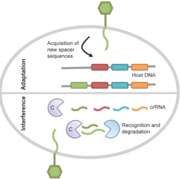
CRISPR interference is the only adaptive immunity system in
prokaryotes. CRISPR loci consist of an array of short direct repeats
separated by highly variable spacer sequences of precise length that
correspond to previously captured foreign DNA from bacteriophages or
plasmids [111,112]. CRISPR associated (cas) genes are located
immediately downstream of the repetitions and encode the protein
machinery in charge of the CRISPR activity. In the adaptation phase
CRISPR loci incorporates additional spacer sequences to enlarge their
activity against invader foreign DNA (Figure 4). Thus, the spacer content
reflects the many different elements that have been encountered by the
bacterial host. Once a spacer is established it can be used for protection
through the interference phase of the CRISPR pathway. This phase
involves the transcription of the CRISPR loci into a single RNA transcript
that is cleaved by the Cas proteins in order to generate CRISPR RNA
(crRNA) units, each containing one targeting spacer. These units will then
interfere with the incoming foreign nucleic acid through hybridization,
leading to its degradation by the bacterial cell degradation machinery
(Figure 4) [112]. Despite CRISPR/Cas adaptive character, bacteriophages
have already stroke back by finding a way to circumvent CRISPR
Figure 4. CRISPR/Cas immunity mechanism. In the adaptation phase of CRISPR
immunity, new spacers from the invading DNA are incorporated into CRISPR loci. During
the interference phase, repeats and spacers are transcribed into a long precursor that is
processed by a complex, made of Cas proteins, to generate CRISPR RNAs (crRNAs). This
complex together with the correspondent crRNA detects the foreign incoming DNA,
targeting the phage genome for degradation. Adapted from [113].
Bondy-Denomy and collaborators described recently several anti-CRISPR
phage-encoded genes mediating phage evasion to CRISPR/Cas system
in Pseudomonas aeruginosa by a yet unknown mechanism [114]. Seed
and collaborators report a Vibrio cholerae phage, ICP1, which uses its
own CRISPR/Cas system to evade a phage immunity system coded on a
host chromosomal island, PLE. Interestingly, and in relation with section
III.2 of this chapter, PLE has been recognized as a phage-related
chromosomal island (PRCI) due to its length, base composition and
genome organization [115]. Upon ICP1 infection, PLE on the bacterial
host circularizes and inhibits ICP1 lytic infection by an unknown
CRISPR/Cas to target PLE for destruction. If ICP1 has not a complete
CRISPR/Cas system, lytic infection is abolished due to the lack of
complementarity between ICP1-CRISPR spacers and the target
chromosomal island [115].
1.2.4 Abortive infection (Abi)
Abortive infection (Abi) system acts later in the phage infectious
process and targets phage replication, transcription, translation or genome
packaging [116]. Abi leads to the premature death of the phage-infected
bacteria, which limits phage replication and favours the surrounding
bacterial population. Although widespread in different bacteria such as E.
coli and B. subtilis, most of Abi systems have been identified in L. lactis
[116]. Abi systems are generally coded by plasmids. Similarly in E. coli
and L. lactis, Lit and AbiD1, respectively, code for an enzyme activated
upon phage infection that promotes cleavage of essential and conserved
components of the bacteria translational apparatus [117,118]. Recently,
the mechanism underlying L. lactis AbiQ abortive infection was unravelled.
AbiQ belongs to the type III toxin-antitoxin systems, which involve an
antitoxic RNA molecule interacting with its cognate toxic protein [119].
This system resembles to another type III toxin-antitoxin system also
involved in an abortive infection mechanism described in Erwinia
carotovora [120]. ABIQ protein has an endoribonuclease activity.
However, the exact mechanism through which it kills phage-infected
bacteria is still unknown [119].
2. Ecological impact
Bacteriophages have a major impact on the ecological balance and
dynamics of microbial life. They are found where bacteria thrive, whether it
is on sediments, aquatic systems or the human body. In nature, phages
they can encounter. The most important conditions are the host availability
and its nutritional and metabolic states that dictate the outcome of viral
infections and proliferation [121]. Regardless of the difficulties,
bacteriophage influence many biogeochemical and ecological processes,
determining biological production and community species composition, as
well as patterns of matter and energy transfer [122]. Although most of the
bacteriophage ecological studies have been performed in the aquatic
environment, the dynamic relationship phage-host can be extended to
other complex microbial ecosystems such as human associated microbial
communities [123]. For example, in the gut, phages are suspected to
shape the functionality and diversity of the human intestinal microbiome
[124]. One of the models that explain how phages control community
species composition is called ‘killing-the-winner’. It stipulates that viruses
control the most abundant or fastest growing population, enabling
less-competitive or slower-growing populations to co-exist with the dominant,
fast-growing hosts. The reduction in size of the dominant host population
gives the opportunity for new hosts to become abundant thus maintaining
the high diversity in the population [125]. Metagenomic studies have
recently revealed the bacteriophage diversity and abundance within the
human gastrointestinal tract [126,127]. Imbalanced composition of the
intestinal microbiota or dysbiosis is associated with many intestinal
diseases, such as inflammatory bowel disease (IBD), irritable bowel
syndrome (IBS) and obesity [128,129]. Given the beneficial or harmful
effects of phages on a bacterial population, phages are suspected to
influence the balance between intestinal symbionts and pathobionts under
certain circumstances [124,130].
2.1 Prophage induction & impact on host fitness
It is largely accepted that temperate phages have a major impact on
evolution of bacteria by shaping their genomes through horizontal gene
transfer [131]. Lysogeny is a common trait among bacteria, suggesting
that prophages provide advantages to their host whether it is a pathogen,
commensal or free living organism. This would compensate the potential
negative aspects of carrying extra DNA, such as metabolic burden from
extra DNA replication and cell lysis after prophage induction. Thus, to
benefit bacterial fitness, evolution would select lysogenic strains with
mutations that inactivate the prophage ability to perform the lytic cycle.
Next, large-scale deletion of prophage genes would decrease the
metabolic burden, while genes that increase host fitness would be
maintained [132]. In support of this theory, it is common to find multiple
prophages in various stages of functionality in the same bacterial
chromosome, as is the case of Streptococcus pyogenes, Lactobacillus
plantarum and different strains of E. coli [133-136]. Indeed, in addition to
fully functional prophages that can be induced to start a lytic cycle and
release phage progeny, bacterial genomes, also harbor non-functional
prophage-like entities: i) defective prophages, that are prophages in a
state of mutational decay. This is the case of the nine E. coli K-12
prophages, CP4-6, DLP12, e14, rac, Qin, CP4-44, CPS-53, CPZ-55 and
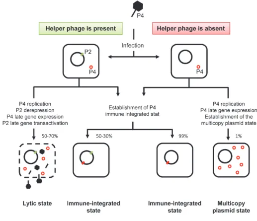
CP4-57 [136]; ii) prophage satellites, that are elements whose genome
has evolved to hijack structural proteins from a helper phage. The best
characterized example of such an interaction occurs between satellite
phage P4 and phage P2 (see section III.1 of this Chapter for details) [137];
and iii) gene transfer agents (GTAs), that are tailed phage like particles
that package random fragments of the bacterial genome. These virion-like
particles can transduce bacterial DNA into another host, in which they can
recombine with the chromosome and be vertically inherited. The best
characterized GTA is encoded on the Rhodobacter capsulatus
When bacterial strains harbor more than one prophage they are called
polylysogenic. Polylysogeny is frequent among pathogens, such as
Escherichia coli O157:H7 strain Sakai (18 prophages) and Streptococcus
pyogenes M3 (6 prophages) (Figure 5) [133,134], but also among
opportunistic pathogens and dairy strains such as E. faecalis V583 (7
prophages) and Lactococcus lactis IL1403 (6 prophages), respectively
[28,139].
Figure 5. Polylysogeny in human bacterial pathogens. Examples of human pathogens
for which the accumulation of prophages has a major impact for pathogenesis. From the
center to periphery: (A) Escherichia coli genomes from O157:H7 Sakai, O157:H7 EDL933,
K12 and CFT073; (B) Salmonella enterica genomes from serovar Thyphimurium LT2 and
serovar Tiphy CT18; (C) Staphylococcus aureus genomes from Mu50, N315, MW2 and
8325; (D) Streptococcus pyogenes genomes from M1, M3 and M18. Prophages are
indicated by dots, which size if proportional to phage genome length. Circles represent
A well studied case of impact of polylysogeny on pathogenesis and
emergence of hypervirulent clones is illustrated by Streptococcus
pyogenes. Serotype M3 strains isolated in the 1920s contains φ315.5 (encoding the SpeA1 exotoxin) while M3 strains causing disease in the
1940s have an additional prophage, φ315.2, encoding the streptococcal superantigen (SSA). At the same time, a nonsynonymous mutation in
SpeA1 on φ315.5 gave rise to SpeA3, a significantly more mitogenic variant. Contemporary serotype M3 strains have one more prophage,
φ315.4, which encodes exotoxin SpeK and streptococcal phospholipase A2, Sla (Figure 6) [141]. Thus, subclone M3 has accumulated prophages
over time resulting in the emergence of a superbug built from a unique
combination of phage-encoded virulence factors [134].
Figure 6. Hypothesis for the emergency of M3 hypervirulent clones. Early in the 20th
century, an ancestral strain, harboring phage 315.5, acquired phage 315.2, harboring SSA.
Subsequently, a single nucleotide mutation resulting in a single amino acid transformed
SpeA1 in SpeA3 variant encoded by phage 315.5. Later, this strain gained phage 315.4
that encodes both Sla and SpeK toxins and disseminated widely in the mid late 1980s.
Adapted from [141].
Prophages can impact bacterial fitness in several ways: introduction of
new fitness factors through lysogenic conversion, gene disruption and
initiate lytic cycle upon induction, frequently due to SOS-response
activation. Antibiotics targeting DNA replication, such as ciprofloxacin and
trimethoprim, can induce the SOS response and consequently prophages.
Frequently, they also enhance the expression of phage-encoded virulence
factors [143]. Some β-lactams, like ampicillin, penicillin, cloxacillin and ceftriaxone, are also able to induce an SOS-response [144].
Antibiotic-induced SOS response and consequent prophage and prophage-related
elements induction, promotes horizontal gene transfer of virulence genes
[145]. β-lactams induced SOS response has been attributed to the formation of reactive oxygen species (ROS) [146,147]. However recent
reports claim that these antibiotics do not induce the production of ROS,
and that their effect is probably due to direct inhibition of cell-wall
assembly, protein synthesis and DNA replication [148,149].
2.1.1 Lysogenic conversion
Lysogenic conversion was first reported in Corynebacterium
diphtheriae, the etiological agent of diphtheria. Freeman reported that
non-lysogenic strains become toxicogenic after infection with a tox+
corynebacteriophage β [150]. This is the typical situation in which bacteriophages encoding virulence factors can convert their bacterial host
from a non-pathogenic strain into a strain with increased virulence. The
tox gene, which codes for diphtheria toxin (DT), is activated by low iron
concentration. After production, DT is secreted and attached to the cell
surface of respiratory epithelium cells where it promotes cell death [151].
Since, the list of lysogenic conversion genes and their mechanisms of
action in many other bacterial pathogens has grown and includes both
Gram-positive and Gram-negative bacteria such as S. aureus, S.
pyogenes, E. coli, S. enterica, Pseudomonas aeruginosa and Vibrio
cholerae (Table 3) [142]. The most important factors for the success of a
acquisition of nutrients and evasion to the innate and adaptive immune
systems. In many of these circumstances, prophage-encoded factors can
provide an advantage to the bacterial host (Table 3) [142,152]. For
example, different phage-encoded proteins promote bacterial evasion
from the immune system through the modification (glucosylation or
acetylation) of the O-antigen of different bacteria, such as S. enterica
[153,154], P. aeruginosa [155,156], Shigella flexneri [153,157] and
Neisseria meningitidis [158]. Other phage-encoded proteins contribute to
bacterial survival to eukaryotic cell environment such as oxidative stress.
Phages Gifsy-2 and Fels-1 from S. enterica code for superoxide
dismutases protecting the bacteria against host produced ROS [159].
Finally, prophages carry an arsenal of extracellular toxins used by their
bacterial host to induce damage in eukaryotic cells. They perform
functions such as cytotoxic (as CTX from P. aeruginosa) [160], cardiotoxic
(as TOX from C. diphtheriae) [150] or neurotoxic (as C1 from C.
botulinum) [161]. Interestingly, two prophages of the E. coli O157:H7
strain and other toxins producing E. coli (STEC) encode
Shiga-toxins, Stx1 and Stx2, which cause severe hemorrhagic colitis and
hemolytic uremic syndrome [162]. Antibiotic treatments of these infections
are deleterious for the patients as they induce prophages, and
consequently increase toxin production [163,164].
2.1.2 Gene disruption
Lysogenization of S. aureus by φ13 results in loss of β-toxin expression due to prophage integration into the 5' end of β-toxin gene (hlb) [165]. In Listeria, φ10403S insertion interrupts comK gene, which corresponds to the master regulator of DNA uptake competence system in
B. subtilis. Prophage excision restores ComK that can activate its target
![Figure 2. Timeline of major milestones in phage history. Adapted from [94].](https://thumb-eu.123doks.com/thumbv2/123dok_br/15763961.640070/50.892.118.652.512.883/figure-timeline-major-milestones-phage-history-adapted.webp)
![Table 2. Overview of prokaryotic virus families [95].](https://thumb-eu.123doks.com/thumbv2/123dok_br/15763961.640070/51.892.263.760.409.799/table-overview-prokaryotic-virus-families.webp)

![Table 3. Phage encoded bacterial virulence factors [142,152].](https://thumb-eu.123doks.com/thumbv2/123dok_br/15763961.640070/65.892.242.771.126.918/table-phage-encoded-bacterial-virulence-factors.webp)
![Figure 11. SaPI life style scenarios [227] (cont.). For A and B see page 54. (C) Co- Co-infection of a SaPI and a helper phage](https://thumb-eu.123doks.com/thumbv2/123dok_br/15763961.640070/84.892.125.658.140.743/figure-sapi-style-scenarios-infection-sapi-helper-phage.webp)

![Figure 14. SaPI and phage 80 α capsid assembly pathways [226]. Phage procapsids are assembled from the major capsid protein (gp47), scaffolding protein (gp46), portal protein (gp42) and a minor capsid protein (gp44)](https://thumb-eu.123doks.com/thumbv2/123dok_br/15763961.640070/87.892.237.736.498.770/figure-assembly-pathways-procapsids-assembled-protein-scaffolding-protein.webp)
![Figure 15. Model for SaPI packaging redirection [226]. pac sites on the concatemeric phage DNA are recognized by the phage encoded TerS and packaged into procapsids through the action of phage TerL](https://thumb-eu.123doks.com/thumbv2/123dok_br/15763961.640070/89.892.249.770.117.308/figure-packaging-redirection-concatemeric-recognized-encoded-packaged-procapsids.webp)
