V
c
9V
d
2 T Cell-Mediated Cytotoxicity
Matilde Todaro1,2, Valentina Orlando2,3, Giuseppe Cicero1, Nadia Caccamo2,3, Serena Meraviglia3, Giorgio Stassi1, Francesco Dieli2,3*
1Dipartimento di Discipline Chirurgiche ed Anatomiche, Universita` di Palermo, Palermo, Italy,2Biomedical Research Centre, Universita` di Palermo, Palermo, Italy,
3Dipartimento di Biopatologia e Biotecnologie Mediche e Forensi, Universita` di Palermo, Palermo, Italy
Abstract
Colon cancer comprises a small population of cancer initiating stem cells (CIC) that is responsible for tumor maintenance and resistance to anti-cancer therapies, possibly allowing for tumor recapitulation once treatment stops. Combinations of immune-based therapies with chemotherapy and other anti-tumor agents may be of significant clinical benefit in the treatment of colon cancer. However, cellular immune-based therapies have not been experimented yet in the population of colon CICs. Here, we demonstrate that treatment with low concentrations of commonly used chemotherapeutic agents, 5-fluorouracyl and doxorubicin, sensitize colon CICs to Vc9Vd2 T cell cytotoxicity. Vc9Vd2 T cell cytotoxicity was largely mediated by TRAIL interaction with DR5, following NKG2D-dependent recognition of colon CIC targets. We conclude that
in vivoactivation of Vc9Vd2 T cells or adoptive administration ofex-vivoexpanded Vc9Vd2 T cells at suitable intervals after
chemotherapy may substantially increase anti-tumor activities and represent a novel strategy for colon cancer immunotherapy.
Citation:Todaro M, Orlando V, Cicero G, Caccamo N, Meraviglia S, et al. (2013) Chemotherapy Sensitizes Colon Cancer Initiating Cells to Vc9Vd2 T Cell-Mediated Cytotoxicity. PLoS ONE 8(6): e65145. doi:10.1371/journal.pone.0065145
Editor:Jacques Zimmer, Centre de Recherche Public de la Sante´ (CRP-Sante´), Luxembourg
ReceivedMarch 5, 2013;AcceptedApril 23, 2013;PublishedJune 6, 2013
Copyright:ß2013 Todaro et al. This is an open-access article distributed under the terms of the Creative Commons Attribution License, which permits unrestricted use, distribution, and reproduction in any medium, provided the original author and source are credited.
Funding:This work has been supported by grants from the Italian Ministry for Instruction, University and Research (contract no. 2008L57JXW to FD), the Italian Ministry of Health (Progetto Ricerca Finalizzata 2007 ‘‘Stem cells in different pathological conditions: innovative therapeutical approches’’ to FD), Istituto Superiore di Sanita` Oncoproteomic Project 2007-527/B/3A/3 (to GS and FD) and the University of Palermo. The funders had no role in study design, data collection and analysis, decision to publish, or preparation of the manuscript.
Competing Interests:The authors declare no financial or commercial conflict of interest. The corresponding author, Francesco Dieli, is an Academic Editor of PLOS ONE. This does not alter the authors’ adherence to all the PLOS ONE policies on sharing data and materials.
* E-mail: francesco.dieli@unipa.it
Introduction
In recent years, novel insights in cancer research have suggested that the capacity to initiate and sustain tumor growth is a unique characteristic of a small subset of cancer cells with stemness properties within the tumor mass, called ‘‘cancer stem cells’’ (CSCs) or ‘‘cancer-initiating cells’’ (CICs) [1]. Chemotherapy remains the primary treatment choice for many advanced cancers and has cytotoxic anti-tumor activity through a range of mechanisms. However, most cancers are resistant to current therapies due to the slow-cycling CICs, the location of these cells within hypoxic niches [2,3], and because the malignant cells have the capacity to develop mechanisms to resist or escape the cytotoxic effects of chemotherapy [4], which include up-regulation of several ATP-binding cassette transporters, active DNA-repair capacity and over-expression of anti-apoptotic molecules that cause changes in the signalling pathways controlling proliferation, differentiation and apoptosis [5].
Several studies have demonstrated that treatment of tumor cells with chemotherapeutic drugs induces or increases their sensitivity to cytotoxicity by NK or T lymphocytes; thus, combinations of cellular immune-based therapies with chemotherapy and other anti-tumor agents may be of significant clinical benefit in the treatment of many forms of cancer [6].
cdT cells are of particular interest for use in such combined therapies due to their potent anti-tumor cytotoxicity and the
relative ease of generationin vitro [7]. Humancd T cells can be
cytotoxic activity toward cancer cells, which is mediated in much the same manner as for CD8 T cells and NK cells, through perforin/granzyme, Fas/FasL, TNF/TNF-R and TRAIL-TRAIL-R pathways [10].
In this study, we have assessed the potential synergy of combining chemotherapy and Vc9Vd2 T cell-mediated cytotox-icity for anti-tumor therapy. Specifically, as colon CICs are resistant to both chemotherapeutic drugs and to Vc9Vd2 T cell-mediated cytotoxicity, we have determined whether chemotherapy can be used to sensitize colon CIC targets to Vc9Vd2 T cell cytotoxicity, based on three lines of evidence: (1) pioneering work by Mattarollo and colleagues [25] has demonstrated high levels of cytotoxicity against solid tumor-derived cell lines with combination treatment utilizing Vc9Vd2 T cells and chemotherapeutic agents; (2) IL-17-producingcdT cells play a decisive role in chemother-apy-induced anti-cancer immune responses in the mouse [26]; (3) treatment of colon CICs with the bisphosphonate zoledronate enhances their sensitivity to Vc9Vd2 T cell killing [27].
We show here that chemotherapeutic drugs currently used for treatment of colon cancer patients, 5-fluorouracyl and doxorubi-cin, are capable to sensitize colon CICs to Vc9Vd2 T cell-mediated killing and we demonstrate that the underlying mechanisms involve NKG2D and TRAIL.
Results
Resistance of Colon CICs to Chemotherapy
We have previously reported that colon cancer comprises a vast majority of differentiated cells and a small population of CICs that are responsible for tumor initiation and maintenance [28]. For this study purposes, we purified and propagated colon cancer spheres from surgical fragments of 5 patients with colon carcinoma. These cancer sphere lines were identified through the expression of CD133 and the epithelial specific antigen ESA, displayed adherence to the culture dishes in the presence of serum and subsequently differentiated into large, polygonal colon cells expressing colon epithelial markers, such as villin, suggesting that colon cancer spheres maintained the ability toin vitrodifferentiate
in enterocyte-like cells. Most importantly, when injected subcuta-neously into NOD/SCID mice, a low number of colon cancer spheres, but not sphere-derived differentiated cells, retained the capacity to form a tumor that closely resembled the human original tumor (Supporting Figure S1).
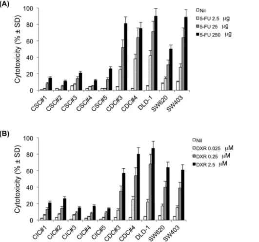
CICs are characterized by high resistance to drugs and general toxins which target rapidly proliferating cells and spare the slow dividing cells, due to an up-regulation of several ATP-binding cassette transporters, active DNA-repair capacity, over-expression of anti-apoptotic molecules that cause changes in the signalling pathways controlling proliferation, differentiation and apoptosis [5]. Accordingly, exposure of 5 different colon CIC lines (CIC#1 to CIC#5) to 5-FU (2.5 and 25mg/ml) (Figure 1A) or DXR (0.025 and 0.25mM) (Figure 1B) for 24–72 hrs had virtually no significant cytotoxic effect, as determined by PI staining. Highest doses of 5-FU (250mg/ml) and DXR (2.5mM) caused low, yet detectable cytotoxicity of CIC lines ranging from 1565% to 2366% (mean 6 SD). Conversely, 5-FU and DXR were fully capable of killing 3 differentiated colon cancer cell lines DLD-1, SW620 and SW403, and 2 differentiated cell lines (CDC#3 and CDC#4) obtained from two patients (P#3 and P#4) where form the CICs lines were also obtained, with a dose-dependent increase in cytotoxicity up to 85%. The viability of untreated cells was all over 90% (Figures 1A and B).
Chemotherapy Sensitizes Colon CICs to Vc9Vd2 T Cell Cytotoxicity
In analogy to their resistance to chemotherapy, the five tested colon CIC lines, were also resistant to Vc9Vd2 T cell-mediated cytotoxicity, even when an E:T ratio of 50:1 was used (Figure 2A). The poor cytotoxic activity against colon CICs was not an intrinsic feature of the Vc9Vd2 T cells, because the differentiated colon cancer cell lines DLD-1, SW620, SW403, CDC#3 and CDC#4 were efficiently killed by two Vc9Vd2 T cell lines COLD2-1 and COLD2-2 obtained from two different colon cancer patients (P#3 and P#4) (Figure 2A), as well as Vc9Vd2 T cell lines obtained from healthy subjects (data not shown). As a control, all the tested Vc9Vd2 T cell lines failed to kill the normal colon cell line CCL-241 (Figure 2A).
In previous studies, we have demonstrated that zoledronate sensitizes colon cancer CICs to Vc9Vd2 T cell cytotoxicity [27]. The capability of Vc9Vd2 T cells to kill colon cancer CICs was then assessed after treatment of the targets with chemotherapy. Representative results obtained with three different CIC lines (CIC#2, CIC#4 and CIC#5) are shown in Figure 2B. Vc9Vd2 T cell cytotoxicity was enhanced in all cases by pre-treatment of target CICs with chemotherapy. In detail, almost complete lysis of CIC lines resulted from the combination of the highest doses of 5-FU (250mg/ml) or DXR (2.5mM) and Vc9Vd2 T cells, with cell death percentages over 90% at an E:T ratio of 20:1. Treatment of targets with lower doses chemotherapy (2.5 and 25mg/ml 5-FU and 0.025 and 0.25mM DXR) resulted in enhanced killing of CIC lines by Vc9Vd2 T cells, indicating that chemotherapy and Vc9Vd2 T cells have additive activity even when used at suboptimal doses.
Chemotherapy Upregulates DR5 (TRAIL-R2) Death Receptor Expression on CICs
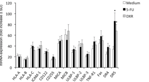
To decipher the molecular mechanisms behind chemotherapy-mediated sensitization of CICs to Vc9Vd2 T cells cytotoxicity, we focused on expression of mRNA encoding for molecules known to be ligands for key activating receptors on Vc9Vd2 T cells and death receptors, before and after exposure of CICs to chemother-apy agents. As shown inFigure 3, all of these molecules were constitutively expressed in CICs, although expression consistently varied amongst different CIC lines; however, no major differences were observed in all tested CIC lines for HLA-class I, ICAM-1, CD155, CD112, MICA/B and ULPBP1–4 expression before and after exposure to chemotherapy agents.
Expression of Fas (CD95), TNF-R1, DR4 (TRAIL-R1) and DR5 (TRAIL-R2) death receptors was increased in the majority of CIC lines following exposure to chemotherapeutic agents (Figure 3), but increased expression of Fas, TNF-R1 and DR4 did not attain statistical significance. The greatest and significant increase was only observed for DR5 expression after exposure of CICs to 5-FU and, although at a lesser extent, DXR (Figure 3). Upregulation of DR5 following 48 hrs exposure of colon CICs to chemotherapy was confirmed by flow cytometry upon staining with specific mAb (Figure 4).
Killing of Chemotherapy-treated CICs by Vc9Vd2 T Cells is Mediated by NKG2D and TRAIL
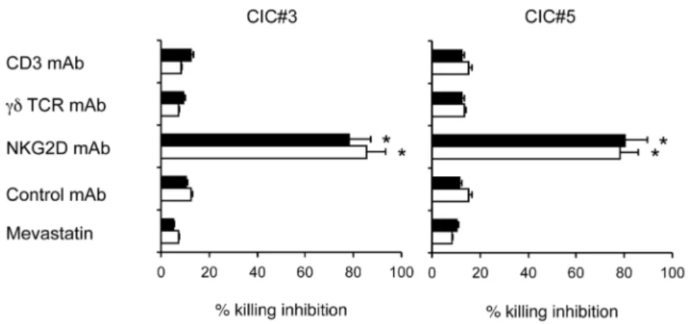
NKG2D receptors. Cytotoxicity of chemotherapy-pretreated colon CIC lines by two different Vc9Vd2 T cell lines was significantly inhibited by anti-NKG2D mAb, while the Vc9Vd2 TCR seems to play a minor role as indicated by the failure of anti-CD3 and anti-pan cd TCR mAbs to inhibit cytotoxicity (Figure 5). In addition, Vc9Vd2 T cell killing of chemotherapy-sensitized targets was assessed in the presence of mevastatin, which inhibits 3-hydroxy-3-methylglutaryl-CoA and prevents zoledronate-mediated accumulation of endogenous phosphoantigens as IPP. Mevastatin failed to inhibit killing of all tested chemotherapy-pretreated colon CIC lines by two different allogeneic Vc9Vd2 T cell lines (Figure 5), thus indicating that chemotherapy-induced sensitization of CICs to Vc9Vd2 T cell cytotoxicity does not rely on production of mevalonate metabo-lites.
To further elucidate the mechanisms of killing of chemother-apy-sensitized colon CICs by Vc9Vd2 T cells, we individually inhibited the granule exocytosis, TNF-a-, TRAIL-, and FasL-mediated pathways. Killing-inhibition experiments revealed that Vc9Vd2 T cell cytotoxicity of chemotherapy-pretreated colon CIC targets was significantly inhibited by anti-DR5 mAb, whereas mAbs against DR4, TNF-a, and FasL, or treatment with CMA to block the granule-exocytosis pathway, all failed to inhibit. Figure 6 shows representative data with two Vc9Vd2 T cell lines and the two colon CIC lines, CIC#2 and CIC#4.
Discussion
It is now emerging that cancer is generated by a population of cells displaying stemness features, named cancer stem cells or cancer-initiating cells (CICs) [1,2]. These cells, which contribute only to a minor fraction of the total tumor mass, undergo long-term expansion with retention of their ability to reproduce the original tumor phenotype, thus providing evidence for self-renewal and tumor-initiating capacity [1,2]. The CIC population is more resistant than differentiated primary cells to conventional chemo-therapy and radiochemo-therapy and to putative innovative therapies such as those based on the use of TRAIL. This refractoriness has been attributed to the fact that CICs express multidrug resistance genes including high levels of anti-apoptotic proteins and ABC (ATP Binding Cassette) transporters which pump out the drugs, but also to the fact that chemotherapy targets dividing cells and consequently fails to kill the slow-cycling CICs [3–5].
Data from recent clinical studies have suggested that combining chemotherapy with immunotherapy has survival benefits than chemotherapy alone [6,29], as outlined for example by the combination of chemotherapy and monoclonal antibodies [30– 32]. Moreover, it is known that chemotherapeutic drugs can sensitize tumor cells to cytotoxicity mediated by CD8, NKT or Vc9Vd2 T cells [33] thorugh several different mechanisms [34]. However, we recently found that colon CICs are resistant to Vc9Vd2 T cell cytotoxicity, unless they are sensitized with zoledronate [35]: similarly, we have now tested the possibility
Figure 1. Cell cytotoxicity following treatment with 5-FU (A) or DXR (B).Colon CICs, differentiated colon cancer cell lines DLD-1, SW620, SW403, CDC#3 and CDC#4 were treated with different concentrations of 5-FU or DXR for 48 hrs. Cytotoxicity (%6SD) was determined by the degree of reduction of viable cells with the ability to retain CFSE and exclude PI (CFSEhighPI2). Shown is a representative experiment out of three.
that chemotherapeutic drugs currently used in the treatment of colon cancer might also sensitize colon CICs to Vc9Vd2 T cell killing.
Initial testing of cytotoxicity revealed that in analogy with our previously reported results [27], many colon CIC lines were resistant to the cytotoxic activity of Vc9Vd2 T cells, but pretreatment with low, sublethal concentrations of chemothera-peutic drugs 5-FU and DXR sensitizes CIC targets to Vc9Vd2 T cell killing, resulting in additive cytotoxicity activity.
Vc9Vd2 T cells interact with and kill tumor targets thorugh several different mechanisms including granule exocytosis, death receptor/ligands interactions with TNF, TRAIL and FasL, and TCR- or NKG2D-mediated recognition of phosphoantigens or stress-inducible molecules, respectively. All tested colon CIC lines constitutively expressed mRNA encoding for HLA-class I, ICAM-1, CD155, CD112, MICA/B, ULPBP1-4, Fas (CD95), TNF-RICAM-1, DR4 (TRAIL-R1) and DR5 (TRAIL-R2) molecules on their
surface, but expression of all these molecules did not render CICs sensitive to Vc9Vd2 T cell killing. However, exposure of colon CICs to 5-FU and, although at a lesser extent DXR, significantly increased DR5 expression.
Several previously published reports in the literature have demonstrated that many chemotherapeutic drugs, including 5-FU and DXR, upregulate DR5 expression on tumor cell lines of distinct tissue origin [36–42]. However, this effect has been reported on differentiated cancer cells, while, to our knowledge, there is no evidence of similar DR5 upregulation on CICs. Whether or not chemotherapy-induced DR5 upregulation is restricted to colon CICs or is a general phenomenon observed on other CICs is actually under study.
Nonetheless, we found that Vc9Vd2 T cells exploited different mechanisms to kill CIC targets, which were strictly dependent on the way of target CICs sensitization. Regardless of whether chemotherapeutic drugs or zoledronate were used to sensitize
Figure 2. Chemotherapy sensitizes resistant colon CICs to Vc9Vd2 cell-mediated cytotoxicity.(A) Cytotoxicity percentage of 2 different to Vc9Vd2 T cell lines, COLD2-1 and COLD2-2 obtained from 2 patients affected by colon cancer, against colon cancer sphere cells from 5 different patients (CIC#1 to CIC#5), differentiated colon cancer cell lines DLD-1, SW620, SW403, CDC#3 and CDC#4, and the normal colon cell line CCL-241, at an E:T ratio of 50:1. (B) Three different target colon CICs (CIC#2, CIC#4 and CIC#5) treated with or without either 5-FU (2.5 to 250mg/ml) or DXR (0.025 to 2.5mM) for 48 hrs were tested for their sensitivity to 2 different to Vc9Vd2 T cell lines, COLD2-1 and COLD2-2 obtained from 2 patients affected by colon cancer and used at an E:T ratio of 20:1. Results indicate cytotoxicity of tumor targets following 6 hrs co-culture with Vc9Vd2 T cell lines. Data are mean percentage6SD of 5 different experiments, each carried out in triplicate.
doi:10.1371/journal.pone.0065145.g002
CICs, Vc9Vd2 T cells killing of these targets was TCR- or NKG2D-mediated: consistent with our previous report [27] chemotherapy-sensitized colon CICs were killed following NKG2D-mediated recognition and TRAIL/DR5 interaction, while both mechanisms were dispensable to the cytotoxicity of zoledronate-sensitized colon CICs, which were almost exclusively killed by TCR-mediated interaction and the perforin/granzyme pathway.
Previous studies have highlighted the importance of NKG2D-MICA/B interactions for tumour cell recognition and effective cytotoxic activity by Vc9Vd2 T cells [35–44]. The difference between NKG2D-mediated recognition of chemotherapy-sensi-tized colon CICs and TCR-mediated recognition of zoledronate-sensitized CIC targets cannot be explained differential expression of MICA/B or ULBPs since neither 5-FU nor DXR changed constitutive expression levels of these molecules. It is likely that phosphoantigens production/expression by colon CICs is very low, below the threshold required for efficient recognition by the reactive Vc9Vd2 TCR, hence target recognition only occurs through NKG2D: the finding that colon CICs become sensitive to Vc9Vd2 T cell cytotoxicity upon exposure to zoledronate [27],
which enhances phosphoantigen accumulation and production, supports this possibility.
We conclude that in vivo activation of Vc9Vd2 T cells or
adoptive transfer ofex vivo-activated Vc9Vd2 T cells, together with
or soon after administration of certain chemotherapeutic drugs may substantially increase their anti-tumor effects. Additional clinical studies are thus needed to assess the efficacy of this combinatory therapy, possibly including the novelcdT cell-based immunotherapeutic approach thatex-vivoexpansion of polyclonal cdT cells followed by introduction of a CD19-specific chimeric antigen receptor render them bispecific and more efficient in killing of CD19+tumor cell linesin vitroand in xenografts [45].
Materials and Methods
Peripheral Blood and Colon Cancer Samples
Human peripheral blood mononuclear cells (PBMC) and colon cancer tissues were obtained in accordance with the ethical standards of the institutional committee of human experimenta-tion from patients undergoing a colon resecexperimenta-tion for colon adenocarcinoma. Histological diagnosis was based on microscopic features of carcinoma cells determining the histological type and grade. PBMC were isolated from colon cancer patients by density gradient centrifugation using Ficoll-Hypaque (Pharmacia Biotech, Uppsala, Sweden) and were cryopreserved in 80% RPMI 1640 (Life Technologies, Monza, Italy), 10% DMSO (Sigma, St. Louis, MO) and 10% heat-inactivated fetal calf serum (FCS, Life Technologies).
According to Italian rules (Article 13 of Legislative Decree no. 196/03), this study did not require authorisation by the local ethical committee. The study was performed in accordance to the principles of the Helsinki declaration and all individuals gave written informed consent to participate.
Purification and Culture of CICs
Cancer tissues were extensively washed in saline buffer containing antibiotics and incubated overnight in DMEM/F12 (Life Technologies) containing penicillin (500 IU/ml), streptomy-cin (500mg/ml) and amphotericin B (1.25mg/ml) (Life
Technol-Figure 3. Colon CICs constitutively express molecules involved in by Vc9Vd2 T cell-mediated cytotoxicity: effect of chemotherapy.
RT-PCR of the expression of mRNA encoding for different surface molecules in colon CICs treated with or without either 5-FU (25mg/ml) or DXR (0.25mM) for 48 hrs. Data represent the mean values6SD of 4 separate experiments, each performed with colon cancer spheres from 5 different patients (CIC#1 to CIC#5).
doi:10.1371/journal.pone.0065145.g003
Figure 4. Chemotherapy upregulates DR5 expression on coloc CICs.Colon CICs were treated with medium, 5-FU (25mg/ml) or DXR (0.25mM) for 48 hrs, washed extensively and stained with anti-DR5 mAb. Flow cytometry histograms show DR5. Mean fluorescence intensity (MFI) for DR5 staining is indicated in the upper right corner of each panel. Dotted lines represent isotype control mAb, while grey filled histogram represent anti-DR5 mAb.
ogies). Enzymatic digestion was performed using collagenase (Life Technologies, 1.5 mg/ml) and hyaluronidase (Sigma, 20mg/ml) in DMEM containing antibiotics/antimycotics for 1 hour. Recov-ered cells were then cultured in serum-free medium (DMEM/F12) supplemented with 6 mg/ml Glucose, 1 mg/ml NaHCO3, 5 mM HEPES, 2 mM L-Glutamine, 4mg/ml Heparin, 4 mg/ml BSA, 10 ng/ml bFGF, 20 ng/ml EGF, 100mg/ml apotrasferrin, 25mg/ml insulin, 9,6mg/ml putrescin, 30 nM sodium selenite anhydrous and 20 nM progesterone (Sigma) to a final concentra-tion of 36105 cells/ml. These culture conditions select for
immature tumor cells that slowly proliferate, giving rise, within 2–3 months, to tumor cell aggregates, called ‘‘spheres’’. Sphere-forming cells can be propagated by enzymatic dissociation of spheres (3 mM EDTA, 50 nM DTT in PBS), followed by re-plating of single cells and residual small cell aggregates in fresh serum-free medium [28,46,47].
Tumorigenicity was evaluated by subcutaneous implantation of either disaggregated colon cancer sphere cells or sphere-derived differentiated cells [27]. Differentiated colon cancer cells lines
DLD-1, SW620 and SW403 (American Type Culture Collection) were obtained from Dr. Ruggero De Maria (‘‘Regina Elena’’ National Cancer Institute, Rome, Italy) and were maintained in DMEM containing antibiotics and 10% FCS. All cell cultures were carried out at 37uC in a 5% CO2humidified incubator.
Anti-tumor Agents, Antibodies and Reagents
The chemotherapeutic agents 5-fluorourcil (5-FU) and doxoru-bicin (DXR) were obtained from Sigma, through the pharmacy of the University Hospital. Drugs were diluted in DMSO and diluted to the required concentrations in PBS prior to use.
The following unconjugated, FITC-, PE-, PE-Cy5- or APC-conjugated monoclonal antibodies (mAbs) were used: anti-TCR Vd2 (B6, BD Biosciences, San Jose´, CA), anti-NKG2D (1D11, eBioscience, San Diego, CA), anti-CD95L (2C101,Vinci Biochem, Firenze, Italy), anti-MICA/B (6D4, BD Biosciences).
Additionally, the following purified mAbs were also used: anti-CD3 (blocking, MEM-57), anti-HLA Class I monomorphic (MEM-147) from Prof. Vaclav Horejsi (Institute of Molecular
Figure 5. Modulation of the cytotoxic activity of Vc9Vd2 T cells by blocking the TCR or NKG2D interactions.The Vc9Vd2 T cell line COLD2-1 was cultured with two chemotherapy-treated colon CICs (CIC#3 and CIC#5) at an E:T ratio of 20:1, in the presence of blocking antibodies to thecdTCR, CD3, NKG2D, or in the presence of mevastatin. Specific cytotoxicity levels achieved by the Vc9Vd2 T cell line COLD2-1 were 65611 for CIC#3 and 7169 for CIC#5. Data are mean6 SD of two experiments carried out in triplicate. Percent inhibition with anti-NKG2D mAb was significantly different than values in all other groups (*p,0.001).
doi:10.1371/journal.pone.0065145.g005
Figure 6. Modulation of the cytotoxic activity of Vc9Vd2 T cells by blocking death receptors interactions. The Vc9Vd2 T cell line COLD2-1 was cultured with two chemotherapy-treated colon CICs (CIC#3 and CIC#5) at an E:T ratio of 20:1, in the presence of blocking antibodies to TNF-a, FasL (CD95L), TRAIL receptors R1 (DR4) or R2 (DR5), or concanamycin A (CMA). Specific cytotoxicity levels achieved by the Vc9Vd2 T cell line COLD2-1 were 6167 for CIC#3 and 65612 for CIC#5. Data are mean6SD of experiments carried out in triplicate. Percent inhibition with anti-DR5 and anti-TRAIL mAbs were significantly different than values in all other groups (*p,0.001).
doi:10.1371/journal.pone.0065145.g006
Genetics, Prague, Czech republic), anti-TCR pancd(IMMU510, a gift of Dr. Marc Bonneville, Institut de Biologie, Nantes, France), anti-TNF-a (Infliximab, a gift of Prof. Giovanni Triolo, Diparti-mento Biomedico di Medicina Interna e Specialistica, Universita` di Palermo, Palermo, Italy), anti-TRAIL receptors TRAIL-R1(DR4), TRAIL-R2 (DR5), TRAIL-R3 (LIT, DcR1) and TRAIL-R4 (TRUNDD, DcR2) all provided by Dr. Henning Walczak (Tumor Immunology Unit, Division of Medicine, Imperial College, London, UK).
Concanamycin A (CMA) and mevastatin were purchased from Sigma, while zoledronate was from Novartis Pharma, Basel, Switzerland.
Generation of Polyclonal Vc9Vd2 T Cell Lines
Polyclonal Vc9Vd2 T cell lines were generated by first enriching PBMC using acd T cell isolation kit (Miltenyi Biotec, Bergisch Gladbach, Germany), followed by sorting single Vc9Vd2 T cells through a FACSAria (BD Biosciences) with specific mAbs. Cells (26103) were then cultured into each well of round-bottom, 96-well plates containing 26104irradiated (40 Gy)
allogeneic PBMC, 26103 irradiated (70 Gy) EBV-transformed
allogeneic B cells, 0.5mg/ml PHA (Sigma), and 200 U/ml recombinant interleukin 2 (Proleukin, Novartis Pharma). Growing lines were expanded in 200 U/ml IL-2 and restimulated every 2 weeks. Usually, cells were collected after 4–6 weeks of culture to be used for functional assaysin vitro.
Cytotoxic Assay
Target colon CIC (105cellsml) were pre-treated with 5-FU (2.5– 250mg/ml), DXR (0.025–2.5mM) or zoledronate (0.5mM) for 24, 48 or 72 hrs. Cells were extensively washed in PBS and stained with CFSE (Merck, Milano, Italy) as follows: 50ml of CFSE were added to 1 ml of target sphere cell suspension (56105cells/ml) in PBS to obtain the final concentration of 2.5mM CFSE. The cells were incubated for 10 minutes at 37uC and gently mixed every 5 min. At the end of incubation, 1 ml of FBS was added to the cell suspension to stop the staining reaction and the cells were centrifuged at 600 g for 5 min at room temperature, washed twice with cold PBS and resuspended in serum-free medium.
Vc9Vd2 T cell lines were resuspended at the final concentra-tions of 106and 2.56106cells/ml, were added to CFSE-stained
target colon CICs (16105) and co-cultures were maintained for 6 hrs a 37uC in presence of 5% of CO2. At the end of the
incubation period, the cells were washed with PBS and stained with 20ml of Propidium Iodide (PI, Sigma, 1mg/ml) for 10– 15 min in ice. Finally 100ml of cold PBS were added before acquisition on a FACSCalibur cytometer (BD Biosciences). The calculation of cytolytic activity was based on the degree of reduction of viable target cells with the ability to retain CFSE and exclude PI (CFSEhighPI2), according to reference [27].
Blocking agents were used to evaluate the mechanisms of Vc9Vd2 T cell-mediated cytotoxicity of colon CICs. To evaluate the contribution of mevalonate metabolites tumor target cells were treated with mevastatin (25mM for 2 h) a selective upstream inhibitor of the mevalonate pathway. After this incubation period, target cells were washed, and Vc9Vd2 T cells added in the
presence of 25mM mevastatin, to maintain a constant concentra-tion of this drug during incubaconcentra-tion because its effect is rapidly reversible [27]. To inhibit perforin-mediated cytotoxicity, Vc9Vd2 T cells were incubated with concanamycin A (CMA, 15 nM) for 30 min at 37uC before co-culture with target CICs, without further washing [27]. To block the relevant cytotoxic pathways, specific or isotype-control mAbs were used at 10mg/ml final concentration just before co-incubation assay [27].
Real-time Quantitative RT-PCR
Total RNA was extracted with the ABI PRISM 6100 Nucleic Acid PrepStation (Applied Biosystems through Life Technologies) according to the manufacturer’s instructions. Random hexamers and an MMLV Reverse Transcriptase kit (Stratagene, La Jolla, CA) were used for cDNA synthesis. Transcripts were quantified by real-time quantitative PCR on an ABI PRISM 7700 Sequence Detector (Applied Biosystems) with Applied Biosystems prede-signed TaqMan Gene Expression Assays and reagents according to the manufacturer’ s instructions. The following probes were used (identified by Applied Biosystems assay identification number): HLA-A, Hs01058806_g1; HLA-B, Hs00818803_g1; HLA-C, Hs00740298_g1; ICAM-1, Mm00516023_m1; CD155,
Hs00197846_m1; CD112, Hs01071562_m1; MICA,
Hs00741286_m1; MICB, Hs00792952_m1; ULBP-1, Mm01180648_m1; ULBP-2, Hs00607609_mH; ULBP-3, Hs00225909_m1; Fas (CD95), Hs00236330_m1; TNF-R1, Mm00441883_g1; DR4 (TRAIL-R1), Hs00269492_m1; DR5 (TRAIL-R2), Hs00366278_m1. For each sample, mRNA abun-dance was normalized to the amount of 18S rRNA.
Statistics
The two-tailed Student’sttest was used to compare significance
of differences between groups. All values are expressed as mean6
standard deviation (SD).
Supporting Information
Figure S1 A low number of colon CIC spheres retain the capacity to form a tumor when injected s.c. into immunodeficient mice. Subcutaneous tumor growth in NOD/SCID mice 10 weeks after injection of 2000 disaggregated cells from colon cancer spheres. One representative experiment of two performed with cells from different donors is shown. (TIF)
Acknowledgments
We thank Ruggero De Maria, Vaclav Horejsi, Marc Bonneville, Giovanni Triolo and Henning Walczak for providing us with the cell lines and reagents.
Author Contributions
Conceived and designed the experiments: GS FD. Performed the experiments: MT VO NC SM. Analyzed the data: GC GS FD. Contributed reagents/materials/analysis tools: GS FD. Wrote the paper: GS FD.
References
1. Vermeulen L, Sprick MR, Kemper K, Stassi G, Medema JP (2008) Cancer stem cells old concepts, new insights. Cell Death Differ 15: 947–958.
2. Koch U, Krause M, Baumann M (2010) Cancer stem cells at the crossroads of current cancer therapy failures–radiation oncology perspective. Semin Cancer Biol 20: 116–124.
3. Baumann M, Krause M, Hill R (2008) Exploring the role of cancer stem cells in radioresistance. Nat Rev Cancer 8: 545–554.
4. Vogelstein B, Fearon ER, Hamilton SR, Kern SE, Preisinger AC, et al. (1988) Genetic alterations during colorectal-tumor development. N Engl J Med 319: 525–532.
5. Dean M, Fojo T, Bates S (2005) Tumour stem cells and drug resistance. Nat. Rev Cancer 5: 275–284.
7. Hannani D, Ma Y, Yamazaki T, De´chanet-Merville J, Kroemer G, Zitvogel L (2012) HarnessingcdT cells in anticancer immunotherapy. Trends Immunol 33: 199–206.
8. Groh V, Porcelli S, Fabbi M, Lanier LL, Picker LJ, et al. (1989) Human lymphocytes bearing T cell receptorc/dare phenotypically diverse and evenly distributed throughout the lymphoid system. J Exp Med 169: 1277–1294. 9. Bonneville M, O’Brien RL, Born WK (2010)cdT cell effector functions: a blend
of innate programming and acquired plasticity. Nat Rev Immunol 10: 467–478. 10. Constant P, Davodeau F, Peyrat MA, Poquet Y, Puzo G, et al. (1994) Stimulation of humancdT cells by nonpeptidic mycobacterial ligands. Science 264: 267–270.
11. Eberl M, Hintz M, Reichenberg A, Kollas AK, Wiesner J, et al. (2003) Microbial isoprenoid biosynthesis and humancdT cell activation. FEBS Lett 544: 4–10. 12. Tanaka Y, Morita CT, Nieves E, Brenner MB, Bloom BR (1995) Natural and synthetic non-peptide antigens recognized by humancdT cells. Nature 375: 155–158.
13. Gober HJ, Kistowska M, Angman L, Jeno P, Mori L, et al. (2003) Human T cell receptorcdcells recognize endogenous mevalonate metabolites in tumor cells. J Exp Med 197: 163–168.
14. Kistowska M, Rossy E, Sansano S, Gober HJ, Landmann R, et al. (2008) Dysregulation of the host mevalonate pathway during early bacterial infection activates humancdTCR cells. Eur J Immunol 38: 2200–2220.
15. Sireci G, Espinosa E, Di Sano C, Salerno A, Fournie´ JJ, et al. (2001) Differential activation of humancdT cells by nonpeptide phosphoantigens. Eur J Immunol 31: 1628–1634.
16. Guo RT, Cao R, Liang PH, Ko TP, Chang TH, et al. (2007) Bisphosphonates target multiple sites in both cis- and trans-prenyltransferases. Proc Natl Acad Sci USA 104: 10022–10027.
17. Vermijlen D, Ellis P, Langford C, Klein A, Engel R, et al. (2007) Distinct cytokine-driven responses of activated blood cd T cells: insights into unconventional T cell pleiotropy. J Immunol 178: 4304–4314.
18. Wesch D, Glatzel A, Kabelitz D (2001) Differentiation of resting human peripheral bloodcdT cells toward Th1- or Th2-phenotype. Cell Immunol 212: 110–117.
19. Caccamo N, La Mendola C, Orlando V, Meraviglia S, Todaro M, et al. (2011) Differentiation, phenotype and function of interleukin-17-producing human Vc9Vd2 T cells. Blood 118: 129–138.
20. Meraviglia S, Caccamo N, Salerno A, Sireci G, Dieli F (2010) Partial and ineffective activation of Vc9Vd2 T cells by Mycobacterium tuberculosis-infected dendritic cells. J Immunol 185: 1770–1776.
21. Eberl M, Roberts GW, Meuter S, Williams JD, Topley N, et al. (2009) A rapid crosstalk of humancdT cells and monocytes drives the acute inflammation in bacterial infections. PLoS Pathog 5: e1000308.
22. Caccamo N, Battistini L, Bonneville M, Poccia F, Fournie´ JJ, et al. (2006) CXCR5 identifies a subset of Vc9Vd2 T cells which secrete IL-4 and IL-10 and help B cells for antibody production. J Immunol 177: 5290–5295.
23. Bansal RR, Mackay CR, Moser B, Eberl M (2012) IL-21 enhances the potential of humancdT cells to provide B-cell help. Eur J Immunol 42: 110–119. 24. Caccamo N, Todaro M, La Manna MP, Sireci G, Stassi G, et al. (2012) IL-21
regulates the differentiation of a humancdT cell subset equipped with B cell helper activity. PLoS One 7: e41940.
25. Mattarollo SR, Kenna T, Nieda M, Nicol AJ (2007) Chemotherapy and zoledronate sensitize solid tumour cells to Vc9Vd2 T cell cytotoxicity. Cancer Immunol Immunother 56: 1285–1297.
26. Ma Y, Aymeric L, Locher C, Mattarollo SR, Delahaye NF, et al. (2011) Contribution of IL-17-producing cd T cells to the efficacy of anticancer chemotherapy. J Exp Med 208: 491–503.
27. Todaro M, D’Asaro M, Caccamo N, Iovino F, Francipane MG, et al. (2009) Efficient killing of human colon cancer stem cells by cd T lymphocytes. J Immunol 182: 7287–7296.
28. Todaro M, Alea MP, Di Stefano AB, Cammareri P, Vermeulen L, et al. (2007) Colon cancer stem cells dictate tumor growth and resist cell death by production of interleukin-4. Cell Stem Cell 1: 389–402.
29. Galon J, Costes A, Sanchez-Cabo F, Kirilovsky A, Mlecnik B, et al. (2006) Type, density, and location of immune cells within human colorectal tumors predict clinical outcome. Science 313: 1960–1964.
30. Feldman EJ, Brandwein J, Stone R, Kalaycio M, Moore J, et al. (2005) Phase III randomized multicenter study of a humanized anti-CD33 monoclonal antibody, lintuzumab, in combination with chemotherapy, versus chemotherapy alone in patients with refractory or first-relapsed acute myeloid leukemia. J Clin Oncol 23: 4110–4116.
31. Linck D, Lentini G, Tiemann M, Fauser AA, Parwaresch R, et al. (2005) Sequential application of chemotherapy and monoclonal CD20 antibody: successful treatment of advanced composite-lymphoma. Leukemia Lymphoma 46: 285–288.
32. Slamon DJ, Leyland-Jones B, Shak S, Fuchs H, Paton V, et al. (2001) Use of chemotherapy plus a monoclonal antibody against HER2 for metastatic breast cancer that overexpresses HER2. N Engl J Med 344: 783–792.
33. Lake RA, Robinson B.W (2005) Immunotherapy and chemotherapy: a practical partnership. Nat Rev Cancer 5: 397–405.
34. Mattarollo SR, Kenna T, Nieda M, Nicol AJ (2006) Chemotherapy pretreatment sensitizes solid tumor-derived cell lines to Va24+
NKT cell-mediated cytotoxicity. Int J Cancer 119: 1630–1637.
35. Corvaisier M, Moreau-Aubry A, Diez E, Bennouna J, Mosnier JF, et al. (2005) Vc9Vd2 T cell response to colon carcinoma cells. J Immunol 175: 5481–5488. 36. Tong HX, Lu CW, Wang QS, Ma LY (2011) Combination of IFNcand chemotherapeutic agents increase TRAIL sensitivity of neuroblastoma cell lines. Eur J Pediatr Surg 21: 304–309.
37. Kang J, Bu J, Hao Y, Chen F (2005) Subtoxic concentration of doxorubicin enhances TRAIL-induced apoptosis in human prostate cancer cell line LNCaP. Prostate Cancer Prostatic Dis 8: 274–279.
38. Yoshida S, Narita T, Koshida S, Ohta S, Takeuchi Y (2003) TRAIL/Apo2L ligands induce apoptosis in malignant rhabdoid tumor cell lines. Pediatr Res 54: 709–717.
39. Evdokiou A, Bouralexis S, Atkins GJ, Chai F, Hay S, et al. (2002) Chemotherapeutic agents sensitize osteogenic sarcoma cells, but not normal human bone cells, to Apo2L/TRAIL-induced apoptosis. Int J Cancer 99: 491– 504.
40. Wen J, Ramadevi N, Nguyen D, Perkins C, Worthington E, et al. (2000) Antileukemic drugs increase death receptor 5 levels and enhance Apo-2L-induced apoptosis of human acute leukemia cells. Blood 96: 3900–3906. 41. Wu XX, Jin XH, Zeng Y, El Hamed AM, Kakehi Y (2007) Low concentrations
of doxorubicin sensitizes human solid cancer cells to tumor necrosis factor-related apoptosis-inducing ligand (TRAIL)-receptor (R) 2-mediated apoptosis by inducing TRAIL-R2 expression. Cancer Sci 98: 1969–1976.
42. Keane MM, Ettenberg SA, Nau MM, Russell EK, Lipkowitz S (1999) Chemotherapy augments TRAIL-induced apoptosis in breast cell lines. Cancer Res 59: 734–741.
43. Groh V, Rhinehart R, Secrist H, Bauer S, Grabstein KH, et al. (1999) Broad tumor-associated expression and recognition by tumor-derivedcdT cells of MICA and MICB. Proc Natl Acad Sci USA 96: 6879–6884.
44. Wrobel P, Shojaei H, Schittek B, Gieseler F, Wollenberg B, et al. (2007) Lysis of a broad range of epithelial tumour cells by humancdT cells: involvement of NKG2D ligands and T-cell receptor- versus NKG2D-dependent recognition. Scand J Immunol 66: 320–328.
45. Deniger DC, Switzer K, Mi T, Maiti S, Hurton L, et al. (2013) Bispecific T-cells expressing polyclonal repertoire of endogenous cd T-cell receptors and introduced CD19-specific chimeric antigen receptor. Mol Ther 21: 638–647. 46. Galli R, Binda E, Orfanelli U, Cipelletti B, Gritti A, et al. (2004) Isolation and
characterization of tumorigenic, stem-like neural precursors from human glioblastoma. Cancer Res 64: 7011–7021.
47. Mazzoleni S, Politi LS, Pala M, Cominelli M, Franzin A, et al. (2010) Epidermal growth factor receptor expression identifies functionally and molecularly distinct tumor-initiating cells in human glioblastoma multiforme and is required for gliomagenesis. Cancer Res 70: 7500–7513.