Submitted20 June 2014 Accepted 25 November 2014 Published11 December 2014
Corresponding authors
Bishoy S. Kamel, bishoyh@gmail.com
M ´onica Medina, mum55@psu.edu
Academic editor
Kenneth De Baets
Additional Information and Declarations can be found on page 10
DOI10.7717/peerj.700
Copyright
2014 Vue et al.
Distributed under
Creative Commons CC-BY 4.0
OPEN ACCESS
Comparative analysis of early ontogeny
in
Bursatella leachii
and
Aplysia
californica
Zer Vue1,4,5, Bishoy S. Kamel1,2, Thomas R. Capo3, Ana T. Bardales3and M ´onica Medina1,2
1School of Natural Sciences, University of California, Merced, CA, USA
2Department of Biology, Pennsylvania State University, University Park, PA, USA
3Rosenstiel School of Marine and Atmospheric Science, Division of Marine Biology and Fisheries,
University of Miami, Miami, FL, USA
4Program in Developmental Biology, Baylor College of Medicine, Houston, TX, USA
5Department of Genetics, University of Texas M.D. Anderson Cancer Center, Houston, TX, USA
ABSTRACT
Opisthobranch molluscs exhibit fascinating body plans associated with the evolution of shell loss in multiple lineages. Sea hares in particular are interesting because Aplysia californicais a well-studied model organism that offers a large suite of genetic tools.Bursatella leachiiis a related tropical sea hare that lacks a shell as an adult and therefore lends itself to comparative analysis withA. californica. We have established an enhanced culturing procedure forB. leachiiin husbandry that enabled the study of shell formation and loss in this lineage with respect toA. californicalife staging.
Subjects Aquaculture, Fisheries and Fish Science, Biodiversity, Developmental Biology, Marine Biology, Zoology
Keywords Shell loss, Sea hares, Biomineralization, Aquaculture, Larvae
INTRODUCTION
The Mollusca has been one of the most successful metazoan lineages in exploiting the advantages of the hard, calcified shell (Lowenstam & Weiner, 1989;Weiner & Dove, 2003). Yet there are several molluscan groups that subsequently evolved to have a highly reduced shell, e.g., squid, or have lost it completely, e.g., sea slugs (Kr¨oger, Vinther & Fuchs, 2011;
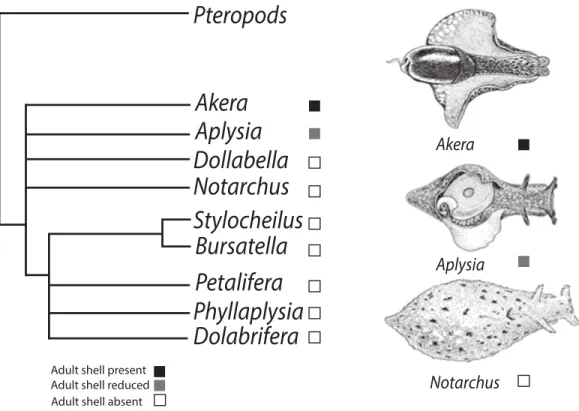
Figure 1 Phylogenetic tree depicting relationships of Anaspidea.Consensus phylogeny of sea hares (Anaspidea) compiled from Medina & Walsh (2000) and Klussmann-Kolb & Dinapoli (2006). Shell character states are depicted by boxes on terminal nodes.
shell-less gastropods (Vermeij, 2013), camouflage (Rudman, 1981;Rudman & Avern, 1989) and mimicry of unpalatable species (Kerstitch, 1989), swimming to escape danger (Gillette & Jing, 2001;Lawrence & Watson III, 2002), and incorporation of defense mechanisms from their diet organisms as their own, e.g., nematocysts (Conklin & Mariscal, 1977;
Greenwood & Mariscal, 1984). Because these adaptations involve anatomical modifications that tend to take place during early development, we consider that differential shell reduction and loss in sea hares provides an excellent opportunity to investigate major transitions in gastropod body plan evolution.
Within the sea hares (Opisthobranchia: Anaspidea), shell reduction or loss has occurred at least twice in adult individuals (Fig. 1) but possibly more times. The order Anaspidea is best known for the work onAplysia californicaas a model system for the study of the cellular basis of behavior (Kandel, 1979) and molecular and genome resources are readily available (Heyland et al., 2011). Transcriptome profiling, combined with whole mount in situ hybridization, has identified differentially expressed genes during shell formation in early developmental stages ofA. californica(Heyland et al., 2011) providing a list of candidate genes involved in the process of shell formation that can now be analysed in other anaspidean taxa.
system (Ghiselin, 1965;Klussmann-Kolb & Dinapoli, 2006;Mikkelsen, 1996;Morton & Holme, 1955), as well as molecular phylogenies (Grande, Templado & Zardoya, 2008;
Klussmann-Kolb & Dinapoli, 2006;Medina & Walsh, 2000;Thollesson, 1999). The current understanding of phylogenetic relationships also enables us to map the evolution of shell reduction and loss within the sea hares. While adults of the genusAplysiaexhibit a reduced shell, the genusBursatellarepresents the derived character state of crown anaspidean taxa where adults lack a shell altogether. Thus the ragged sea hare,Bursatella leachiiexhibits some developmental differences relative toA. californicaproviding a good comparative system to study shell evolution in this gastropod lineage. Both species undergo two distinct periods of shell growth separated by cessation during the metamorphic process. Following theA. californicalife cycle staging (Kriegstein, 1977), characteristic veliger spiral shell growth commences during the encapsulated embryonic phase and continues to the end of the planktotrophic larval phase, stage 6. Growth resumes post metamorphosis at stage 10, when the shell changes from a spiral to a planar shell growth pattern.A. californicahas an internalized shell in adulthood, whereasB. leachiiundergoes post-metamorphic shell growth followed by shell loss soon after metamorphosis (Paige, 1988).
Aplysia californicais one of a few invertebrate species with long-lived planktotrophic larvae that can be successfully cultured in the lab (Carefoot, 1987;Kriegstein, 1977). Today, after optimized short generation times and developmental inducers, a large number ofA. californicacan be grown in the laboratory under controlled hatchery conditions. High fecundity and quick growth provide abundant experimental stock of multiple life stages (Capo et al., 2009). With the success ofA. californicacultures year-round, having additional hatchery populations of other anaspidean species is an attainable goal given our understanding of the ecology and evolution of related taxa (Carefoot, 1987). Habitat and dietary preferences inB. leachiiare now well-known, facilitating animal husbandry.B. leachiilives in tropical subtidal waters (Ramos, Lopez Rocafort & Miller, 1995) feeding on cyanobacterial biofilms found on sandy substrates (Paige, 1988;Ramos, Lopez Rocafort & Miller, 1995).
In this study we report a more detailed description of theB. leachiilife cycle than previously available, normalized to theA. californicahatchery culturing procedures currently in place at the NationalAplysiaResource facility (Capo et al., 2009). We also report new optimal culture conditions forB. leachii. We conclude by describing the most apparent differences in the developmental program of both species, with emphasis on metamorphic stages during which shell reduction and loss take place with discussion of potential biomineralization proteins involved in shell formation in sea hares.
METHODS
Broodstock and eggs/larval rearing
and Atmospheric Science (RSMAS) as previously described (Capo et al., 2009;Capo et al., 2002). The animals were fed a daily ration of the following laboratory-cultured seaweeds: Gracilaria ferox(forA. californica) and a mixture of blue–green algae and epiphytes (for B. leachii). The light cycle of both species was maintained at 12 h light: 12 h dark. The seawater temperature was 13–15◦C forA. californicaand 22–26◦C forB. leachii. In year 1
of the study, cultures were maintained at the same temperature (22◦C) but theB. leachii cultures died. In the subsequent trial, parallel cultures were maintained at 22◦C and 25◦C
forA. californicaand at 25◦C forB. leachii. Mating pairs were monitored throughout the day for active egg-laying. During oviposition, a 10 cm portion of egg strand was collected, rinsed immediately with 0.45µm filtered seawater, placed in a 2l flask to which Na2EDTA
(0.25 mg/l) was added to bind heavy metals in the natural seawater that may deleteriously affect development (Capo et al., 2002). The eggs and seawater were vigorously aerated until one day prior to hatching in a temperature-controlled incubator at 22◦C and 25◦C for A. californicaand 25◦C B.leachiiin the last trial of the culturing experiments. Hatching
occurred 7–8 days after the eggs were deposited and the cordon (egg strand) was inspected under a dissecting microscope at six days post-oviposition to validate normal and synchro-nized development of embryos. Strands not meeting these standards were discarded.
The number of larvae/mm of cordon was estimated by cutting three portions of known length, using an ocular micrometer. Each segment was dissolved in 2% sodium hypochlorite and the shells were counted. Day 0 shell length (SL) for both species was determined by measuring 25 individuals from each portion of the cordon using an ocular micrometer at 50X magnification. The appropriate initial larvae density was provided by aseptically cutting the appropriate cordon length, immediately rinsing with 2µm filtered
seawater, and directly transferring it into the larvae culture vessel.
Seawater was collected from Bear Cut, Virginia Key, FL and prepared by prefiltration through a 15µm glass media filter. The salinity was adjusted to 32 ppt with deionized
water, and aerated with chloramphenicol (2.5 mg/l), Na2EDTA (0.25 mg/l). Eighteen to 24
h later the seawater was vacuum filtered through a 2µm prefilter (Millipore AP2504700)
(Kriegstein, Castellucci & Kandel, 1974;Nadeau et al., 1989). The desired concentration of microalgae and estimated length of egg mass were added to filtered seawater in 2 L roller bottles (Corning). The vessel was sealed with Parafilm®and plastic wrap to eliminate the air-water interface (Capo, Perritt & Paige, 1987;Paige, 1986;Tamse, Kuzirian & Capo, 1990). The cultures were incubated on a continuously rotating (1 rpm) roller bottle apparatus (Wheaton), with a 24 h fluorescent light regime (∼0.001µE/cm2/s) at a constant
temperature of 22◦C (Kriegstein, Castellucci & Kandel, 1974;Nadeau et al., 1989;Tamse, Kuzirian & Capo, 1990). Roller apparatus positions were randomly assigned to each culture vessel and remained fixed throughout the experiment.
After hatching, larvae were measured and the culture media was changed every 7 days. The larvae were collected on a 74µm mesh screen, rinsed with filtered seawater (FSW) and
complex (Sigma) and 2.0 mg/ml pH 8.3 fish-grade Trizma (Sigma) solution for 5 min to inhibit bacterial growth. This treatment also acted to suppress larval swimming behavior and provided a non-lethal method to facilitate shell length measurements. Weekly SL of 25 larvae was measured and the larval stage for bothA. californicaandB. leachiiwas determined through Kriegstein’s staging scheme forA. californica(Kriegstein, 1977). Once the exposure period ended, the iodine concentration was reduced by the incremental addition of a 0.4% sodium thiosulfate solution to the treatment bath until the characteristic iodine color had disappeared. The larvae were rinsed in FSW and transferred to a clean, acid-washed roller bottle with FSW containing the appropriate amount of microalgae and sealed (250×103cells/ml,Isochrysis sp.—CCMP1324). The bottles were then returned to the previously assigned roller bottle apparatus and position.
For imaging of each stage ofB. leachii, the larvae were placed in filtered seawater (0.22µm) containing 340 mM of magnesium chloride. Once animals were narcotized,
photographs were taken with an Olympus BX51 microscope or a Leica MZ16F stereoscope. Scanning Electron Microscopy was performed on a limited numbers of larval shells from both species.
RESULTS
Post-hatching larval development and shell growth
The life cycle staging ofB. leachiimentioned here is equivalent to the staging scheme that was described forAplysia californica(Kriegstein, 1977) and currently in use at the University of Miami’sAplysiahatchery (Rosenstiel School, 2012). Stage 1 is characterized by a newly hatched veliger containing a Type 1 shell (Thompson, 1961). InB. leachii, Stage 1 larvae have a maximum shell diameter of 141.1±6.9µm(N=25)and the veliger’s shell
grows rapidly—an average of 21µm per day (Fig. 2). Stage 2, defined by the appearance
of the eyes, and is reached within 4 days post-hatching. By day 5, the shell length is 264.6±13.9µm(N =25)with the presence of 1.5 whorls. After 6 days post-hatching,
the larval heart appears (Stage 3). By day 7, the maximum shell size (Stage 4) is reached at 284.2±19.0µm(N =25)(Supplemental Information 1). Almost at the same time
the foot expands to form a well-developed propodium (Stage 5). On day 9, the larvae reach competency and settle (Stage 6) when exposed to a substratum. A morphological pigmented spot on the shell, similar toA. californica(Kriegstein, 1977), is also present in B. leachii.Paige (1988)andPaige (1986)failed to observe and report pre-metamorphic pigmentation most likely due to the use of artificial seawater. Pigmentation is a clear indicator of competency to metamorphose, and can be reached as early as 9 days post-hatching.A. californicalarvae grown at 22◦C and 25◦C showed that there was no
difference in growth. A two-way repeated measures ANOVA reflects that there was no difference in the size ofA. californicagrown at 22◦C vs. 25◦C (Supplemental Information 1andSupplemental Information 2). In 2006, total mean shell length(n=25)forA. californicagrown at 22◦C averaged 134.6µm (s=3.7µm) for Stage 1, 227.6µm (s=
15.0µm) for Stage 2, 337.7µm (s=20.8µm) for Stage 3 and 392.8µm (s=10.0µm)
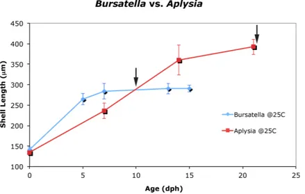
Figure 2 Larval and juvenile growth ofBursatella leachiiandAplysia californicain laboratory set-tings.Veliger shell length ofA. californicaandB. leachiilarvae grown at 25◦C in 2006. Shell length was measured weekly from day of hatching until 80% competency; error bars represent±1 standard devia-tion. Arrow indicates timing of competency: 9 days post-hatching inB. leachiiand 22 days post-hatching inA. californica.Previous attempts to cultureB. leachiilarvae at 22◦C were unsuccessful (not shown).
134.6µm (s=3.7µm) for Stage 1, 236.1µm (s=18.6µm) for Stage 2, 360.6 (s=36.9µm)
for Stage 3 and 392.3µm (s=18.9µm) for Stage 5 (Supplemental Information 2).
Metamorphic larvae development ofBursatella leachii
Metamorphic development and post-larval development ofBursatella leachiiis similar to other previously described sea hares (Kriegstein, 1977;Paige, 1988;Switzer-Dunlap, 1978;
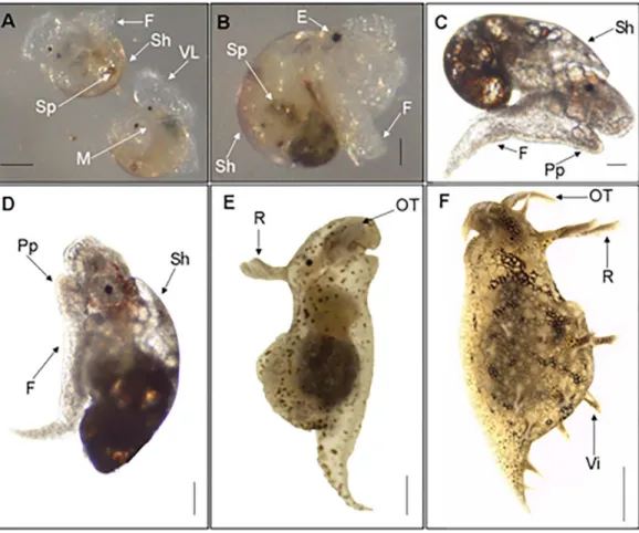
Figure 3 Metamorphic development ofBursatella leachii.Metamorphic competence of the veliger larvae (stage 6, A) correlates with many morphological characteristics (i.e., red spots, propodium, etc.). Once settled, the larvae will attach and shed their velar lobes, becoming benthic (stage 7, B). Stage 8 (not shown) marks the end of metamorphosis, characterized by the fusion of the two halves of the velum lobe and the loss of the larval heartbeat. Stages 9–10 marks the development of specific morphological structures of juveniles, such as the elongation of the juvenile or post metamorphic shell (stage 9, C; stage 10, D). Adult characteristics, such as the complete shedding of the shell, rhinophores, villae and oral tentacles, will start to appear in late stage 11 (E) and the adult (F). VL, Velar Lobe; Sh, Shell; Sp, Spot; M, Mouth; F, Foot; E, Eye; Pp, Propodium; R, Rhinophores; OT, Oral Tentacles; Vi, Villae. Scale bar in A: 100µm, in B: 67µm, in C: 108µm, in D: 134µm, in E: 254µm, in F: 1mm.
similar in early embryonic stages but diverges as juvenile development takes place leading to shell loss inB. leachii. We examined by SEM both whole shells and cross-sections of larval shells (Supplemental Information 3). Despite some noticeable similarities between the two species, unfortunately due to the small size of the larval shells, we either did not have enough replicates per stage or missed stages altogether to raise clear conclusions about larval shell shape and internal structure.
DISCUSSION
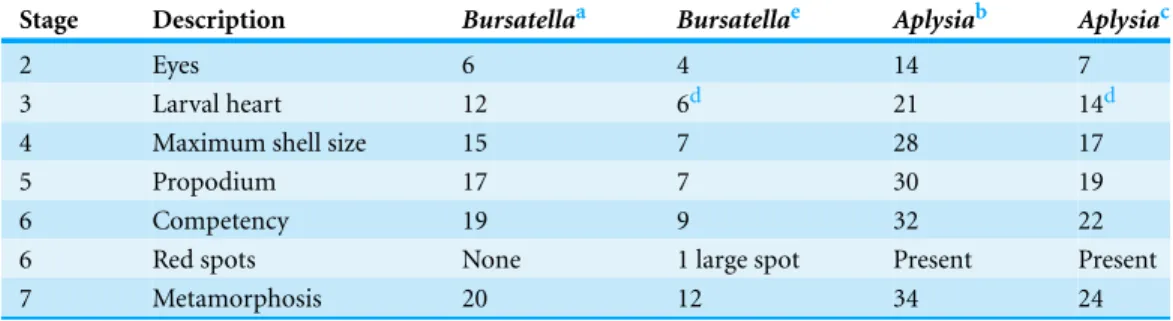
Table 1 Comparison of developmental schedules ofAplysia californicaandBursatella leachii. Com-parison of morphological development schedules ofA. californicalarvae as reported byKriegstein (1977) compared toCapo et al. (2009)and comparison ofB. leachiilarvae as reported byPaige (1988)compared to the present study. Values are the number of days post-hatching until the specified developmental stage was observed.
Stage Description Bursatellaa Bursatellae Aplysiab Aplysiac
2 Eyes 6 4 14 7
3 Larval heart 12 6d 21 14d
4 Maximum shell size 15 7 28 17
5 Propodium 17 7 30 19
6 Competency 19 9 32 22
6 Red spots None 1 large spot Present Present
7 Metamorphosis 20 12 34 24
Notes.
aPaige, 1988.
bKriegstein, 1977. cCapo et al., 2009.
d50 beats/minute not taken into consideration. ePresent study.
we describe the life cycle ofB. leachiiin the context of the development of the larval shell and its subsequent loss in the post-metamorphic stages.
Bursatella leachii development
The embryonic development ofBursatella leachiihas been described previously ( Beb-bington, 1969;Paige, 1988) and thus will not be further discussed here. The larval developmental sequence ofB. leachiiis similar to other sea hares (Switzer-Dunlap, 1978)—a hatched veliger with a hyperstrophically coiled shell, a reddish tint, and bilobed velum.B. leachiilarvae differ both in size and growth rate relative toA. californica, being both larger (approximately 10µm) and faster growing, though the larval development
follows the staging sequence previously devised in the literature (Kriegstein, 1977;Paige, 1988). Similar toKriegstein (1977), our study demonstrated the presence of one prominent Stage 6 pigmentation spot inB. leachii.
Initial stages of post-metamorphic development of sea hares with a planktonic larval form are also similar,Table 1summarizes the larval development ofB. leachii(Paige, 1988) relative toA. californica(Capo et al., 2009;Kriegstein, 1977). Recent advances in larval culture techniques provide the tools for life cycle comparisons. The need for readily available developmental stages is important for experimental developmental biology studies such as metamorphic transitions. In the particular case of sea hares, hatchery populations provide an ideal supply of samples for the study of larval shell loss.
at Stage 11 when the shell is discarded. At this stage inA. californica, the shell becomes overgrown by folds of the mantle, causing the shell to be internalized. Given that both species follow a similar developmental program through metamorphosis, it seems quite plausible that the underlying mechanism of larval shell formation is also quite similar, only differing during settlement/post-metamorphosis. We conclude that larval shell formation appears to be homologous in these two species, which makes this process amenable to comparisons such as the examination of spatio-temporal gene expression of genes involved in the formation of the shell in both species. It seems plausible that the evolution of shell loss is the consequence of modifications to the regulatory machinery of shell formation genes, as most molluscs have the ability to make shells at least in the embryonic stages.
Shell development in Anaspidea
of the fascinating phenomenon of biomineralization and evolution of shell loss in opisthobranchs.
B. leachii husbandry
We present an improved strategy to cultureB. leachiiin larger numbers than previously reported. We attempted to rear both species under similar conditions butA. californicais a temperate species from the Western North America, where coastal upwelling is prevalent and water temperatures low relative to tropical waters whereB. leachiiis common. Therefore we decided to use a slightly higher temperature (25◦C) for the second year the cultures were established in the lab. The primary goal of this study was to produce individuals from comparable stages, however, despite small sample sizes and limited controls, our efforts have lead to an improved culturing method forB. leachiiwith larger larval yields than previously reported (Paige, 1988).
CONCLUSION
We have established a reliable culturing technique forB. leachiithat makes this species amenable to experimentation at all developmental stages (Capo et al., 2009). Transcrip-tome data and whole mountin situhybridization available forA. californica(Heyland, 2006) have enabled developmental genetics research (Heyland et al., 2011) in anaspideans. While comparative studies of biomineralization genes in sea hares are in their infancy, with developmental homology clearly established and an improved cultivation protocol, we are primed to shed light on how the genetic toolkit that controls shell formation and subsequent reduction or loss differs betweenA. californicaandB. leachii.
ACKNOWLEDGEMENTS
We thank Phillip Gillette for his efforts in culturingB. leechii; Benoˆıt Dayrat for help with Fig. 1; Alice Hudder for training. We also acknowledge the support of Michael R. Dunlap and the Imaging and Microscopy Facility (IMF) at the University of California, Merced. This manuscript was prepared by Zer Vue in partial fulfilment of requirements for the master’s program in Quantitative Systems Biology at UC Merced. We thank Chris Voolstra, Michael DeSalvo and Shini Sunagawa for providing feedback on an earlier version of this manuscript.
ADDITIONAL INFORMATION AND DECLARATIONS
Funding
Support for this project was provided by NSF DEB-0542330 and IOS 0926906. The funders had no role in study design, data collection and analysis, decision to publish, or preparation of the manuscript.
Grant Disclosures
The following grant information was disclosed by the authors: NSF: DEB-0542330.
Competing Interests
M ´onica Medina is an Academic Editor for PeerJ.
Author Contributions
• Zer Vue, Bishoy S. Kamel, Thomas R. Capo, Ana T. Bardales and M ´onica Medina conceived and designed the experiments, performed the experiments, analyzed the data, contributed reagents/materials/analysis tools, wrote the paper, prepared figures and/or tables, reviewed drafts of the paper.
Supplemental Information
Supplemental information for this article can be found online athttp://dx.doi.org/ 10.7717/peerj.700#supplemental-information.
REFERENCES
Bebbington A. 1969.Bursatella leachiiguineensis subsp. nov. (Gastropoda, Opistobranchia) from Ghana.Proceedings of the Malacological Society of London38:323–341.
Capo TR, Bardales AT, Gillette PR, Lara MR, Schmale MC, Serafy JE. 2009.Larval growth, development, and survival of laboratory-rearedAplysia californica: effects of diet and veliger density.Comparative Biochemistry and Physiology Part C: Toxicology & Pharmacology 149:215–223.
Capo TR, Fieber LA, Stommes DL, Walsh PJ. 2002.The effect of stocking density on growth rate and maturation time in laboratory-reared california sea hares.Contemporary Topics in Laboratory Animal Science41:18–23.
Capo T, Perritt S, Paige J. 1987.The mass culture ofAplysia californica. In:Fifty-third annual meeting of the American malacological union, 18.
Carefoot T. 1987.Aplysia: its biology and ecology. In: Barnes M, ed.Oceanography and marine biology anuual review. London: CRC Press, 167–284.
Conklin EJ, Mariscal RN. 1977.Feeding behaviour, Ceras structure, and nematocyst storage in the aeolid Spurilla neapolitana (Mollusca).Bulletin of Marine Science27:658–667.
Ghiselin MT. 1965.Reproductive function and the phylogeny of opisthobranch gastropods. Malacologia3:327–378.
Gillette R, Jing J. 2001.The role of the escape swim motor network in the organization of behavioral hierarchy and arousal in pleurobranchaea.American Zoologist41:983–992 DOI 10.1668/0003-1569(2001)041[0983:TROTES]2.0.CO;2.
Gosliner TM. 1985.Parallelism, parsimony and the testing of phylogenetic hypothesis: the case of opistobranch gastropods. In: Vrba ES, ed.Species and Speciation. Pretoria: Museum monograph No. 4, Transvall Museum, 105–107.
Gosliner TM. 1991. Morphological parallelism in opistobranch gastropods.Malacologia 32:313–327.
Grande C, Templado J, Zardoya R. 2008.Evolution of gastropod mitochondrial genome arrangements.BMC Evolutionary Biology8:61DOI 10.1186/1471-2148-8-61.
Heyland A. 2006.Signaling mechanisms underlying metamorphic transitions in animals. Integrative and Comparative Biology46:743–759DOI 10.1093/icb/icl023.
Heyland A, Vue Z, Voolstra CR, Medina M, Moroz LL. 2011.Developmental transcriptome ofAplysia californica’.Journal of Experimental Zoology Part B: Molecular and Developmental Evolution316:113–134DOI 10.1002/jez.b.21383.
Jackson DJ, McDougall C, Green K, Simpson F, Worheide G, Degnan BM. 2006.A rapidly evolv-ing secretome builds and patterns a sea shell.BMC Biology4:40DOI 10.1186/1741-7007-4-40. Jackson DJ, McDougall C, Woodcroft B, Moase P, Rose RA, Kube M, Reinhardt R, Rokhsar DS,
Montagnani C, Joubert C, Piquemal D, Degnan BM. 2010. Parallel evolution of nacre building gene sets in molluscs. Molecular Biology and Evolution27:591–608 DOI 10.1093/molbev/msp278.
Kandel ER. 1979.Behavorial biology of Aplysia. San Francisco: W. H. Freeman and Company. Kempf SC. 1984.Symbiosis between the zooxanthellae Symbiodinium (=Gymnodinium)
microadriaticum (Freudenthal) and four species of nudibranchs.The Biological Bulletin 166:110–126DOI 10.2307/1541435.
Kerstitch A. 1989.Sea of Cortez marine invertebrates, a guide for the Pacific Coast, Mexico to Ecuador. Monterey, California: Sea Challengers.
Klussmann-Kolb A, Dinapoli A. 2006. Systematic position of the pelagic Thecosomata and Gymnosomata within Opisthobranchia (Mollusca, Gastropoda)—revival of the Pteropoda. Journal of Zoological Systematics and Evolutionary Research44:118–129 DOI 10.1111/j.1439-0469.2006.00351.x.
Kriegstein AR. 1977.Stages in the post-hatching development ofAplysia californica.Journal of Experimental Zoology199:275–288DOI 10.1002/jez.1401990212.
Kriegstein AR, Castellucci V, Kandel ER. 1974.Metamorphosis of Aplysia californicain laboratory culture.Proceedings of the National Academy of Sciences of the United States of America71:3654–3658DOI 10.1073/pnas.71.9.3654.
Kr¨oger B, Vinther J, Fuchs D. 2011. Cephalopod origin and evolution: a congruent picture emerging from fossils, development and molecules.Bioessays 33:602–613 DOI 10.1002/bies.201100001.
Lawrence KA, Watson III WH. 2002.Swimming behavior of the Nudibranch Melibe leonina. Biological Bulletin203:144–151DOI 10.2307/1543383.
Lowenstam HA, Weiner S. 1989.On biomineralization. New York: Oxford University Press. Medina M, Walsh PJ. 2000.Molecular systematics of the order Anaspidea based on mitochondrial
DNA sequence (12S, 16S, and COI). Molecular Phylogenetics and Evolution15:41–58 DOI 10.1006/mpev.1999.0736.
Mikkelsen PM. 1996. The evolutionary relationships of Cephalaspidea s. l. (Gastropoda: Opisthobranchia): a phylogenetic analysis.Malacologia37:375–442.
Morton JE. 1960.Molluscs—an introduction to their form and function. Harper & Brothers. Morton JE, Holme NA. 1955.The occurrence at Plymouth of the opisthobranch Akera bullata
with notes on its habits and relationships.Journal of the Marine Biological Association of the United Kingdom34:101–112DOI 10.1017/S0025315400008638.
Paige JA. 1986. The laboratory culture of 2 aplysiids, aplysia-brasiliana rang, 1828, and
bursatella-leachii-plei (Rang, 1828) (gastropoda, opisthobranchia) in artificial seawater.Veliger 29:64–69.
Paige JA. 1988.Biology, metamorphosis and postlarval development ofBursatella leachii pleirang (Gastropoda: Opistobrachia).Bulletin of Marine Science42:65–75.
Ramos LJ, Lopez Rocafort JL, Miller MW. 1995.Behavior patterns of the aplysiid gastropod Bursatella leachiiin its natural habitat and in the laboratory.Neurobiology of Learning and Memory63:246–259DOI 10.1006/nlme.1995.1029.
Rosenstiel School. 2012.National resource facility forAplysia.Available athttp://aplysia.miami. edu/(accessed 4 December 2012).
Rudman WB. 1981.Polyp mimicry in a new species of aeolid nudibranch mollusc.Journal of Zoology193:421–427DOI 10.1111/j.1469-7998.1981.tb03454.x.
Rudman WB, Avern G. 1989. The genus Rostanga (Nudibranchia: Dorididae) in the
Indo-West Pacific.Zoological Journal of the Linnean Society96:281–338DOI 10.1111/j.1096-3642.1989.tb01832.x.
Rumpho ME, Summer EJ, Manhart JR. 2000.Solar-powered sea slugs. Mollusc/algal chloroplast symbiosis.Plant Physiology123:29–38DOI 10.1104/pp.123.1.29.
Sarashina I, Yamaguchi H, Haga T, Iijima M, Chiba S, Endo K. 2006.Molecular evolution and functionally important structures of molluscan dermatopontin: implications for the origins of molluscan shell matrix proteins.Journal of Molecular Evolution62:307–318 DOI 10.1007/s00239-005-0095-2.
Switzer-Dunlap M. 1978.Larval biology and metamorphosis of aplysiid gastropods. In: Chia F-S, Rice ME, eds.Settlement and metamorphosis of marine envertebrate larvae. New York: Elsevier, 197–206.
Switzer-Dunlap M, Hadfield MG. 1977. Observations on development, larval growth and metamorphosis of four species of Aplysiidae (Gastropoda: Opistobranchia) in laboratory culture. Journal of Experimental Marine Biology and Ecology29:245–261 DOI 10.1016/0022-0981(77)90069-7.
Tamse C, Kuzirian A, Capo T. 1990.Roller culture system: the fitness machine forHermissenda crassicornislarvae. In:Amer malacol union abstr 56th ann mtg, vol. 61.
Thollesson M. 1999.Phylogenetic analysis of Euthyneura (Gastropoda) by means of the 16s rRNA gene: use of a fast gene for higher-level phylogenies.Proceedings of the Royal Society of London. Series B: Biological Sciences266:75–83DOI 10.1098/rspb.1999.0606.
Thompson TE. 1961.The importance of the larval shell in the classification of the Sacoglossa and the Acoela (Gastropoda: Opisthobranchia).Proceedings of the Malacological Society of London 34:233–258.
Vermeij GJ. 1993.Evolution and escalation: an ecological history of life. Princeton, New Jersey: Princeton University Press.
Vermeij GJ. 2013. On escalation.Annual Review of Earth and Planetary Sciences41:1–19 DOI 10.1146/annurev-earth-050212-124123.
W¨agele H, Klussmann-Kolb A. 2005.Opisthobranchia (Mollusca, Gastropoda)—more than just slimy slugs. Shell reduction and its implications on defence and foraging.Frontiers in Zoology 2:1–18DOI 10.1186/1742-9994-2-3.