op-iap
in Cotton (
Gossypium hirsutum
L.) Enhances
Tolerance to Verticillium Wilt
Juan Tian1., Xueyan Zhang2., Benguo Liang3
, Shanwei Li4, Zhixia Wu2, Qianhua Wang2, Chunxu Leng1, Jiangli Dong1*, Tao Wang1
1State Key Laboratory of Agrobiotechnology, College of Biological Sciences, China Agricultural University, Beijing, China, 2Key Laboratory of Cotton Genetic Improvement, Ministry of Agriculture, Institute of Cotton Research, Chinese Academy of Agricultural Sciences, Anyang, China,3Horticulture Department, Xinyang Agricultural College, Xinyang, China,4State Key Laboratory of Crop Biology, Shandong Agricultural University, Taian, China
Abstract
Background:Programmed cell death plays an important role in mediating plant adaptive responses to the environment such as the invasion of pathogens. Verticillium wilt, caused by the necrotrophic pathogenVerticillium dahliae, is a serious vascular disease responsible for great economic losses to cotton, but the molecular mechanisms of verticillium disease and effective, safe methods of resistance to verticillium wilt remain unexplored.
Methodology/Principal Findings:In this study, we introduced baculovirus apoptosis inhibitor genesp35andop-iapinto the genome of cotton via Agrobacterium-mediated transformation and analyzed the response of transgenic plants to verticillium wilt. Results showed thatp35andop-iapconstructs were stably integrated into the cotton genome, expressed in the transgenic lines, and inherited through the T3generation. The transgenic lines had significantly increased tolerance to
verticillium wilt throughout the developmental stages. The disease index of T1–T3 generation was lower than 19,
significantly (P,0.05) better than the negative control line z99668. After treatment with 250 mg/L VD-toxins for 36 hours, DNA from negative control leaves was fragmented, whereas fragmentation in the transgenic leaf DNA did not occur. The percentage of cell death in transgenic lines increased by 7.11% after 60 mg/L VD-toxin treatment, which was less than that of the negative control lines’s 21.27%. This indicates thatp35andop-iapgene expression partially protects cells from VD-toxin induced programmed cell death (PCD).
Conclusion/Significance:Verticillium dahliaecan trigger plant cells to die through induction of a PCD mechanism involved in pathogenesis. This paper provides a potential strategy for engineering broad-spectrum necrotrophic disease resistance in plants.
Citation:Tian J, Zhang X, Liang B, Li S, Wu Z, et al. (2010) Expression of Baculovirus Anti-Apoptotic Genesp35andop-iapin Cotton (Gossypium hirsutumL.) Enhances Tolerance to Verticillium Wilt. PLoS ONE 5(12): e14218. doi:10.1371/journal.pone.0014218
Editor:Haibing Yang, Purdue University, United States of America
ReceivedMay 30, 2010;AcceptedNovember 14, 2010;PublishedDecember 3, 2010
Copyright:ß2010 Tian et al. This is an open-access article distributed under the terms of the Creative Commons Attribution License, which permits unrestricted use, distribution, and reproduction in any medium, provided the original author and source are credited.
Funding:This work was supported by the major projects of genetically modified organisms breeding of China (2008ZX08005-003 and 2008ZX08009-002). The funders had no role in study design, data collection and analysis, decision to publish, or preparation of the manuscript.
Competing Interests:The authors have declared that no competing interests exist. * E-mail: dongjl@cau.edu.cn
.These authors contributed equally to this work.
Introduction
Verticillium wilt of cotton caused byVerticillium dahliaeis one of the most serious diseases among cotton-producing regions worldwide. Verticillium dahliae is one subset of necrotrophic soilborne pathogens that form microsclerotia as surviving structures in the soil [1]. Once induced by root exudates, hyphae germinate and grow toward nearby roots, thought to be attracted by a nutrient gradient [2]. After penetrating the root cortex through wounds or by penetrating epidermal cells and crossing the endodermis, the fungus invades the vascular tissue. From there, it produces a large number of conidia and migrates through the xylem to the aerial parts of the plant, resulting in disease symptoms such as leaf vein browning and chlorosis, wilting, vascular discoloration, premature defoliation, and most severely, plant
proteins (VdNEPs), which acted as elicitors in inducing phyto-alexin production and programmed cell death in cotton suspension-cultured cells.
Programmed cell death (PCD) is an essential process not only for plant growth and development; it is also responsible for cell death in response to pathogen attacks and various abiotic stressors such as wounding, salt, cold, UV light, and herbicide (e.g., Paraquat) treatment [10,11,12]. Although the general understand-ing of plant PCD has progressed, its regulation and execution are still poorly understood, especially compared to the understanding of animal apoptosis. In animal cells, a core component of the PCD machinery is a family of cysteine-dependent, aspartate-specific proteases called ‘‘caspases.’’ During apoptosis, the initiator caspase is activated by pro-apoptotic signals and cleaves inactive pro-forms of effector caspases, thereby activating them. Effector caspases in turn cleave numerous cellular proteins, eventually leading to regulated apoptosis [13,14]. The absence of caspase ortholog sequences in plants has been demonstrated by sequencing Arabidopsisand rice genomes [15]. However, previous reports have shown that animal caspase inhibitors can block plant PCD and most instances of plant PCD are associated with the induction of caspase-like events. Del Pozo and Lam [16] found that specific inhibitors of caspase-1 YVAD-CMK) and caspase-3 (Ac-DEVD-CHO) have been observed to inhibit the occurrence of tobacco hypersensitive response (HR) induced by bacteria and Tobacco mosaic virus (TMV). Furthermore, they demonstrated caspase-1 and capase-3 participation in plant HR responses. The same caspase inhibitors also have been critically implicated in apoptosis of tobacco cells induced by isopentyladenosine and menadione [17,18,19].
Baculoviruses encode two mechanistically distinct apoptotic suppressors, inhibitor of apoptosis (IAP) and P35. Both viral proteins prevent premature insect cell death and thereby promote virus multiplication. The baculovirus anti-apoptotic proteins P35 and IAP are also effective in preventing plant PCD induced by bacterial, fungal, and viral infections [10,19,20,21,22,23].
Baculovirus P35 was first isolated from Autographa californica nuclear polyhedrosis virus(AcMNPV) and is a broad-spectrum caspase inhibitor in nematode, Drosophila, and mammalian cells [24,25,26,27]. The crystal structure of P35 is similar to a teapot. The ‘‘handle’’ of the teapot has a reactive site loop with a caspase cleavage site DQMD, which is recognized and cleaved by caspase. After cleavage, caspase and P35 form stable complexes between them via a covalent thioester bond, leading to caspase inactivity [28]. Derived from baculovirus Orgyia pseudotsugata multicapsid polyhedrosis virus (OpMNPV), Op-IAP is a potent inhibitor of apoptosis with two baculovirus IAP repeat (BIR) domains at the N-terminus and a really interesting new gene (RING)-finger domain at the C-terminus; it can inhibit the activity of caspases through the binding of its conserved BIR domains to the active sites of caspasesin vitroandin vivo[29].
The transgenic maize embryogenic callus with baculovirusp35 andop-iapinhibitsAgrobacterium tumefaciens-induced PCD [19]. The transgenic tomatoes expressing p35 can effectively inhibit AAL mycotoxin-induced cell apoptosis and reduce disease symptoms after inoculation with several necrotrophic tomato pathogens[20]. The narrow-leafed lupin containing thep35gene had significantly reduced necrotrophic disease symptoms [22].
Cotton is one of the most important cash crop in the world, the average annual cotton production loss as a result of Verticillium wilt is tremendous. In recent years, breeding new lines by genetic engineering to improve resistance to Verticillium wilt is one of important approaches for disease control. For example, transgenic coloured cotton plants over-expressing foreignGastrodiaanti-fungal
protein were found to display robust resistance againstV. dahliaein the field[30]. The crude leaf extracts from the transgenic cotton lines containing bean chitinase gene orD4E1gene (the synthetic antimicrobial peptide) inhibited the growth of V. dahliae in vitro[31,32]. Transgenic cottons expressing glucose oxidase dem-onstrated significant increase in tolerance to V. dahliae, but vegetative growth, seed production and seedling vigour were also reduced[33]. In this study, we conducted the first attempt to introduce baculovirus apoptosis inhibitor genesp35andop-iapinto the genome of cotton throughAgrobacterium-mediated transforma-tion and analyzed the response of transgenic plants to verticillium wilt. Results from this work reveal that caspase inhibitors can block plant cell death associated with cotton host–V. dahliaeinteractions. These results add to the growing evidence for pathogenesis-associated cell death in plants sharing common regulatory and mechanistic features with PCD in animals and evoke breeding strategies for verticillium wilt control.
Materials and Methods
Plasmid construction
The cDNA clones of baculovirus p35 (900 bp) and op-iap (807 bp) in the insect expression vector pIE were a gift from Dr. Qianjun Li (University of Alabama at Birmingham, USA; GenBank Acc. Nos.: L22858 and L22564, respectively). PCR products were obtained using the following primers: p35, 59 -atgtgtgtaatttttccggtag-39and 59-ttatttaattgtgtttaatatt -39, andop-iap, 59-atgagctcccgagcaattgg-39 and 59-ttacacttggtacatgcgcacc-39, which were sequenced and subsequently cloned into pMD-18T (Takara Bio, Tokyo, Japan). Thep35gene replaced the reporter gene of pBI121 through restriction withBamHI andSacI enzymes to construct an intermediate vector pBI121–p35, with thep35gene under the 35SCauliflower mosaic virus (CaMV) promoter and the nopaline synthase (nos) terminator. The GUS gene in the binary vector pCAMBIA 1301 (CAMBIA, Canberra, ACT, Australia) was replaced by theop-iapgene by restriction withBglII andBstEII, under the 35S CaMV promoter and the nos terminator, termed p1301–iap. Thep35gene expression cassette obtained byPstI and EcoRI restriction enzyme digestion was inserted into the p1301–iap vector of multiple cloning sites to construct the co-expression vector p1301–iap–p35.The plasmid was transformed into compe-tent cells ofA. tumefaciens strain EHA105 using the freeze–thaw method.
Plant materials and plant transformation
seedling medium (MSB containing 0.1 mg/L IBA, 30 g/L glucose, 2 g/L Gelrite, pH 5.8) to form regenerated plants. The regenerated plants from independent resistant calluses were grafted onto the rootstock seedlings[34] and then moved to greenhouse cultivation. Hygromycin at 80 mg/L was used to paint leaves of the regenerated plants to evaluate hygromycin resistance. Leaves of susceptible seedlings will change color from green to yellow after 5–7 days. After 3 repeated applications, hygromycin-resistant plants were kept for further molecular analysis and disease essay. After self-pollination, T1 generation seeds were collected, and self-pollinated T2 and T3 generation seeds were harvested over the next 2 years, respectively.
Expression analysis of transgenes
To determine the integration of cassettes in the genome, polymerase chain reaction (PCR) was conducted using specific primer pairs (p35: 59-cccacagatggttagagagg-39 and 59 -gtag-tagtcgttgcgttcgt-39 and op-iap: 59-atgagctcccgagcaatt-39 and 59 -acgcctcagtcatcaccc -39) to amplify the CaMV 35S promoter p35 region andop-iapfrom T1to T3transgenic cotton plants. Genomic DNA (gDNA) was extracted and purified from young leaves using the method described by Paterson et al. [35]and Li et al. [36]. PCR of thep35gene was conducted in a Touchgen thermal cycler (Techne, Cambridge, UK) using the following program: initial denaturing at 95uC for 5 minutes followed by 35 cycles of denaturing at 95uC for 50 seconds, annealing at 55uC for 50 seconds, extension at 72uC for 1 minute 30 seconds, and final extension at 72uC for 10 minutes. For theop-iap gene, the same program was used except for the annealing step, which was 58uC for 50 seconds, extension at 72uC for 1 minute. PCR products obtained were analyzed by 1% agarose gel electrophoresis followed by ethidium bromide staining.
For Southern blot analysis, 30mg gDNA of T3transgenic cotton plants and non-transformed negative controls was digested with EcoRI. The restriction fragments were size-fractionated by 0.8% (w/v) agarose gel electrophoresis and transferred to a Hybond-N+ nylon membrane (Amersham Pharmacia Biotech, UK). Probes were prepared from purified PCR products of the p35 (870 bp) and op-iap (583 bp) coding region. The labeling of probe, prehybridization, hybridization and detection were performed by the protocol of DIG High Prime DNA Labeling and Detection Starter KitI (catalog no. 11745832910; Roche Applied Science, Mannheim, Germany).
The transcription of p35 and op-iap was detected by semi-quantitative reverse transcription (RT)-PCR. RNA was extracted from young leaves of cotton following Li et al. [36] and all RNA samples were treated with DNaseI (Takara Bio) to prevent contamination with gDNA. One mg of the treated RNA was reverse-transcribed into cDNA using M-MLV Reverse Transcrip-tase following the manufacturer’s protocol (Takara code: D2639A). PCR of 1ml first-strand cDNA was used to amplify genes by using specific primer pairs (p35:59 -gtagaaatcgacgtgtccca-gac -39 and 59-tgagcaaacggcacaataactt -39 ;op-iap: 59 -atgagctccc-gagcaatt-39 and 59-acgcctcagtcatcaccc -39) and the following programs: 95uC for 5 min, 28 cycles of denaturing at 95uC for 40 seconds, annealing at 49uC (p35) or 58uC (op-iap) for 40 seconds, extension at 72uC for 40 seconds and final extension at 72uC for 10 minutes. The RT-PCR for the cotton house-keeping gene histone-3 (GenBank Acc. Nos.: AF024716) was performed with specific primer (59-ataccgtcctggaactgttgctc-39and 59 -aacatat-cagacgccccacttca-39) under the same conditions as described above to determine whether the equal amounts of total RNA were used in the RT-PCR reactions among samples. All RT-PCR expression
assays were independently performed and analyzed three times under identical conditions.
Soluble proteins were extracted with 0.5 ml of an extraction buffer [23]. Protein concentration was measured according to the method of Bradford [37] using a Bio-Rad reagent (Bio-Rad, Hercules, CA) with bovine serum albumin (BSA; Sigma, St. Louis, MO) as a standard. After boiling for 10 minutes in the sample buffer, 10ml of proteins was loaded in a 12% SDS-polyacrylamide
gel, and the proteins were subsequently transferred onto a Hybond-N membrane (Bio-Rad) via a semidry trans-electroblot-ter. After blocking for 1 hour in TBST buffer [20 mM Tris (pH 7.5), 150 mM NaCl, and 0.1% Tween 20] with 5% nonfat dry milk at room temperature, the membrane was probed with the anti-p35(1:1500, catalog no. IMG-5740; IMGENEX, San Diego, CA) and the anti-iap antibodies (1:1000, catalog no. GTX23930; GeneTex, Irvine, CA). Goat anti-rabbit IgG alkaline phosphatase conjugate (1:1000, catalog no. bsap-0295G; Beijing Boaosen Biotechnology, Ltd., Beijing, China) was used as a secondary antibody and the hybridization membrane was washed three times using NBT/BCIP chromogenic stain.
Pathogen cultivation and inoculation
The techniques used for disease assessment in the field were those previously reported by Wang et al. [30].The transgenic cotton lines and non-transformed negative controls with three replications were grown in the cotton verticillium wilt artificial nursery at the Institute of Cotton Research of the Chinese Academy of Agricultural Sciences.Verticillium dahliaewith moder-ate virulence can cause the susceptible control (ji11) infection rmoder-ate of 80%, or a disease index above 50. Disease severity was surveyed respectively in the seedling stage, flowering stage, and boll stage. According to the standard protocol developed by Wang et al. [30], the degree of infection by pathogenic fungi was divided into five grades with disease scores ranging between 0 and 4 (0: healthy plants, no fungal infection; 1: ,25% of the leaves showing yellowing or abnormal yellow spots; 2:#25 to,50% of the leaves showing yellow spots and curled leaf edges; 3:#50 to,75% of the leaves showing brown spots and curled leaf edges with some leaves dropping; and 4: $75% of the leaves produce yellow or yellow irregular spots between the main vein of leaves). The infection rate and disease index were calculated according to the following formula:
infection rate (%) = (the number of total infected plants/the number of total checked plants)6100
disease index = [(Sdisease scores6number of infected)/total
checked plants6highest grade disease (4)]6100.
The mean and standard deviation (SD) were calculated based on three replications. The statistical significance (P,0.05) between the wild-type plant and each transgenic lines was tested through student’s t-test using Statistical Analysis System (SAS) (SAS Institute Inc., USA).
Preparation of crude VD-toxin fromV. dahliae
Verticillium dahliae Kleb (V229), a strong pathogenic and non-defoliating strain originally isolated from diseased cotton tissue and obtained from Prof. Yingzhang Li (College of Biological Sciences, China Agricultural University), was first activated in potato dextrose agar medium at 22uC in the dark for 5 days. The fungal mycelia were cultured in Czapek’s medium (containing 30.0 g sucrose, 3.0 g NaNO3, 1.0 g K2HPO4, 1.0 g MgSO4?7H2O, 1 g KCl, and 0.01 g FeSO4?7H2O in 1 L of distilled water)[7] by shaking at 130 rpm and 28uC for 14 days. The cultures were centrifuged at 10,0006gfor 30 minutes to remove spores and the
lyophilization, the freeze-dried filtrate was then dissolved in distilled water; the solution was filtered through a 0.45-mm filter (Millipore, Billerica, MA) and used as a crude VD-toxin extract [38,39,40]. Protein content in crude extracts was determined according to Bradford [37] with BSA (Sigma) as a standard.
Nuclear DNA fragmentation
The leaves from 6-week-old transgenic lines (KB1) and non-transformed negative control seedlings were treated with 250 mg/ L VD-toxin for 24 and 36 hours, then frozen in liquid nitrogen and ground into a fine powder. DNA was isolated as described by Paterson et al. [35]and Li et al. [36]. Fourmg DNA treated with
25mg/ml DNase-free RNase A (Takara) for 1 hour at 37uC electrophoreses on a 2% agarose gel. After electrophoresis, the gel was stained with 0.5mg/ml ethidium bromide for 30 minutes, de-stained in 1 mM MgSO4 for 1 hour, and observed and photographed on a UV light box [41,42].
Flow cytometry analysis of cell death
Cotton protoplasts were isolated from the transgenic lines (KB1) and non-transformed negative controls. The washed 4-week-old leaves were briefly dried and cut into small leaf strips (0.5 mm) with a razor blade; then the strips were incubated in an enzyme solution [3% (w/v) cellulose R-10 (Yakult Honsha, Tokyo, Japan), 1.5% Macerozyme R-10 (Yakult Honsha), 0.4 M mannitol, 20 mM MES, 10 mM CaCl2, 0.2 mM KH2PO4, 1 mM KNO3, 1 mM MgSO4, 0.1mM CuSO4, 10mM KI, pH 5.8] in the dark by gentle shaking. The protoplasts were separated from undigested materials using 75-mm stainless steel mesh and the crude protoplast filtrates were sedimented by centrifugation for 2 minutes at 700 rpm. Finally, the purified protoplasts were resuspended in W5 solution (154 mM NaCl, 125 mM CaCl2, 5 mM KCl, 5 mM glucose, 1.5 mM MES-KOH, pH 5.8) [43]. Protoplasts were counted using a hemocytometer.
Cotton protoplasts were treated with 30 mg/L and 60 mg/L VD-toxin for 3 hours and then the number of dead protoplastes was determined using a propidium iodide (PI) fluorescent probe. Loss of plasma membrane integrity around apoptotic cells can be demonstrated using fluorogenic compounds such as PI, which is excluded by an intact membrane. Once the cell membrane permeability increases, PI enters the cell and binds stoichiomet-rically to nucleic acids. Fluorescence emission is proportional to the degree of cell death. Protoplasts were incubated with 30mg/ ml PI in culture medium for 5–10 minutes [44] and PI staining was quantified in the FL-2 channel of a FACSCalibur instrument (Becton, Dickinson and Company, Franklin Lakes,
NJ) using the CELL Quest program (Becton, Dickinson and Company). Each sample for flow cytometry analysis contained 10,000 protoplasts.
Results
Cotton transformation
The transformation binary vector carrying an anti-apoptotic baculovirus p35 gene cassette (CaMV 35S promoter-p35 ORF-Nos terminator) and the op-iap gene cassette (CaMV 35S promoter-op-iap ORF-Nos terminator) was designated p1301– p35-iap (Fig. 1). Agrobacterium-mediated transformation of G. hirsutumvar. Z99668 using the co-expression vector p1301–p35– iap(Fig. 1) yielded 19 independent T0transformation events after several rounds of selection. Putative transformed shoots from 17 transformant events were successfully rooted by grafting methods and matured. All 17 putative transgenic cotton plants had similar phenotypes as non-transformed negative controls with respect to growth, leaf shape, and flowering. After 3 years of self-pollination, T1, T2 and T3 seeds were harvested. All offspring seeds had comparable sizes and appearances to the non-transformed negative controls (F1, F2and F3). The analyses of three putative transformed plants labeled KB1, KB2, and KB3, which were randomly chosen from 17 putative transgenic cotton plants, are provided in this report.
Integration and expression ofp35andop-iap in transgenic lines
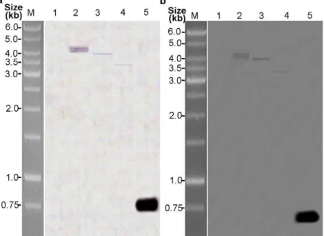
The integration of two cassettes into the genomes of the three transgenic plants for each generation was confirmed by PCR and southern blot using genomic DNA of the T3 transgenic plants. Using a primer internal to the CaMV 35S promoter and top35, and a primer internal to the op-iap gene, PCR products of the expected sizes of 1266 bp (CaMV 35S-p35) and 583 bp (op-iap) were obtained from all three T1–T3transgenic samples for each transgenic plant (Fig. 2). The product sizes were identical to amplification from the p1301–p35–iap co-expression vector, whereas no PCR products were obtained from the amplification of genomic DNA in non-transformed negative controls. The DNA of three transgenic T3plants and the untransformed control plant was digested withEcoRI, and respectively hybridized with DIG-labeled p35 and op-iap gene probes. All three transgenic plants exhibited hybridization signals with two probes while the non-transformed control did not. Withp35probe, plant KB1 showed two hybridization signals, while KB2 and KB3 had one band of hybridization respectively (Fig. 3a). The hybridization signals with
Figure 1. Schematic map of the T-DNA region ofp35andop-iap.Thep35gene replaced the reporter gene of pBI121 through restriction with
BamHI and SacI enzymes to construct an intermediate vector pBI121–p35. Theop-iap gene replaced the reporter gene of pCAMBIA 1301 by restriction withBglII andBstEII termed p1301–iap. Thep35gene expression cassette obtained byPstI andEcoRI restriction enzyme digestion was inserted into p1301-iapof the multiple cloning sites to construct the co-expression vector p1301-iap-p35.
op-iapprobe displayed the same patterns and the copy numbers as p35(Fig. 3b). The results of this analyses indicated the presence and stable integration of thep35andop-iapgenes into the genome of all three transgenic plants. PCR analysis of the progeny of these transgenic lines of cotton suggested thatp35andop-iapwere able to be stably inherited into the progeny.
To detect accumulation of p35 and op-iap transcripts, semi-quantitative RT-PCR of isolated total RNA from the three T3 transgenic plants and the non-transformed negative controls was
performed to demonstrate expression of thep35andop-iapgenes. PCR amplification of cDNA from all three T3transgenic plants and the plasmid control template yielded the expected products of 533 bp or 583 bp in length, respectively, while no corresponding products were amplified from non-transformed negative controls (Fig. 4). DNA sequence analysis showed that the DNA sequences integrated in the three T3transgenic plants were identical to that of the region amplified from the p1301–p35–iap co-expression vector control.
Figure 2. PCR analysis ofp35andop-iapgenes in T1-T3generations of the transgenic cotton lines.PCR products from genomic DNA of
the three transgenic T1–T3generation and non-transformed negative control plants. (A) the presence ofp35in T1generation. (B) the presence ofp35
in T2generation. (C) the presence ofp35in T3generation. (D) the presence ofop-iapin T1generation. (E) the presence ofop-iapin T2generation. (F)
the presenceop-iapin T3generation. Lane 1 the negative control plant; lanes 2, 3, 4 the three transgenics lines (KB1, KB2, KB3); lane 5 the p1301-p35 -iapco-expression vector control.
doi:10.1371/journal.pone.0014218.g002
Figure 3. Southern blot analysis of three T3transgenic cotton lines.Southern blot hybridization analysis of genomic DNA extracted from
leaves of three transgenic cotton plants and non-transformed negative control plants. (a) DNA from transgenic cotton plants and non-transformed negative control plants was digested withEcoRI and hybridized with a DIG-labeledp35probe (870 bp); (b) DNA from three transgenic cotton plants and non-transformed negative control plants was digested withEcoRI and hybridized with a DIG-labeledop-iapprobe (583 bp); Lane 1 the negative control plant; lanes 2, 3, 4 the three transgenics lines (KB1, KB2, KB3); lane 5: positive control [purified PCR products of thep35(870 bp) andop-iap
To further confirm the protein expression ofp35andop-iapin transgenic plants, total soluble protein was extracted from the three T3transgenic plants and non-transformed negative controls and analyzed by Western blotting. Figure 5 shows that using anti-p35and anti-iap antibodies revealed the presence of 35-kDa and 30-kDa protein bands, which are the same size as P35 and OP-IAP reported by Birnbaum et al. [45]and Wang et al. [23] in transgenic cotton lines KB1, KB2, and KB3. The non-trans-formed negative controls showed no positive signal for P35 and IAP proteins.
Disease resistance survey of the T1, T2, and T3
generations
To accurately evaluate disease resistance in transgenic cottons, we used an artificial nursery to evaluate resistance to verticillium wilt of cotton. Disease severity was expressed as a disease index (DI). Analysis of verticillium wilt resistance or susceptibility of the transgenic cotton lines was based on visual assessment of chlorosis and necrosis symptoms of leaves. The infection rate and disease index were calculated for each plant using the formula stated earlier. Disease evaluation showed that the disease index of T1, T2, and T3generations of the three transgenic cotton lines (KB1, KB2, and KB3) throughout the developmental stages were lower than 19 and the resistance levels were between highly resistance (HR) to disease and disease-resistant (R), which was significantly (P,0.05) better than those of the negative control line z99668 (Table 1, Fig. 6). The results of the pathogenicity assay were consistent with that of the molecular analyses, which demonstrated that the strong resistance of the KB1, KB2, and KB3 lines to verticillium wilt was due to the heterologous expression ofp35andop-iap.
DNA laddering in fungus-infected plants
To test whether VD-toxins induce PCD in cotton, genomic DNA was isolated from T3transgenic (KB1) and non-transformed negative control leaves after being treated with 250 mg/L VD-toxin for 24 and 36 hours. DNA ladders are the hallmark feature of PCD, as a result of the activation of cell death-specific endonucleases such as DNases that cleave the DNA at the linker regions between oligonucleosomes to produce nucleosomal and oligonucleosomal DNA fragments (180 bp and multiples of 180 bp) that generate a characteristic ‘‘ladder’’ pattern during agarose gel electrophoresis. As shown in Fig. 7 (lane 3), the isolated DNA from non-transformed negative control leaves after treat-ment with 250 mg/L VD-toxin for 36 hours was fragtreat-mented and formed characteristic ladders that differed by just under 200 bp, whereas the transgenic line treated with 250 mg/L VD-toxin for 36 hours had no such ladder formation (Fig. 7, lane 4). That VD-toxins trigger plant cells to die by induction of a PCD mechanism involved in pathogenesis and that the expression ofp35andop-iap genes can inhibit such mechanism seem reasonable.
Flow cytometry analysis of cell death
To determine whetherp35andop-iapgene expression protects cells from VD-toxin-induced PCD, protoplasts isolated from the T3transgenic lines (KB1) and non-transformed negative controls were treated with 30 mg/L and 60 mg/L VD-toxin for 3 hours to induce cell death and then analyzed by flow cytometry after PI staining. After VD-toxin treatment, the percentage of protoplast death for the transgenic lines (KB1) and non-transformed negative controls all displayed an increase, but the percentage of dead protoplasts for non-transformed negative controls had a signifi-cantly higher increase than the transgenic lines (KB1). As shown in Fig. 8, in the 30 mg/L VD-toxin treatment, the percentage of dead protoplasts for the non-transformed negative controls and the transgenic lines (KB1) increased by 10.1% and 1.27%, respective-ly. In the 60 mg/L VD-toxin treatment, the percentage of dead protoplasts increased by 21.27% and 7.11%, respectively. These results indicate that p35 and op-iap gene expression partially protects cells from VD-toxin-induced PCD.
Discussion
PCD plays an important role in mediating plant adaptive responses to several stimuli, such as senescence, or in response to pathogens and environmental stresses. In this study, we introduced Figure 4. Semi-quantitative RT-PCR analysis ofp35andop-iap
expression in three T3transgenic cotton lines.Semi-quantitative
RT-PCR products from RNA extracted from the young leaves of three T3
transgenic plants and non-transformed negative controls. (a) p35
expression. (b) op-iap expression. (c) house-keeping gene histone-3
expression. Lane 1 the negative control plant; lanes 2, 3, 4 the three T3
transgenic lines (KB1, KB2, KB3); lane 5 the p1301-p35-iapco-expression vector control; lane M molecular mass marker is GeneRulerTM1 kb DNA
ladder (MBI Fermentas, Maryland, USA). doi:10.1371/journal.pone.0014218.g004
Figure 5. Western blot assays of expression of P35 and OP-IAP protein in T3transgenic cotton lines. The total soluble protein
extracted from the young leaves of the T3transgenic lines and
non-transformed negative controls. (A) P35 expression. (B) OP-IAP expression. Lane 1 the negative control plant; lane 2, 3, 4 the three T3transgenic lines (KB1, KB2, KB3); lane M molecular mass marker is
PageRuler TM prestained protein ladder (MBI Fermentas, Maryland,
USA).
anti-apoptotic p35 and op-iap genes from baculovirus into the genome of cotton and analyzed the response of transgenic plants to verticillium wilt. The results showed that the construct containing p35 and op-iap was not only stably integrated into the cotton genome and expressed in the transgenic lines, but also able to be
inherited through to the T3 generation. Three transgenic lines (KB1, KB2, and KB3) performed markedly (P,0.05) better than the negative control line z99668 when inoculated withV. dahliaein the verticillium wilt nursery room. After treatment with 250 mg/L VD-toxins for 36 hours, DNA from the non-transformed negative
Figure 6. The disease resistance phenotype of T3transgenic cotton lines in the verticillium wilt artificial nursery.A. Disease symptoms
of verticillium wilt at the cotton flowering stage. The leaf shows yellow or brown spots and curled leaf edges. B. The disease resistance phenotype of the T3transgenic cotton lines (KB1, KB2, KB3) and the negative control lines (z99668) at the flowering stage in the cotton verticillium wilt artificial
nursery. Three T3transgenic cotton lines did not exhibit disease symptoms, while the leaves of the negative control lines (z99668) showed yellow or
brown spots and curled leaf edges, some leaves dropped. doi:10.1371/journal.pone.0014218.g006
Table 1.Analyses of disease resistance of transgenic cotton lines.
plant number
Infection rate(%)
Disease Index(DI)
Disease resistant(R)/Tolerance(T) /Susceptible(S)
Transplant T1/F1 T2/F2 T3/F3 T1/F1 T2/F2 T3/F3 T1/F1 T2/F2 T3/F3
KB1 16.7* 62.3
54.2* 60.96
38.1* 61.49
14.6* 61.05
17.9* 60.87
13.5* 60.45
R R R
KB2 24.1* 60.75
47.3* 61.04
44.1* 60.82
16.9* 60.45
15.1* 60.89
18.8* 60.56
R R R
KB3 10.7* 61.32
53.6* 60.62
39* 60.66
8.8* 60.66
17.1* 60.46
15.9* 60.56
HR R R
Control Z99668 35.8
60.85
49.6
60.7
56.4
60.95
33.8
60.66
20.6
60.7
25.5
60.95
T T T
Disease evaluation standards: HR, highly resistant plants with a DI value between 0 and 10.0; R, resistant plants with a DI value between 10.1 and 20.0; T, disease tolerance plants with a DI value between 20.1 and 35.0; S, susceptible plants with a DI value.35.0. The means standard deviations (SD) were calculated based on three replications. Asterisks indicate statistical significance (P,0.05) between the control plant(z99668) and each transgenic lines (KB1, KB2 and KB3).
control leaves was fragmented, whereas that from the transgenic lines was not. The percentage of dead protoplasts from the non-transformed negative controls was higher than that from the transgenic cotton lines after VD-toxin treatment, indicating that p35 and op-iap gene expression in transgenic lines enhances the tolerance to verticillium wilt and partially protects cells from VD-toxin-induced cell death.
Previous studies have reported results consistent with our observations. Hansen [19] showed that transgenic maize embryo-genic callus with the baculovirus apoptosis inhibitory genes p35 andop-iapcan inhibitAgrobacterium-induced PCD. Tobacco plants
expressing the human anti-apoptotic genesbcl-2,bcl-xL, nematode Ced-9, and baculovirusOp-iap showed increased resistance to the tomato spotted wilt virus (TSWV), with fewer local lesions and prevention of systemic spread of the virus [10]. Lincoln et al. [20]and Wang et al. [23]showed that expression of the baculovirus p35 gene effectively blocks apoptotic cell death as induced by either a host-selective toxin or any of several necrotrophic pathogens, leading to protection against the disease caused by these pathogens. p35 transgenic tobacco, passion-fruit, and narrow-leafed lupin all have increased tolerance to necrotrophic fungal pathogens and herbicides [21,22,23]. As noted above, a unifying aspect of these results is the fact that the plants with expression of anti-apoptotic genes are all resistant to necrotrophic pathogens, which kill plant cells and use the plant as a nutrient source for growth, colonization, and reproduction. Therefore, the inhibition of apoptotic pathways is a potential strategy for engineering necrotrophic disease resistance in plants.
The mechanism of action of these anti-apoptotic genes in plants, at present, is poorly understood. In this paper, the baculovirus anti-apoptotic protein P35, an IAP known to inhibit animal caspase, was effective in preventing PCD induced by VD-toxins. Although plants have no homologs of animal caspase genes and no identified caspase-dependent pathways, several identified proteases involved in plant PCD show caspase-like activities. The two serine proteases in Avena sativa exhibit caspase-like activity and have aspartate specificity, but contain a Ser-active site; they were involved in a PCD-induced protease cascade leading to the activation of another protease targeted to the chloroplast and cleaved Rubisco [46]. The vacuolar processing enzymes showed caspase-1 activity was essential for PCD induced by TMV [47] and fumonisin B1 [48], as well as for developmental cell death in the formation of the seed coat [49]. Chichkova et al. [50,51] identified a novel PCD-related subtilisin-like protease that possessed caspase specificity and activity essential for PCD-related responses to TMV and abiotic stresses. Cell death-related proteins may exist that are functional equivalents with little or no sequence homology to animal counterparts, providing the functional domains to interact with the P35 and Op-IAP products.
Verticillium dahliae, which comprises a subset of necrotrophic pathogens, enters the plant by way of the root system and invades the protoxylem vessels. The molecular mechanisms of verticillium diseases are unclear. In this study, after VD-toxin treatment, cotton DNA was fragmented and formed characteristic ladders, and the percentage of dead protoplasts for the untransformed and transformed cotton all displayed an increase. These results were consistent with those of Wang et al. [9] who purified VdNEPs that induced DNA laddering of cotton suspension-cultured cells, and Palmer et al.[52]who purified an 18.5-kDa phytotoxic protein that induced more dead protoplasts of susceptible cotton cultivars than resistant cultivars. In addition, Chu et al. [40]reported that the secreted glycoproteins ofV. dahliae play an important role in the wilting symptom and that native glycoproteins at concentrations higher than 5 mg/L resulted in cell death. Meyer et al. [7,53]showed that cotton plasma membranes have high-affinity, specific binding sites for the protein lipopolysaccharide complex (PLPC) fromV. dahliae, and that the PLPC induced symptoms of wilting and necrosis and inhibited the H+
-ATPase activity of plasma membranes, stimulating phenylalanine ammonia-lyase activity. All this serves to illustrate that the secreted substances ofV. dahliaeare the key biochemical factors that cause the plant disease, which can trigger plant cells to die by induction of a PCD mechanism involved in pathogenesis.
Taken together, this work represents the first attempt to show heterologous expression of the baculovirus anti-apoptoticp35and Figure 7. DNA laddering in cotton lines after treatment with
VD-toxins. Ethidium bromide-stained agarose gel (2%) of DNA extracted from T3 transgenic (KB1) leaves after treatment with
250 mg/L VD-toxin for 24 hours (lane 2) and 36 hours (lane 4), DNA extracted from the negative control leaves after treatment with 250 mg/L VD-toxin for 24 hours (lane 1) and 36 hours (lane 3). lane M molecular mass marker is GeneRuler TM 100 bp DNA ladder (MBI
Fermentas, Maryland, USA).
op-iap genes in cotton plants. The results demonstrated that p35 andop-iapgene expression partially protects cells from VD-toxin-induced PCD and enhances resistance to verticillium wilt disease. These results provide a potential strategy for engineering broad-spectrum disease resistance in plants and a good system for further study on plant PCD, as well as providing further evidence that pathogenesis-associated cell death in plants shares common regulatory and mechanistic features with PCD in animals.
Acknowledgments
We thank Dr. Qianjun Li for the generous gift ofp35andop-iapgene, and Prof. Yingzhang Li for providingVerticillium dahliaestrain V229.
Author Contributions
Conceived and designed the experiments: JD TW. Performed the experiments: JT XZ BL SL QW CL. Analyzed the data: JT XZ ZW. Contributed reagents/materials/analysis tools: ZW JD TW. Wrote the paper: JT XZ JD TW.
References
1. Fradin EF, Thomma B (2006) Physiology and molecular aspects of Verticillium wilt diseases caused by V-dahliae and V-albo-atrum. Molecular Plant Pathology 7: 71–86.
2. Huisman OC (1982) Interrelations of Root-Growth Dynamics to Epidemiology of Root-Invading Fungi. Annual Review of Phytopathology 20: 303–327. 3. Klosterman SJ, Atallah ZK, Vallad GE, Subbarao KV (2009) Diversity,
pathogenicity, and management of Verticillium species. Annual Review of Phytopathology 47: 39–62.
4. BejaranoAlcazar J, BlancoLopez MA, MeleroVara JM, JimenezDiaz RM (1997) The influence of verticillium wilt epidemics on cotton yield in southern Spain. Plant Pathology 46: 168–178.
5. Pegg GF (1965) Phytotoxin Production by Verticillium albo-atrum Reinke et Berthold. Nature 208: 1228–1229.
6. Buchner V, Nachmias A, Burstein Y (1982) Isolation and Partial Characteriza-tion of a Phytotoxic Glycopeptide from a Protein-Lipopolysaccharide Complex Produced by a Potato Isolate of Verticillium-Dahliae. Febs Letters 138: 261–264. 7. Meyer R, Slater V, Dubery IA (1994) A Phytotoxic Protein-Lipopolysaccharide Complex Produced by Verticillium-Dahliae. Phytochemistry 35: 1449–1453. 8. Gour HN, Dube HC (1985) Effects of Ouabain and Phytotoxic Metabolites from
Verticillium-Dahliae on the Cell-Membranes of Cotton Plants. Physiological Plant Pathology 27: 109–118.
9. Wang JY, Cai Y, Gou JY, Mao YB, Xu YH, et al. (2004) VdNEP, an elicitor from Verticillium dahliae, induces cotton plant wilting. Applied and Environ-mental Microbiology 70: 4989–4995.
10. Dickman MB, Park YK, Oltersdorf T, Li W, Clemente T, et al. (2001) Abrogation of disease development in plants expressing animal antiapoptotic genes. Proceedings of the National Academy of Sciences of the United States of America 98: 6957–6962.
11. Khurana SMP, Pandey SK, Sarkar D, Chanemougasoundharam A (2005) Apoptosis in plant disease response: A close encounter of the pathogen kind. Current Science 88: 740–752.
12. Mitsuhara I, Malik KA, Miura M, Ohashi Y (1999) Animal cell-death suppressors Bcl-x(L) and Ced-9 inhibit cell death in tobacco plants. Current Biology 9: 775–778.
13. Boatright KM, Salvesen GS (2003) Mechanisms of caspase activation. Current Opinion in Cell Biology 15: 725–731.
14. Thornberry NA, Lazebnik Y (1998) Caspases: Enemies within. Science 281: 1312–1316.
15. Bonneau L, Ge Y, Drury GE, Gallois P (2008) What happened to plant caspases? Journal of Experimental Botany 59: 491–499.
16. del Pozo O, Lam E (1998) Caspases and programmed cell death in the hypersensitive response of plants to pathogens. Current Biology 8: 1129–1132. Figure 8. Flow Cytometry Analysis of Cell Death Induced by VD-toxins.The protoplasts isolated from the T3transgenic lines (KB1) and
non-transformed negative controls were treated with 30 mg/L and 60 mg/L VD-toxin for 3 hours and analyzed by flow cytometry after PI staining. Flow cytometry analysis was conducted using 10,000 protoplasts per sample. Percentages of dead protoplasts are indicated. Vertical axis refers to PI fluorescence intensity and the abscissa refers to the number of protoplast. Data are representative of three independent experiments.
17. Mlejnek P, Prochazka S (2002) Activation of caspase-like proteases and induction of apoptosis by isopentenyladenosine in tobacco BY-2 cells. Planta 215: 158–166.
18. Sun YL, Zhao Y, Hong X, Zhai ZH (1999) Cytochrome c release and caspase activation during menadione-induced apoptosis in plants. Febs Letters 462: 317–321.
19. Hansen G (2000) Evidence for Agrobacterium-induced apoptosis in maize cells. Molecular Plant-Microbe Interactions 13: 649–657.
20. Lincoln JE, Richael C, Overduin B, Smith K, Bostock R, et al. (2002) Expression of the antiapoptotic baculovirus p35 gene in tomato blocks programmed cell death and provides broad-spectrum resistance to disease. Proceedings of the National Academy of Sciences of the United States of America 99: 15217–15221.
21. de Freitas DS, Coelho MCF, Souza MT, Marques A, Ribeiro BM (2007) Introduction of the anti-apoptotic baculovirus p35 gene in passion fruit induces herbicide tolerance, reduced bacterial lesions, but does not inhibits passion fruit woodiness disease progress induced by cowpea aphid-borne mosaic virus (CABMV). Biotechnology Letters 29: 79–87.
22. Wijayanto T, Barker SJ, Wylie SJ, Gilchrist DG, Cowling WA (2009) Significant reduction of fungal disease symptoms in transgenic lupin (Lupinus angustifolius) expressing the anti-apoptotic baculovirus gene p35. Plant Biotechnology Journal 7: 778–790.
23. Wang Z, Song J, Zhang Y, Yang B, Wang Y, et al. (2008) Mechanism analysis of broad-spectrum disease resistance induced by expression of anti-apoptotic p35 gene in tobacco. Sheng Wu Gong Cheng Xue Bao 24: 1707–1713. 24. Sugimoto A, Friesen PD, Rothman JH (1994) Baculovirus P35 Prevents
Developmentally Programmed Cell-Death and Rescues a Ced-9 Mutant in the Nematode Caenorhabditis-Elegans. Embo Journal 13: 2023–2028.
25. Davidson FF, Steller H (1998) Blocking apoptosis prevents blindness Drosophila retinal degeneration mutants. Nature 391: 587–591.
26. Hisahara S, Araki T, Sugiyama F, Yagami K, Suzuki M, et al. (2000) Targeted expression of baculovirus p35 caspase inhibitor in oligodendrocytes protects mice against autoimmune-mediated demyelination. Embo Journal 19: 341–348. 27. Viswanath V, Wu ZJ, Fonck C, Wei QZ, Boonplueang R, et al. (2000) Transgenic mice neuronally expressing baculoviral p35 are resistant to diverse types of induced apoptosis, including seizure-associated neurodegeneration. Proceedings of the National Academy of Sciences of the United States of America 97: 2270–2275.
28. Means JC, Clem RJ (2008) Evolution and function of the p35 family of apoptosis inhibitors. Future Virology 3: 383–391.
29. Wei YB, Fan TJ, Yu MM (2008) Inhibitor of apoptosis proteins and apoptosis. Acta Biochimica Et Biophysica Sinica 40: 278–288.
30. Wang YQ, Chen DJ, Wang DM, Huang QS, Yao ZP, et al. (2004) Over-expression of Gastrodia anti-fungal protein enhances Verticillium wilt resistance in coloured cotton. Plant Breeding 123: 454–459.
31. Tohidfar M, Mohammadi M, Ghareyazie B (2005) Agrobacterium-mediated transformation of cotton (Gossypium hirsutum) using a heterologous bean chitinase gene. Plant Cell Tissue and Organ Culture 83: 83–96.
32. Rajasekaran K, Cary JW, Jaynes JM, Cleveland TE (2005) Disease resistance conferred by the expression of a gene encoding a synthetic peptide in transgenic cotton (Gossypium hirsutum L.) plants. Plant Biotechnology Journal 3: 545–554. 33. Murray F, Llewellyn D, McFadden H, Last D, Dennis ES, et al. (1999) Expression of the Talaromyces flavus glucose oxidase gene in cotton and tobacco reduces fungal infection, but is also phytotoxic. Molecular Breeding 5: 219–232. 34. Wang W, Chen WX, Zhi Z, Xu HL, Gao YF, et al. (1999) Studies on highly
efficient planting of transgenic cotton. Acta Botanica Sinica 41: 1072–1075.
35. Paterson AH, Brubaker CL, Wendel JF (1993) A rapid method for extraction of cotton (Gossypium spp.) genomic DNA suitable for RFLP or PCR analysis. Plant Molecular Biology Reporter 11: 122–127.
36. Li XB, Cai L, Cheng NH, Liu JW (2002) Molecular characterization of the cotton GhTUB1 gene that is preferentially expressed in fiber. Plant Physiology 130: 666–674.
37. Bradford MM (1976) Rapid and Sensitive Method for Quantitation of Microgram Quantities of Protein Utilizing Principle of Protein-Dye Binding. Analytical Biochemistry 72: 248–254.
38. Zhen XH, Li YZ (2004) Ultrastructural changes and location of beta-1,3-glucanase in resistant and susceptible cotton callus cells in response to treatment with toxin of Verticillium dahliae and salicylic acid. Journal of Plant Physiology 161: 1367–1377.
39. Zhang YS, Wang JX, Liu JF, Fang ZD (1989) Studies on the isolation, purification and bioassay of toxin from Verticillium dahliae Kleb. Acta Mycologica Sinica 8: 140–147.
40. Chu ZQ, Jia JW, Zhou XJ, Chen XY (1999) Isolation of glycoproteins from Verticillium dahliae and their phytotoxicity. Acta Botanica Sinica 41: 972–976. 41. Stein JC, Hansen G (1999) Mannose induces an endonuclease responsible for
DNA laddering in plant cells. Plant Physiology 121: 71–79.
42. Ryerson DE, Heath MC (1996) Cleavage of nuclear DNA into oligonucleosomal fragments during cell death induced by fungal infection or by abiotic treatments. Plant Cell 8: 393–402.
43. Sun YQ, Zhang XL, Nie YC, Guo XP, Jin SX, et al. (2004) Production and characterization of somatic hybrids between upland cotton (Gossypium hirsutum) and wild cotton (G-klotzschianum Anderss) via electrofusion. Theoretical and Applied Genetics 109: 472–479.
44. Kim M, Lim JH, Ahn CS, Park K, Kim GT, et al. (2006) Mitochondria-associated hexokinases play a role in the control of programmed cell death in Nicotiana benthamiana. Plant Cell 18: 2341–2355.
45. Birnbaum MJ, Clem RJ, Miller LK (1994) An Apoptosis-Inhibiting Gene from a Nuclear Polyhedrosis-Virus Encoding a Polypeptide with Cys/His Sequence Motif. Journal of Virology 68: 2521–2528.
46. Coffeen WC, Wolpert TJ (2004) Purification and characterization of serine proteases that exhibit caspase-like activity and are associated with programmed cell death in Avena sativa. Plant Cell 16: 857–873.
47. Hatsugai N, Kuroyanagi M, Yamada K, Meshi T, Tsuda S, et al. (2004) A plant vacuolar protease, VPE, mediates virus-induced hypersensitive cell death. Science 305: 855–858.
48. Kuroyanagi M, Yamada K, Hatsugai N, Kondo M, Nishimura M, et al. (2005) Vacuolar processing enzyme is essential for mycotoxin-induced cell death in Arabidopsis thaliana. Journal of Biological Chemistry 280: 32914–32920. 49. Nakaune S, Yamada K, Kondo M, Kato T, Tabata S, et al. (2005) A vacuolar
processing enzyme, delta VPE, is involved in seed coat formation at the early stage of seed development. Plant Cell 17: 876–887.
50. Chichkova NV, Kim SH, Titova ES, Kalkum M, Morozov VS, et al. (2004) A plant caspase-like protease activated during the hypersensitive response. Plant Cell 16: 157–171.
51. Chichkova NV, Shaw J, Galiullina RA, Drury GE, Tuzhikov AI, et al. (2010) Phytaspase, a relocalisable cell death promoting plant protease with caspase specificity. Embo Journal 29: 1149–1161.
52. Palmer CS, Saleeba JA, Lyon BR (2005) Phytotoxicity on cotton ex-plants of an 18.5 kDa protein from culture filtrates of Verticillium dahliae. Physiological and Molecular Plant Pathology 67: 308–318.