Osseointegration and Cell Response on Titanium Implant Surface
Treatments
Universidade Fernando Pessoa
Faculdade de Ciências da Saúde
Osseointegration and Cell Response on Titanium Implant Surface
Treatments
Universidade Fernando Pessoa
Faculdade de Ciências da Saúde
Osseointegration and Cell Response on Titanium Implant Surface
Treatments
Dissertação apresentada à
Universidade Fernando Pessoa
como parte dos requisitos para a obtenção do
grau de Mestre em Medicina Dentaria
Introdução:
A osteointegração de um implante é essencial para garantir êxito clínico. Vários tratamentos de superfície foram introduzidos para melhorar a osseointegração e a biocompatibilidade de um implante.
Objectivos:
O objectivo de esta revisão bibliográfica é explorar as diferentes propriedades de Titanio e como á modifica-las, depois de introduzir um implante de Titanio num tecido de osso vivo, a osteointrgração, a resposta celular e a biocompatibilidade do implante podem ser afectadas.
Materiais e métodos:
Para esta revisão bibliográfica, 96 artigos foram usados, tombados das seguintes bases de dados: Pubmed, Science direct and Scielo. Através das seguintes palavras chave: "Surface treatments", "Titanium dental implants", "osseointegration", "anodization", "anodic oxidation", "plasma spraying", "HA coatings", "hydroxyapatite coatings", "acid etching", "grit blasting”, “tribocorrosion" AND “biotribocorrosion”.
Conclusão:
Introduction:
The osseointegration of an implant is essential in order to achieve clinical success. Various surface treatments have been introduced in order to achieve a better osseointegration and biocompatibility of the implant.
Objectives:
The objective of this review was to explore the different properties of titanium and how by changing them, after a titanium implant being introduced to living bone tissue, osseointegration, cellular response and biocompatibility of the implant can be affected.
By this it can be understood what to look for in an implant surface, concluding to the ideal surface characteristics a Ti implant should possess.
Materials and Methods:
For this review, they were used 96 articles, retrieved from the following search engines: Pubmed, Science direct and Scielo. Through the following key words: "Surface treatments", "Titanium dental implants", "osseointegration", "anodization", "anodic oxidation", "plasma spraying", "HA coatings", "hydroxyapatite coatings", "acid etching", "grit blasting”, “tribocorrosion" AND “biotribocorrosion”.
Conclusion:
I Introduction……….1
II Development………...2
1)Materials and Methods………...2
2) Biology of wound healing………3
3) Dental implant materials……….…….6
4) Tribocorrosion……….8
5) Implant surface character………...11
5.1) Topography……….11
5.2) Surface Roughness………..12
5.3) Surface Charge………...……….13
5.4) Surface Energy………14
5.5) Wettability………..15
5.6) Surface Chemistry……...………17
5.7 Atomic Copmposition………..18
6) Methods of surface modifications of implants………20
6.1) Grit Blasting/Sand Blasting……….21
6.2) Acid Etching………...23
6.3) Grit Blasting and Acid Etching………26
6.4) Plasma Spraying………...29
7) Discussion……….39
III Conclusion………...42
Figure 1 - Drawings to show the initiation of distance osteogenesis……….5
Figure 2 - Particles of Titanium and aluminium encrusted in a sandblasted titanium surface………....………23
Figure 3 - Microtopography of an acid etched Titanium dental implant………..24
Figure 4 - SEM image of SLA/modSLA surface on c.p. titanium………..27
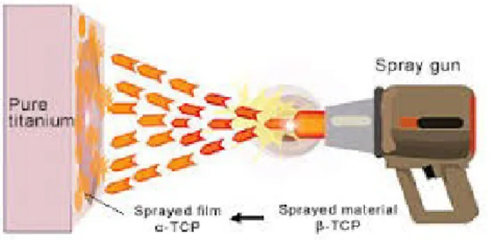
Figure 5 - A spraying raw material powder is passed through the plasma flame for spraying
on pure titanium as a base material..………....30
Figure 6 - Scanning electron microscope (SEM) image of titanium plasma–sprayed surface implant with rough surface characteristics. A, Implant with titanium plasma–sprayed
surface (×40)………...…...32
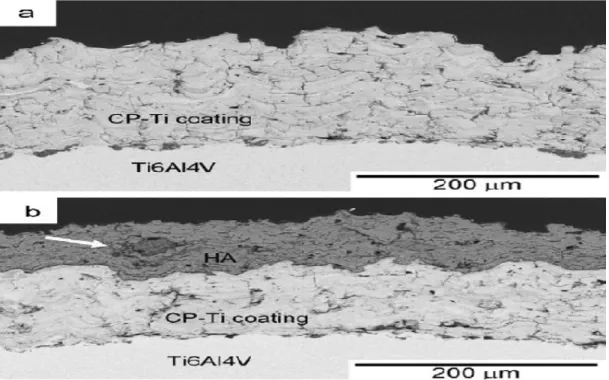
Figure 7 - Cross-sectional BEI images of the (a) CP-Ti and (b) HA coating on the CP-Ti.
(Image retrieved from the article (Yang & Yang, 2013)……….…35
Figure 8 - SEM micrographies (5000x) of the Ti anodic films produced in 1.0M H2SO4/150V,
HA- Hydroxyapatite
PDGF- Platelet-Derived Growth Factor
TGF- Transforming Growth Factor beta
Cp-Ti- Commercially Pure Titanium
TiO2- Titanium Oxide
RANKL- receptor activator of nuclear factor kappa-B ligand
OPG- osteoprotegerin
TNF-a- Tumor Necrosis Factor alpha
Sa- Surface Roughness
CA- Contact Angle
CaP- Calcium Monophosphide
Al2O3- Aluminium Oxide
SiO2- Silica Oxide
BIC- Bone to Implant Contact
BCP- Biphasic Calcium Phosphate
HCL- Hydrogen Chloride
H2SO4- Sulfuric Acid
HNO3- Nitric Acid
HF- Hydrofluoric Acid
Sdr- Developed Interfacial Area Ratio
Sds- Summit Density
SLA- Sand-blasted, Large grit, Acidetched
AE- Acid Etched
RT – Room temperature
ROS- Reactive Oxygen Species
NO(•)- Nitric Oxide
H2O2- Hydrogen Peroxide
HOCl- Hypochlorous Acid
NO3- Nitrate
CAT- Catalase
SOD- Superoxide Dismutase
PTTS- Plasma Treated Surface
SIT- Sandblasted and Ion Implanted
PS-Plasma Sprayed
PSIT- Plasma Sprayed and Ion Implanted
SEM- Scanning Electron Microscope
VPS- Vapor Plasma Spraying
I. Introduction
The oral rehabilitation of missing teeth by dental implants is one of the most commonly
used surgical procedures nowadays. Beginning in the late 1960s the focused efforts of P.I.
Branemark led to the detailed microscopic characterization of interfacial bone formation
at machined titanium endosseous implants. This process, of bone formation at the
endosseous implant surface, known as osseointegration was considered a positive
outcome that was contrasted to fibrous encapsulation, a negative and undesired result that
leads to failure of the treatment. Osseointegration gives stability able to resist forces and
distribute them uniformly to the bone making possible its loading. So, it is clear that
clinical success is dependent on the direct structural and functional connection between
ordered, living bone and the surface of a load carrying implant. About 50-80%
bone-implant contact is found sufficient to have a clinically successful bone-implant (Lian et al.,
2010).
To improve osseointegration and biocompatibility of Ti implants, the nature of the
implant itself has to be changed. In order to do that, various surface treatments have been
introduced to the market that change the mechanical, physical and chemical properties of
titanium.
II. Development
1) Materials and Methods:
The bibliographic review of this study was realized between the months of May, June and
July of 2016.
References 1985 to 2015were accepted in this review.
For this review 96 articles were used out of the initial 129 that were selected from
abstract reading.
Criteria of inclusion were: indexed articles relevant to the theme of the dissertation,
articles written in Greek, English or Portuguese and articles that were of scientific
interest.
Criteria of non-inclusion were: articles irrelevant to the theme of dissertation, articles that
by further inspection provided no further insight to the theme, and articles that did not
provide conclusions.
Keywords used for this review were: "Surface treatments", "Titanium dental implants",
"osseointegration", "anodization", "anodic oxidation", "plasma spraying", "HA
coatings", "hydroxyapatite coatings", "acid etching", "grit blasting”, “tribocorrosion"
2) Biology of wound healing following implant placement
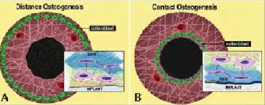
Peri-implant osteogenesis can be in distance and in contact from the host bone. In
distance osteogenesis, new bone is formed on the surfaces of old bone in the peri-implant
site. The bone surfaces provide a population of osteogenic cells that lay down a new
matrix until reaching the implant. An essential observation here is that new bone is not
forming on the implant, but the latter does become surrounded by bone. Thus, in these
circumstances, the implant surface will always be partially obscured from bone by
intervening cells. Distance osteogenesis is typical of cortical bone healing. In contact
osteogenesis, new bone starts forming directly on the implant surface. Since, by
definition, no bone is present on the surface of the implant upon implantation, the implant
surface has to become colonized by bone cells before bone matrix formation can begin
(Davies J. E., 2008).
Bone matrix is synthesized by only one cell: the osteoblast. Since each osteoblast may
become a completely entombed osteocyte, the osteoblast is incapable of migration away
from the bone surface, and the only method by which this surface can receive further
additions is by the recruitment of more osteogenic cells to the surface, which then
differentiate into secretorily active osteoblasts. Bone matrix mineralizes and has no
inherent capacity to “grow.” Once bone formation has been initiated, the matrix and the
cells that have synthesized that matrix have almost no ability to govern the ongoing
pattern of bone growth on the implant surface. The only way for new bone to be formed
on an implant, is by osteogenic cells to migrate to its surface. Then, if we require that
bone “grows” around the implant to establish functional endosseous integration, this too
can only be achieved by the continued recruitment around, and migration of osteogenic
cells to the implant surface. Thus, it can be concluded that the most important stages of
endosseous healing precede bone formation (Davies J. E., 2008).
During surgery, dental implant surfaces interact with blood components from ruptured
blood vessels. Within a short period of time, various plasma proteins such as fibrin get
and kinin systems become activated. As in fracture healing, the migration of bone cells in
peri-implant healing will occur through the fibrin of a blood clot. Since fibrin has the
potential to adhere to almost all surfaces, it can be anticipated that the migration of
osteogenic cell populations towards the implant surface will occur. However, as the
migration of cells through fibrin will cause retraction of the fibrin scaffold, the ability of
an implant surface to retain this fibrin scaffold during the phase of wound contraction is
critical in determining whether the migrating cells will reach the implant surface.
Activation of platelets occurs as a result of interaction of platelets with the implant
surface as well as the fibrin scaffold and this leads to thrombus formation and blood
clotting (Anil et al., 2011).
Moreover, platelets are a rich source of many growth and differentiation factors which
play a key role in the wound healing process by acting as signaling molecules for
recruitment and differentiation of the undifferentiated mesenchymal stem cells at the
implant surface. Platelet degranulation releases a number of growth factors, such as
platelet-derived growth factor (PDGF) and transforming growth factor beta (TGF-b),
together with vasoactive factors such as serotonin and histamine, factors that play an
important role in the regulation of the wound-healing cascade, (JY, Gemmel and Davies,
2001)
Absorption of proteins such as fibronectin and vitronectin on the surface of dental
implants could promote cell adhesion and osseointegration. During the initial remodeling,
a number of immune cells mediate early tissue response followed by migration of
phagocyte macrophages. These cells initially remove the necrotic debris created by the
drilling process and then undergo physiological changes which lead to expression of cell
surface proteins and production of cytokines and pro-inflammatory mediators. This
cytokine-regulated cellular recruitment, migration, proliferation and formation of an
extracellular matrix on the implant surface can be influenced by the macrophages. These
cells express growth factors such as fibroblast growth factor (FGF-1, FGF-2, FGF-4),
transforming growth factors, epithelial growth factor as well as bone morphogenetic
process that includes angiogenesis. (Anil S, 2011)
Thus, as in fracture healing, the migration of osteogenic cells in peri-implant healing will
occur through the transient three-dimensional biological matrix formed as a product of
the coagulation cascade—the fibrin of the blood clot—and may be both potentiated and
directed, either directly or indirectly through knock-on stimulatory events involving
leukocytes, (Bromberek et al., 2002) by the release of cytokines, growth factors, and
microparticles from platelets activated by contact with the implant surface. Finally, when
the osteogenic cells reach the implant surface, they can initiate bone matrix secretion, by
osteogenic cells, of the cement line matrix. This is a collagen-free, mineralized interfacial
matrix laid down between old bone and new bone (Davies J., 1996).
Figure 1: Drawings to show the initiation of distance osteogenesis (A) and contact
osteogenesis (B) where differentiating osteogenic cells line either the old bone or implant
surface respectively. The insets show the consequences of these two distinctly different
patterns of bone formation. In the former the secretorily active osteoblasts, anchored into
their extracellular matrix by their cell processes, become trapped between the bone they are
forming and the surface of the implant. The only possible outcome is the death of these cells.
On the contrary, in contact osteogenesis, de novo bone is formed directly on the implant
surface, with the cement line in contact with the implant (insert) and is equivalent to the
osteonal interface illustrated in Figure 1 (can be viewed at www.ecf.utoronto.ca/ ~bonehead/
3) Dental Implant Materials
When a material is inserted into an organism, an interaction between the two is
inevitable, leading to three possible outcomes, integration, incorporation, or rejection.
The outcome depends on various factors, such as the nature of the material, that can be
classified as: bioinert, biotolerated, or bioactive; osteogenic, osteoconductive or
osteoinductive. These properties influence the biological response that will be induced
pos-implantation (Albrektsson and Johansson, 2001).
Bioinert material, is a material that doesn't induce a foreign body reaction when
introduced to the organism and establishes direct contact with the surrounding tissues
without interposition of fibrous tissue. A biotolerated material is moderately accepted
from the organism; that means that there is no total direct contact and fibrous tissue can
be involved in the healing process. Bioactive material, is a material that not only forms a
direct contact with the surrounding tissues but also establishes a chemical bond.
Generally, these materials present calcium and phosphate ions at their surface, that help
establish a chemical bond (Puleo and Nanci, 1999).
An osteogenic material is capable to mobilize cells of the osteoblastic cell line, to a
determined place, and promote the formation of bone tissue. An osteoconductive
material, guides the formation of bone to its surface when implanted in such tissue by
mobilizing osteogenic cells. An osteoinductive material, promotes formation of bony
tissue, when implanted out of such tissue, by mineralizing undifferentiated cells, and
inducing their differentiation to osteoblastic cells. An ideal material would possess all of
the three abovementioned properties (Albrektsson and Johansson, 2001).
One of the most frequently used biomaterials for dental implants is commercially pure
titanium (Cp-Ti) and its alloys. An implant is a structure that is surgically inserted in the
alveoli and through means of osseointegration establishes a tight connection with the
bone. The reason behind its commercial popularity among dentists is its optimal
resistance and thermoelectric conduction. Its corrosion resistance and biocompatibility
but also its osseointegration are consequence to an oxide layer (TiO2) with a width of
2-10nm that Ti forms immediately when exposed to air (Bordji, et al., 1996).
The most used Ti alloy is Ti6Al4V. Aluminum is added to increase the mechanical
resistance of the implant while Vanadium stabilizes the phase maintaining resistance to
corrosion. Commercially pure titanium and Ti6Al4V are materials with a very similar
elasticity to the bone and so, forces and tensions are distributed homogenously from the
cervical area of the implant to the bone (Anusavice, Shen and Rawls, 2013).
There are 4 grades of commercially pure Ti (1,2,3 and 4) with grade 1 being the purest
with a 99.8% of Ti, grade 4 being the most used commercially. Elements that can be
found in Cp-Ti are considered impurities that are permitted from American Society for
Testing and Materials. Cp-Ti is a light metal with high resistance to corrosion and a good
relation between mechanical resistance and resistance to fracture when subjected to loads
similar to occlusal forces. Ti6Al4V on the other side presents a higher mechanical
4) Tribocorrosion
The oral cavity is a dynamic environment, that sometimes can be hostile for an implant.
Biofilm, acids produced by bacteria or even substances that can be found in saliva such as
fluoride and chloride when in contact with the Ti can induce the chemical process known
as corrosion to its surface impairing its mechanical resistance (Souza et al., 2013; Souza
et al.,2015).
Tribocorrosion is the phenomenon which occurs when two surfaces that are in contact
suffer from simultaneous corrosion and degradation. When this happens in a biological
environment it is known as biotribocorrosion. Biotribocorrosion of a material is
dependent on its chemical and mechanical properties as well as the medium that is
inserted into. For example: the oxide layer that protects Ti from corrosion can be
damaged by mechanical wear, and in return be susceptible to chemical corrosion (Celis,
Pontiaux, and Wegner, 2006; Souza et al., 2015).
The presence of biofilm in a Ti surface causes a decrease of pH and so turns the environ
-ment acidic, promoting chemical reactions that result in corrosion. Dental implants are
situated in an area where oxygen is scarce or non-existent, so, only anaerobic bacteria can
survive in this environment. Porphyromonas gingivalis, Aggregatibacter
actinomycetem-comitans, Prevotela intermedia are examples of anaerobic bacteria that can cause
peri-odontal and periimplantary diseases (Sissons, Wong and Shu, 1998; Barbour et al.,2007;
Cruz et al., 2011).
When corrosion or wear occurs metallic particles and ions are released to the surrounding
tissues. The immune system recognizes them as foreign objects and an inflammatory
response mediated by cytokins occurs in the periimplantary tissues, leading to
reabsorption of the bone and periimplantary diseases (Renvert,Persson, 2009; Cruz et al.,
Metallic ions can accumulate in surrounding tissues or can be spread through systemic
circulation to organs like the lungs, spleen, lymph nodes and the liver. This accumulation
can alter the expression of the receptor activator of nuclear factor kappa-B ligand
(RANKL) and to the osteoprotegerin (OPG) in osteoblastic cells, which contribute to the
osteoclastic activity of pathologic osseous remodeling (Urban et al., 2000; Triplett et al.,
2003).
A study elaborated by Wachi T. et al. (2015) showed that Ti ions with a concentration of
9ppm synergize with the bacteria P. gengivalis-LPS to increase the expression of cytocin
ligant2 (CCL2)n RANKL and OPG at the gingival tissues, initiating inflammation and
reabsorption of the bone. A concentration of 13ppm is enough to provoke necrosis of the
cell.
Ti particles with a size of 10μm or less cause inflammatory responses and are considered
cytotoxic, while particles of 1-3μm can be phagocytized by neutrophils or macrophages.
Phagocytation of these particles causes an inflammatory response to the tissue, an
increase in tumor necrosis factor alpha (TNF-a) and the phagocyte itself undergoes
degenerative morphologic changes. TNF-a is a pre-inflammatory cytokin that capable of
causing cellular death (Kumazawa et al., 2002).
It is well known that in high concentration metallic particles are harmful for fibroblasts,
altering their proliferation and viability. In vitro experiments proved that Ti particles with
a concentration of 0.001% in a saline solution can increase the proliferation of
fibroblasts, but higher concentrations can decrease it. Recent studies showed as well that
nanometric TiO2 particles, that are released during biotribocorrosion, can induce
cytotoxicity and genotoxicity. It was shown that in cellular cultures containing these
particles, cell viability decreased and mutations increased. A culture of alveoli
macrophages showed that when in contact with TiO2 particles, the cells were under
oxidative stress, proving their genocytotoxicity (Maloney et al., 1993; Wang, Sanderson
But not only the TiO2 particles can cause damage to the cells, Vanadium and Aluminum
particles have also proven to be cytotoxic, impairing cellular growth, as shown by a study
elaborated by Okazaki (2001), Vanadium is potentially genotoxic as well (Manaranche,
5) Implant surface character
Implant surface character is one implant design factor affecting the rate and extent of
osseointegration.
The process of osseointegration is now well described both histologically and at the
cellular level. Precisely how much of the implant surface directly contacts bone, how
rapidly this bone accrual occurs, and the mechanical nature of the bone/implant
connection is influenced by the nature of the implant surface itself (Mendonca 2008).
The surface characteristics of an implant which influence the speed and strength of
osseointegration include surface chemistry, topography, wettability, charge, surface
energy, crystal structure and crystallinity, roughness, strain hardening, the presence of
impurities, thickness of titanium oxide layer, and the presence of metal and non-metal
composites(Anil et al., 2011).
5.1 Topography
Implant surface topography refers to macroscopic and microscopic features of the
implant surface. Surface topography plays an important role in the osseointegration of
titanium implants (Le Guehennec, 2007).
It is not clear whether the height of surface irregularities is more important than the
distance between them, and which combination of these factors could improve
osseointegration. Although the increase in surface roughness promotes greater
mechanical anchorage, the implant bone interface strength will not increase with the
5.2 Surface Roughness
The surface roughness of the implants can significantly alter the process of
osseointegration because the cells react differently to smooth and rough surfaces.
Fibroblasts and epithelial cells adhere more strongly to smooth surfaces, whereas
osteoblastic proliferation and collagen synthesis are increased on rough surfaces (Boyan
et al., 2001). Investigators have demonstrated that while the adhesion of fibroblasts is
lesser on rough surfaces, the adhesion and differentiation of osteoblastic cells are
enhanced (Wennerberg and Albrektsson, 2000).
Smooth surfaces as well as excessive roughness induce osteoblasts into a fibroblast
phenotype. Smooth surfaces induce it because of the lack of space for osteoblasts to grow
while excessive roughness induce it due to large spaces between the irregularities. In
vitro and in vivo studies have shown that titanium surface roughness influences a number
of events in the behavior of cells in the osteoblastic lineage, including spreading and
proliferation, differentiation, and protein synthesis (Sammons et al., 2005; Zhao, 2006).
The organization of the cytoskeleton, special orientation of the cells as well as synthesis
and mineralization of the bone matrix are favored by an increased surface roughness. It
has been shown that titanium implants with adequate roughness may influence the
secondary stability of implants, enhance bone-to-implant contact, and may increase
removal torque force (Wennerberg and Albrektsson, 2009).
Based on the average surface roughness (Sa) surfaces with an average Sa≤1μm are
considered smooth and those with a Sa>1μm are considered rough. e.g. Machined
Titanium is a smooth surface with Sa values of 0.53 to 0.96 μm (Sykaras et al., 2000).
High roughness results in mechanical interlocking between the implant surface and the
bone ingrowth. However, a major risk with high surface roughness may be an increase in
may limit these two parameters (Wennerberg and Albrektsson, 2000).
The microtopographic profile of dental implants is defined for surface roughness as being
in the range of 1-10μm. This range of roughness maximizes the interlocking between
mineralized bone and the surface of the implant. A theoretical approach suggested that the
ideal surface should be covered with hemispherical pits approximately 1.5μm in depth
and 4μm in diameter. Implant surface roughness is divided, depending on the dimension
of the measured surface features into macro, micro, and nano-roughness (Anil et al.,
2011).
Macro roughness comprises features in the range of millimeters to tens of microns. This
scale directly relates to implant geometry, with threaded screw and macro porous surface
treatments. Micro roughness is defined as being in the range of 1–10 μm. This range of
roughness maximizes the interlocking between mineralized bone and implant surface.
Studies supported by some clinical evidence suggest that the micron-level surface
topography results in greater accrual of bone at the implant surface. The use of surfaces
provided with nanoscale topographies are widely used in recent years. Nanotechnology
involves materials that have a nano-sized topography or are composed of nano-sized
materials with a size range between 1 and 100 nm. Nanometer roughness plays an
important role in the adsorption of proteins, adhesion of osteoblastic cells and thus the
rate of osseointegration (Anil et al., 2011).
5.3 Surface Charge
It has been shown that the surface charge of a dental implant affects its osseointegration.
A negative charge or a positive charge has been found to be more promising than a
neutral charge since a charge on an implant surface promotes hydrophilicity. (Boyan,
1996).
multiple layers of cells and enlarged colonies of osteoblast-like cells can be also
observed. In contrast, cell adhesion and proliferation on positively charged biomaterial
were found to be subdued (Yan Guo, Matinlinna and Tang, 2012).
An implant treated with acid etching, using NaOH as the acidic solution, forms a
bioactive sodium titanium oxide (sodium titanate) layer, on the titanium surface, that is
charged negatively. The negatively charged layer, attracts positively charged calcium ions
that begin to accumulate on the biomaterial surface, turning it to a positive charge; hence,
the surface starts to attract negatively charged phosphate ions, which react with the
calcium ions to form a calcium phosphate (i.e. a type of apatite) layer. This calcium
phosphate layer takes an amorphous structure after its formation, and it subsequently
transforms into more stable crystalline apatite (Hamouda, et al., 2012; Yan Guo,
Matinlinna and Tang, 2012).
A charged implant surface can induce electrical attraction or repulsion between the
implant surface and the surrounding chemical species, depending on their polarity.
Besides the effect on crystal nucleation, another significant role of Ca2+ is to attract
cell-adhesion proteins (e.g., integrins, fibronectin, and osteonectin), which are characterized
by their capacity to interact with a specific ligand. These proteins significantly affect the
attachment, adhesion, and spreading of osteoblasts. Consequently, osteoblasts attach and
proliferate on a matrix grown on the bone-like apatite layer formed with Ca2+ ions, which
may result in faster and stronger bone-to-implant bonding. In contrast, a positively
charged implant surface attracts anionic groups which act as antiadhesive molecules,
which negatively affect osteoblast adhesion (Ohgaki et al., 2001).
5.4 Surface Energy
Surface charge influences surface energy which is a measure of the extent to which the
bonds are unsatisfied at the surface. The surface energy of an implant is an important
surface free energy on the implant surface enhances the hydrophilicity of its surface,
which in turn promotes the adhesion of blood components such as proteins, as well as
osteoblasts. Studies have shown though, that an extremely high surface energy while
promoting the adhesion of cells, can hinder their motility and their subsequent functions
(Yan Guo, Matinlinna and Tang, 2012; Gittens et al., 2014).
A high surface free energy initiates the absorption of proteins such as fibronectin, which
is an extracellular matrix protein responsible for osteoblast cell differentiation, cell-cell
interactions and cell-matrix interactions (Jia et al., 2015).
A high surface energy results in a high degree of wettability; thus when an implant is
exposed to blood, the entire surface will almost immediately be covered by it, stimulating
the blood proteins to attach to the surface, to start the bone healing process, however
from a clinical point of view, a recent overview failed to find convincing evidence of the
effectiveness of increasing surface energies (Wennerberg, Galli, and Albrektsson, 2011).
Wetting of high and low energy substrates: The energy of the bulk component of a solid
substrate is determined by the types of interactions that hold the substrate together. High
energy substrates are held together by bonds, while low energy substrates are held
together by forces. Covalent, ionic, and metallic bonds are much stronger than forces
such as van der Waals and hydrogen bonding. High energy substrates are more easily wet
than low energy substrates. In addition, more complete wetting will occur if the substrate
has a much higher surface energy than the liquid (De Gennes, 1985).
5.5 Wettability
Wettability is influenced by the surface energy of an implant and is one of the most
important factors for bone to implant contact. It governs the degree of contact that the
surface of the implant will have with the physiological environment. It can be described
contact with it such that it spreads over the surface and wets it (Kilpadi and Lemons,
1994; Zhao, Liu, & Ding, 2005). Wettability and surface energy influence the adsorption
of proteins, and increase adhesion of osteoblasts on the implant surface. An increased
wettability enhances the biocompatibility of an implant, promoting interactions between
the implant and the biological environment (Sartoretto et al., 2015). A hydrophilic surface
is better for blood coagulation than a hydrophobic surface. The expressions of
bone-specific differentiation factors for osteoblasts are higher on hydrophilic surfaces.
Consequently, dental implants manufacturers have developed high hydrophilic and rough
implant surfaces which in turn exhibited better osseointegration than implants with
smooth surfaces (Anil et al., 2011).
Contact angle (CA): A way to experimentally determine wetting is to look at the contact
angle (θ)
If θ=0 the liquid completely wets the substrate.
If 0<θ<90, high wetting occurs.
If 90<θ<180, low wetting occurs.
If >180, the liquid does not wet the substrate at all.
Water CAs very close to 0 are termed as superhydrophilic and above 150 degrees as
superhydrophobic (Gittens et al., 2014).
The Young Equationrelates the contact angle to interfacial energy:
cosθ= (
γsv-γsl) / γlv
energy between the substrate and the liquid, γlv is the interfacial energy between the
liquid and gas phases, and θ is the contact angle between the gas and the
solid-liquid interface.
As seen from Young's equation, the contact angle is directly coupled to the surface
energy, where a surface with high surface energy has a low contact angle and is easily
wetted in contrast to a low-energy surface with a high contact angle.
5.6 Surface Chemistry
Surface chemistry can be roughly defined as the study of chemical reactions at interfaces.
It is closely related to surface engineering, which aims at modifying the chemical
composition of a surface by incorporation of selected elements or functional groups that
produce various desired effects or improvements in the properties of the surface or
interface. Surface chemistry involves adhesion of proteins, bacteria, and cells on
implants. Surface chemistry has the potential to alter ionic interactions, protein
adsorption, and cellular activity at the implant surface (Schliephake et al., 2005). These
modifications may subsequently influence conformational changes in the structures and
interactive natures of adsorbed proteins and cells. Furthermore, within the complexities
of an in vivo environment containing multiple protein and cellular interactions, these
alterations may differentially regulate biologic events. Modifications to the implant
surface chemistry may lead to alterations in the structure of adsorbed proteins and have
cascading effects that may ultimately be evident at the clinical level (Anil et al., 2011).
Surface chemistry is highly dependent on surface topography and vice versa, since a
treatment that produces a certain topography will change the chemistry of the implant’s
surface. While overlooked oftenly, surface chemistry plays an important role in the
absorption of proteins, the cell attachment, the behavior of the attached cells as well as
biocompatibility of the implant itself. While topography kept constant, changes in surface
to fully cytocompatible (Cassinelli et al., 2003; Thevenot, Hu and Tang, 2008)
In vivo evidence has supported the use of alterations in surface chemistry to enhance
osseointegration (Sartoletto et al., 2015).
5.7 Atomic Composition
Atomic composition plays an important role in osseointegration since cellular
connections are established initially in an atomic level through chemical components
present at the surface, including O2 and Ti. Titanium is a very reactive material, forming
an oxide layer immediately after contacting O2. Because of this layer the maximum
percentage of Ti in its surface is 33%, with the rest 66% being oxygen. Realistically
though, as described by many authors, Ti implants present a percentage of Ti less than
20% due to contamination with hydrocarbons and carbides (Morra et al., 2003).
Machined implants are the ones to contain more contaminants due to the oils and
lubricants used during production and polishing processes.
Even chemical sterilization can increase the percentage of contaminants found in the
implant surface, leading many companies to use radiations as the sterilization process
(Morra and Cassinelli, 1997; Sakai et al., 1998; Morra et al., 2003).
Machined implants are found to contain a toxic element, Pb. Probably this element
contaminates machined surfaces through the machines used to create this type of surface.
Elements such as nitrogen, fluoride, aluminum, silicon, phosphorus and calcium have
been found in plasma treated and acid surfaces, though in very small quantities. Nitrogen
and fluoride can be found in surfaces that were treated with acids, due to the contact with
the acids themselves, while nitrogen can be explained in Plasma treated surfaces from the
Ti particles that are projected, nitrogen could bond to these particles. All these particles
6) Methods of surface modifications of implants
It has been shown that the possibility of osseointegration failure due to fibrous connective
tissue development between implant surface and bone is increased when the latter has not
undergone any treatment (Le Guehennec et al., 2007, Von Wilmowsky et al., 2014). The
importance of interface in the microscopic and ultramicroscopic structure between
implant-bone and implant-soft tissues needs to be stressed. In order to amplify and
accelerate osseointegration, various implant modifications have been presented seeking to
higher bone to implant contact.
The methods employed for surface modifications of implants can be broadly classified
into 3 types-mechanical; chemical; and physical. These different methods can be
employed to change the implant surface chemistry, morphology, and structure. The main
objective of these techniques is to improve the bio-mechanical properties of the implant
such as stimulation of bone formation to enhance osseointegration, removal of surface
contaminants, and improvement of wear and corrosion resistance (Anil et al., 2011).
The mechanical methods include grinding, blasting, machining, and polishing. These
procedures involving physical treatment generally result in rough or smooth surfaces
which can enhance the adhesion, proliferation, and differentiation of cells. The physical
methods of implant surface modification include plasma spraying, sputtering, and ion
deposition. Plasma spraying includes atmospheric plasma spraying and vacuum plasma
spraying. This is used for creating titanium and CaP coatings on the surfaces of titanium
implants. Sputtering has been used to deposit thin films on implant surfaces to improve
their biocompatibility, biological activity, and mechanical properties such as wear
resistance and corrosion resistance. Methods of surface modification of titanium and its
alloys by chemical treatment are based on chemical reactions occurring at the interface
between titanium and a solution. The chemical methods of implant surface modifications
include chemical treatment with acids or alkali, hydrogen peroxide treatment, sol-gel,
chemical vapor deposition, and anodization. Chemical surface modification of titanium
wettability/surface energy (Bagno and Di Bello, 2004; Anil et al., 2011).
6.1 Grit Blasting/Sand Blasting
Blasting is explained as the use of abrasive particles against another material under high
pressure in order to make it smoother, remove contaminants, or to roughen the surface.
Titanium surfaces can be grit blasted with hard ceramic/metallic particles in order to
roughen them. The particles are projected through a nozzle at high velocities by means of
compressed air to the titanium implant surface and depending on the size and shape of the
ceramic particle, which is polyhedral with sharp corners (Barriuso et al., 2014), and on
the velocity of the blasting, erosion and material tearing of the titanium surface, is
inflicted. The result is different surface roughness levels that can be produced on the
implant's surface. The blasting material should be chemically stable, biocompatible and
should not hamper the osseointegration of the titanium implants. The most common
particles that are used are Alumina (Al2O3), titanium oxide (TiO2) and calcium phosphate
(Ca2P2O7) (Parekh, Shetty and Tabassum, 2012).
Sand blasting, besides roughening the surface to increase the surface area, it also is a
method that cleans surface contaminants and produces beneficial surface compressive
residual stress. As a result, such treated surfaces demonstrate higher surface energy,
indicating higher surface chemical and physical activities and enhancing fatigue strength
as well as fatigue life (Oshida and Daly, 1990).
Topographic variations of the order of 10nm and less may become important because
microroughness on this scale length consists of material defects such as grain boundaries,
steps and vacancies, which are known to be active sites for absorption and thus may
influence the bonding of biomolecules to the implant surface. There is evidence that
surface roughness on a micron scale allows cellular adhesion that alters the overall tissue
response to biomaterials. Microrough surfaces allow early better adhesion of mineral ions
or atoms, biomolecules and cells form stronger fixation of bone or connective tissue,
prevent microorganism adhesion and plaque accumulation, when compared with the
smooth surfaces (Oshida, 2007).
Alumina (Al2O3) or silica (SiO2) particles are most frequently used as a blasting media,
but because alumina oxide is insoluble in acid, its residues may have a negative effect on
bone formation, principally caused by the release of cytotoxic silicon or aluminum ions in
the peri-implant tissue (Aliofkhazraei, 2015).
Titanium oxide is also used for blasting titanium dental implants. Titanium oxide particles
with an average size of 25μm produce a moderately rough surface in the 1–2μm range on
dental implants. An experimental study using microimplants in humans has shown a
significant improvement for bone-to-implant contact (BIC) for the TiO2 blasted implants
in comparison with machined surfaces. Other experimental studies confirmed the
increase in BIC for titanium grit-blasted surfaces. Other studies have reported high
clinical success rates for titanium grit-blasted implants, up to 10 years after implantation.
Comparative clinical studies gave higher marginal bone levels and survival rates for TiO2
grit-blasted implants than for machined turned implants. Wennerberg, Albrektsson
and Laussma, (1996) demonstrated with a rabbit model that grit-blasting with TiO2 or
Al2O3 particles gave similar values of bone–implant contact, but drastically increased the
biomechanical fixation of the implants when compared to smooth titanium. These studies
have shown that the torque force increased with the surface roughness of the implants
while comparable values in bone apposition were observed.
Using biphasic calcium phosphate (BPC) particles to roughen a surface by means of grit
blasting, has been found to produce a more biocompatible surface, when compared to
TiO2 and Al2O3 surfaces, and with an average surface roughness of 1.57μm. These
particles have been proven to not cause cytotoxicity to mouse osteoblastic cells of the
MC3T3-E1 cell line. BCP treated implants have been proven also to promote an earlier
cell differentiation and bone apposition when compared to alumina grit-blasted and
machined surfaces. These particles can be also removed by means of acid-etching,
Basiuk, 2015)
The particles that are projected with grit-blasting, while roughening the implant’s surface,
have been found to impair the mechanical performance of the implant. The projected
par-ticles can cause notch-like superficial defects, evidence of erosion and material tearing.
Fine cracks can also be observed around particles that are firmly attached to the craters
they created in the metal. These observations are evidence of a reduced long term
me-chanical performance. Grit blasted surfaces were found to have a reduced endurance limit
of 25% when compared to polished surfaces (Shemtov-Yonan, Rittel, and Dogoroy, 2014)
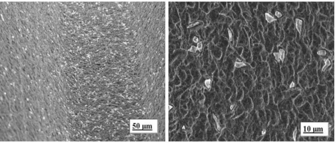
Figure 2: Particles of Titanium (left) and aluminium (right) encrusted in a sandblasted
titanium surface (can be viewed at:
http://www.intechopen.com/books/implant-dentistry-a-rapidly-evolving-practice/factors-affecting-the-success-of-dental-implants)
6.2 Acid Etching
Chemical etching with strong acids such as HCl, H2SO4, HNO3 and HF is another method
for roughening the surface of dental implants. Acid etching of titanium removes the oxide
layer and parts of the underlying material producing micro pits on the implant surface
with sizes ranging from 0.5 to 3μm and larger pits of approximately 6 to 10μm in
treatment time. The micro pits that are formed result in an enlarged active surface area
that subsequently increases the retention and biomechanical interlocking between implant
and bone as well as enhancing osteoblast activity with quicker formation of bone at the
interface. This yields low surface energy and reduces the possibility of contamination
since no particles are encrusted in the surface, having a positive effect on bone
apposition, a higher percentage of contact surface area when comparing with a grit
blasted surface and strong implant anchorage. This type of surface not only facilitates
retention of osteogenic cells, but also allows them to migrate towards the implant surface
(Wong et al., 1995; Juodzbalys, Sapragoniene, Wennerberg, 2003; Cho and Park, 2003).
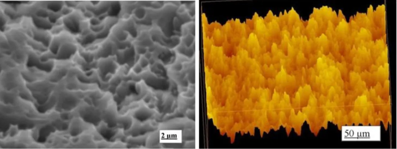
Figure 3: Microtopography of an acid etched Titanium dental implant (can be viewed at:
http://www.intechopen.com/books/implant-dentistry-a-rapidly-evolving-practice/factors-affecting-the-success-of-dental-implants)
Each manufacturer has its own acid etching method regarding concentration, time and
Etching time and etching temperature
A recent study made by Bruno Ramos Chrcanovic, Ann Wennerberg et al. (2015)
evaluated how etching time and temperature influence the roughness parameters of a
specific acid solution (HCl/ H2SO4). After treating the surface of 30 Ti discs with the
same solution but with different etching temperatures and times it was observed that
irregularities on the surface of the discs became deeper with increased etching
temperature and increased in size and depth with increasing etching time. The increase in
temperature changed the irregularity pattern from linear grooves with sharp edges to
micro-pits and finally to deeper valleys, removing the grooves produced by the polishing
process. An etching temperature of 60 or 90°C will provide a moderate roughness only
after at least 15 minutes of etching time. But an etching temperature of 120°C is too high
when the etching time is of 15' or more as the irregularities that the treatment produced
were visible with the naked eye and in some sites peeled when scratched.
When comparing etching time and etching temperature it can be clearly observed from
this study that temperature plays a more important role in the variance of the surface
roughness parameters. Specifically, it was observed that 43.2% of the variance in Sa
(mean roughness) is being explained by the temperature and 11.5% by the time. The
numbers for Sdr and Sds were 34.6% and 31.3%, respectively for the temperature and
10.9% and 0.06% for the time (Chrcanovic, Wennerberg and Martins, 2015).
Acid Etching roughness and topography
Acid etching produces a minimally rough surface of Sa values around 0.5-3μm,
depending on the acid solution, acid time and temperature, the bulk material and surface
microstructure. Due to the presence of hydrogen ions in the acid, there is a speculation of
a hybrid layer. The oxide layer has been found to be amorphous and with a thickness of
Various studies have shown that this surface treatment provides a better osseointegration
when compared to as-machined implants. It was found that bone to implant contact was
higher 12 months after implantation and a higher removal torque after 1,2 and 3 months
(Ballo et al., 2011).
In order for the acidic solutions to change the properties of titanium and roughen its
surface they have to come in direct contact with it. But before attacking the metallic
titanium, the acids must first dissolve the protective titanium oxide layer. During the
course of the corrosion process of titanium, native hydrogen ions (H+) are released. These
small ions diffuse rapidly into the metal because the latter is left without its dense
protective oxide layer; the sub-surface is therefore enriched with hydrogen (Aronsson et
al., 2001). When saturation in hydrogen is reached, titanium hydride is formed. Titanium
hydride may be biologically important because a hydride layer is much better suited as a
template for binding biomolecules chemically into a titanium surface (Videm et al.,
2008). GIRXD analysis have been shown that Ti hydride is present in Ti implants treated
with acid etching.
Each acid etching treatment produces a unique surface topography with distinct surface
properties that are dependent on various parameters, such as acid mixture composition,
time of the treatment, temperature and prior treatments. A weak acid might not affect the
morphology and low etching temperature might produce small micropits, for whereas a
combination of strong acids in high concentration, high etching temperatures and treated
for a considerable amount of time will likely produce a rough surface with numerous
micro pits (Chrcanovic and Martins, 2014).
6.3 Grit blasting and Acid Etching
Soaking in acidic solutions implants that were previously grit blasted serves many
purposes. The acid solution reduces the highest peaks, smoothening the irregularities
caused by the blasting particles, reducing the average surface roughness to typically
contaminants, and creates a titanium hybrid intermediate to the implant and the titanium
oxide layer (Conforto et al., 2004).
By rinsing the SLA implant in a nitrogen atmosphere and storing in saline solution until
installation, the amount of carbon contamination could be reduced, improving the
hydrophilicity of the implant surface (Rupp et al., 2006). The result of this procedure is a
new hydrophilic surface (SLActive). Several studies have shown that SLActive implants
achieve a better bone contact, earlier stability and reduce the healing time from 12 to 6
weeks when compared to SLA implants (Buser et al., 2004; Schwarz et al., 2007).
SLA implants have shown in in vivo tests as well as in in vitro tests a superior and faster
osseointegration when compared with other surfaces, especially in the initial healing
period, this could be explained to the higher production of cytokines and growth factors
that were observed by Kieswetter et al., (1996).
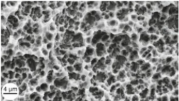
Figure 4: SEM image of SLA/modSLA surface on c.p. titanium (can be viewd
at:http://pocketdentistry.com/sandblasted-and-acid-etched-implant-surfaces-with-or-without-high-surface-free-energy-experimental-and-clinical-background/)
Sand blasted and acid etched surfaces have a hydrophobic surface and the new SLActive
implants have a hydrophilic surface which shows a stronger bone response. These have a
are seen in the SLA implants. The different etching processes also may lead to the
formation of Titanium hydrides (TiH2, TiH3, TiH4 or a combination of them) and the
replacement of hydride by oxygen results in the slow transformation of the implant
surface, resulting in nanometer sized particles of titanium on the surface. The nano
roughness may be important in the protein adhesion, immediately after the implant
placement. Sand blasting and etching can increase the rate and amount of the bone
formation. The alkaline phosphatase specific activity was enhanced and osteocalcin
production, the latent transforming growth factor beta and prostaglandin E2, all which
were involved in the bone formation were found to be increased (Garg, Bedi and Garg,
2012).
M. Herrero-Climent et al. (2013) performed studies in vitro and in vivo with 4 different
implant treatments. One control group (as-machined), one acid etched group (0.35M HF
acid, 15'', RT), a grit blasted (Al particles 600μm in size, 0.25Mpa pressure) and a grit
blasted and acid etched group. The in vitro results showed a high surface roughness for
the grit blasted and SLA groups (average roughness Ra= 4.74 and 4.23 respectively) and
a moderate for the AE group (Ra=1.69). It was found that the roughest surfaces showed
the highest number of adhered cells, with the Gblasted and Gblast+AE surfaces showing
almost double figures in comparison with the control and the AE group, which in turn,
did not improve the cell adhesion. Ti samples were implanted into white rabbits and
retrieved after 1, 3 and 10 weeks of implantation in order to test osseointegration in vivo.
After histological analysis it was found that only Gblast and Gblast+AE samples
presented new immature bone at the periimplant area. The other groups presented only
the originally-machined bone during surgery which was in contact with threads of the
implants. This bone provided good initial stability. Results from this study confirm that
an increase in roughness translates to a higher cell adhesion, and that acceleration of
osseointegration at short-terms of implantation can be achieved by Gblast and Gblast+AE
implants. Roughness and topographical features are the most relevant of surface
6.4 Plasma Spraying
Biomaterial is any material, substance or combination of substances, of natural or
synthetic source that interacts with biological systems to stimulate the growth or replace
any tissue or organ for any period of time (Binyamin et al., 2006). Titanium is the most
widely used biomaterial in the manufacture of implants for several uses, especially as
bone replacement. This is because it has showed excellent mechanical properties as well
as resistance to corrosion and biocompatibility (Tsaryk et al., 2007).
Though biocompatible, exposure to Ti in a living system has been linked with increased
H2O2 and other reactive oxygen species (ROS). It is well known that ROS are now
appreciated to play several important roles in a number of biological processes and
regulate cell physiology and function. ROS are a heterogeneous chemical class that
includes radicals, such as superoxide ion O2(•-), OH (•) and NO(•), and non-radicals,
such as H2O2, singlet oxygen ((1)O2), HOCl, and NO3 (-) (Vara, Pula, 2014). Despite
their physiological roles, ROS can also damage several biomolecules and all aerobic
organisms have developed defenses against them, these include antioxidant enzymes such
as catalase (CAT), superoxide dismutase (SOD) and glutathione peroxidase, which
regulate the oxidative stress of the cells. Unbalanced ROS and reactive nitrogen species
(RNS) generation can cause lipid peroxidation, protein oxidation and DNA damage,
which could potentially lead to genotoxicity (Freires de Queiroz, et al., 2014).
Different surface treatments show different genotoxicity, one that has gained attention
due to its lower genotoxic and cytotoxic results is plasma spraying.
Plasma is obtained through an electric gas discharge, applying a potential difference
between two electrodes inserted into a chamber at pressures below 100Pa. The ions
produced are accelerated on the cathodically polarized electrode doing several effects
Figure 5: A spraying raw material powder is passed through the plasma flame for spraying
on pure titanium as a base material.
Argon plasma spraying produces a surface with good mechanical properties without
changing its chemical composition. It was shown as well from studies made by Freires de
Queiroz et al., (2013) that argon plasma spraying induces lower genotoxicity than an
untreated surface. For this study Ti disks were bombarded with pure argon atmosphere,
using a plasma energy source, generating a plasma treated surface (PTTS). The treatment
produced an uneven surface with a roughness of (Ra) 0.11μm. PTTS were found to be
more hydrophilic when compared to control. Cytotoxic assays showed that though the
cells on both surfaces (control and PTTS) were under oxidative stress conditions, as H2O2
production was higher when compared to the negative control, H2O2 levels of the
untreated surface were significantly higher and that decreased the viability of cells on
those implants as was confirmed by viability assays. Results showed that despite the
PTTS surfaces inducing oxidative stress to the adhered cells, the higher hydrophilicity of
those surfaces protected partially the cells from suffering DNA damage, thus proving to
be less cytotoxic than untreated surfaces.
A decrease of corrosion resistance of the plasma treated surfaces indicated a thin oxide
layer film and propensity of oxygen evolution reaction, causing the increase of
superoxide anion radicals at the extracellular medium that in turn stimulate the release of
SOD3. SOD3 catalyzes the dismutation of superoxide anion radicals into H2O2 and O, in
doesn't happen in untreated surfaces, the increased stress on these surfaces was attributed
to their low hydrophilicity, and to the higher DNA damage when compared to PTTS
(Freires de Queiroz et al., 2013).
It has been shown that a high oxidative stress increases the activity of antioxidant
enzymes (Mates, 2000; Husain et al., 2003). Another study though showed that it didn’t
affect, or reduced it slightly their activity, but increased the gene expression of them,
leading to an excessive consumption or degradation of those enzymes and in succession
adhered cells can't cope with the accumulation of ROS resulting to oxidative damage of
the cell (Lino-dos-Santos et al., 2011).
A thick oxide layer is important in Ti implants because it improves corrosion resistance,
avoiding the release of particles, ions and unstable ions during the electrochemical
process of corrosion and oxidative stress, improving the biocompatibility (Tavares et al.,
2009)
Yang, Ong and Tian, (2002) performed in vivo tests on beagles with 4 different surface
treatments (sandblasted Ti, PS porous Ti, sandblasted and ion implanted (SIT) and PS and
ion implanted (PSIT). After retrieving the implants in different time periods (2, 4 and 8
weeks) it was found that after 2 weeks there was already newly formed bone around the
implant and at 8 weeks the new bone was identical to the preexisting bone and
concentrated on the Haversian canal, indicating a complete healing of the drilled bone
area. At a higher magnification the newly formed bone was observed to grow within the
pores of PSIT 100-200μm, suggesting mechanical interlocking between the implant and
the bone. It was also observed that when Ti concentration was decreased, Calcium and
Phosphate concentrations increased, confirming the presence of a Ca-P layer at the PSIT
implant. That layer increased from 4-6μm to 30μm in a period of 4 weeks (from 4th week
to 8th week). A significant increase in the concentration of Ca and P in the SIT and PSIT
implant was observed at the implant surface from 4 to 8 weeks suggesting a tendency for
gradual mineralization to occur into the pores of the implants. Fracture tests performed on
mechanical interlocking was successful. TiO2 was present in PSIT, confirming a thick
oxide layer. The porous surface provided an increased contact surface that benefited
implant fixation but at the same time did not result in an increased release of Ti ions that
could harm the cells. From these results it was concluded that ion implantation helps
plasma sprayed implants to improve osseointegration by means of favoring the
mineralization process when compared to a PS treatment.
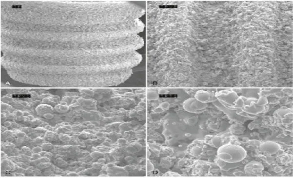
Figure 6: Scanning electron microscope (SEM) image of titanium plasma–sprayed surface
implant with rough surface characteristics. A, Implant with titanium plasma–sprayed
surface (×40). B, notably complex macrotopography on titanium plasma–sprayed surface
(×100). C, Titanium plasma–sprayed surface with 1- to 25-µm particles (×500). D, Titanium
plasma–sprayed surface with 1- to 25-µm particles (×1000). (image can be viewed at:
6.5 Hydroxyapatite Coatings
A plasma spraying gun can be used in order to obtain a coat. Through a powder feeder
Titanium or HA particles can be fed to the plasma spraying gun and projected at very
high temperatures and speeds onto the substrate which is positioned at a controlled
distance, roughening the implant's surface while at the same time forming a coat, with a
composition expected to be similar to the initial powder (Denmati et al., 2011).
An HA coat has been reported to improve the osseointegration of a cementless metallic
implant. HA is chemically similar to the bone, and is a source of calcium and phosphate
to the bone-HA interface. Osteoblasts form osteoid directly on the HA surface coating,
suggesting that the bone-implant interface is bonded both chemically and biologically to
the HA (Goodman et al., 2013).
Traditionally, HA coatings have been thought of as osteoconductive. However, calcium
phosphate biomaterials with certain 3-dimentional geometries have been shown to bind
endogenous bone morphogenetic proteins, and therefore some have designated these
ma-terials with osteoinductive properties (Le Geros, 2002).
In studies performed in canine models, formation of new bone was found even at
dis-tances of 400μm from the HA surface, suggesting the gradient effect of osseoconductive
properties of HA. Huang et al., (2015) evaluated the effects of HA coating in Ti implants
on the in vivo biological performance of porous Ti alloy (Ti6Al4V). Vapor plasma
spray-ing (VPS) is reported to produce a higher crystallinity than the conventional atmospheric
plasma spraying (APS), VPS was used to produce HA coated and uncoated implants for
this study. Implants were retrieved after 2 or 4 months pos-implantation. Analysis
showed that the HA coating was scattered in the internal area of the pores and some
inte-rior regions were not covered, nevertheless bone formation was supeinte-rior to that in the
un-coated group at both time intervals. Reconstructed 3D images showed that newly formed
bone was distributed into the peripheral region of the Ti at both time points, however
surface at 4 months while at this time period the uncoated implant was filled with fibrous
tissue at the same area, which may impede new bone ingrowth. Under higher
magnifica-tion it was observed that newly generated bone was tightly connected to the HA coat of
the HA-TI implant, without interposition of non-bone tissue, but the uncoated surface
showed fibrous tissue, indirect bone-implant contact and gaps. It was observed as well
that in the coated substrate numerous osteoblasts were distributed in a linear fashion
along the exterior surface of the immature bone, which indicates a favorable bone
forma-tion. Based on these findings it was concluded that HA-coated implants provide a better
osseointegration, bone-implant integration abilities and bone ingrowth than the uncoated
one. Such an anchoring effect could improve the fixation strength of the implant, which
may reduce the risk of surface coating delamination.
The bioresorption of HA coatings is still a matter of controversy. The two main methods
of resorption include one that is solution mediated (dissolution), and another that is cell
mediated via phagocytosis. The HA coatings undergo variable resorption which is
dic-tated by numerous chemical, biological and mechanical factors including the composition
and physico-chemical properties of the coating, the anatomical location, and whether
mi-cromotion is present at the interface with bone. Increased crystallinity appears to slow
re-sorption of HA, and decrease bone ingrowth (Goodmanet al., 2013).
During the clinical use of HA-coated implants, failure may occur at the coating-substrate
interface. Hydroxyapatite coatings delaminate from the Ti because of an insufficient
adhesion to its surface. Compressive stresses as well as mechanical and thermal
mismatch between Ti and hydroxyapatite decrease the adhesion as well (Yang, 2011).
In order to strengthen the interface bonding and reduce stress between the Ti substrate
and the HA, a titanium bond coat (Cp-Ti) can be used. The bond coat was found to
increase adhesion at such level that failure test analysis after push out tests showed that
failure occurred between the bone itself and not at the implant-HA interface or HA-bone
interface. This strong fixation of the HA coat to the implant was attributed to the rough