Samantha Al-Dujayli
Mechanical stability of Morse taper implant – abutment
connection ; A focused review
Universidade Fernando Pessoa
Faculdade de Ciências da Saúde
Samantha Al-Dujayli
Mechanical stability of Morse taper implant – abutment
connection ; A focused review
Universidade Fernando Pessoa
Faculdade de Ciências da Saúde
Samantha Al-Dujayli
Mechanical stability of Morse taper implant – abutment
connection ; A focused review
Universidade Fernando Pessoa
Faculdade de Ciências da Saúde
V
Resumo:
O objetivo principal deste estudo foi realizar uma revisão sistemática sobre a integridade
mecânica das conexões cone Morse de implante em relação à presença de microgap e
manutenção do torque.
Uma ampla pesquisa eletrônica foi realizada nos bancos de dados PubMed, Embase e
Medline com as palavras chaves: dental implant” e “dental abutment” e (“conical” ou “taper”
ou “cone”). “removal torque” e “Morse Taper”, “torque loss” e “micro gap size”
Estudos in vitro demonstraram que os pilares cônicos têm melhor desempenho, no que diz
respeito ao selamento bacteriano e a manutenção do torque e estabilidade do pilar, do que os
pilares não cônicos.
Estudos in vivo mostraram que as taxas de sucesso e sobrevivência dos sistemas de implantes
cone Morse comparadas aos outras, são quase comparáveis; porém os implantes com conexão
cone Morse mostraram menor perda ossea marginal.
Conclui-se , com esta revisão sistemática, que o uso de implantes com conexões cone Morse
parece ser mais vantajoso, pois de mostraram ter melhor desempenho em termos de
estabilidade mecânica e selamento bacteriano.
VI
Abstract:
The main aim of this study was to carry out a systematic review on the mechanical integrity
of Morse taper implant-abutment connections in relation to the presence of micro gap and
torque maintenance.
A broad electronic search was conducted using PubMed, Embase, and Medline databases
with the logical operators: “dental implant” AND “dental abutment” AND (“conical” OR
“taper” OR “cone”). “Removal torque” and “Morse Taper”, “Torque” and “micro gap size”
In vitro
studies demonstrated that conical abutments are more advantageous than non-conical
abutments, and appeared to be superior in terms of bacterial seal performance, torque
maintenance, and abutment stability.
In vivo
studies showed that the success and survival rates for conical and non-conical
implant-abutment systems are almost comparable; however, the results indicated that, conical
connection implants are more favourable as the majority of cases showed less marginal bone
loss around.
This systematic review points out that the use of conical implant–abutment connections seem
to be more advantageous as they clearly showed better performance in terms of mechanical
stability and bacterial seal.
VII
Acknowledgement:
I must express my very profound gratitude to my thesis advisor Professor Jorge Pereira for
the continuous encouragement and support throughout the process of researching and writing
this thesis.
I would also like to specially thank my parents for providing me with unfailing support; this
accomplishment would not have been possible without them. Thank you.
Samantha Al-Dujayli
VIII
Main Contents
I. Introduction: ... 10
II. Methodology:... 13
III. Development / Results: ... 14
A. In Vitro studies:... 14
1. Microgap size and seal performance: see table I / Attachment ... 14
2. Loading / fatigue performance and Torque maintenance: ... 17
B. In vivo studies: ... 20
1) Animal studies: ... 20
2) Human studies: ... 21
IV. Discussion ... 22
V. Conclusion ... 25
A.
In vitro
studies: ... 25
B.
In vivo
studies: ... 25
VI. References: ... 50
IX
Tables Contents
Table I / Seal Performance ... 23
The presence of Microgap and seal performance after fatigue test ... 23
The effect of different tightening torque values on the seal performance ... 24
Table II / The Mechanical integrity ... 32
The effect of the presence of Biofilm on the removal torque value ... 32
The effect of Biofilm on the removal torque... 34
Stress/ loading Performance ... 35
Tightening / loosening torque, cold welding ... 40
Table III / stress/ loading performance ... 42
Bending moment/ maximal load resistance ... 42
Table IV stress/ strain distribution ... 45
Stress/ loading Performance ... 45
Table V / Studies conducted in Animals... 53
Marginal bone loss ... 53
Table VI / Studies conducted in Humans ... 54
10 Introduction:
Dental implants have achieved long-term success due to the osseointegration of the highly
biocompatible titanium integrating to the surrounding bone,
(Branemark Pl et al. 1977;
Albrektsson et al. 2012).
Following the establishment of osseointegration, the success of implant system depends on
the mechanical and biological stability of the contacting metal components (Implant
abutment interface) (Branemark Pl et al. 1977; Albrektsson et al. 2012; Zarb GA, Schmitt
A2010).
The implant-abutment interface has been reported to be a significant factor in terms of load
transfer, adverse biological responses, and technical complications.
In every implant system, the efficiency of the implant-abutment connection (interface) system
depends on several factors, such as component design, connection geometry between implant
and abutment, mechanical fit or contact between the implant and its set inner surface on the
abutment, component mechanical and physical properties, and torque application (Carotenuto
G, Palumbo et al. ( 1999).
Implant systems differ in terms of the design of the implant
–
abutment interface with
particular differences between both conical and non-conical connection systems (indexed
external or internal connections). The implant
–
abutment connection represents the weakest
point of dental endosseous implant fixtures, as it must be strong enough in order to withstand
maximal and permanent masticatory forces as well as penetration by bacteria.( Schmitt CM1,
et al. 2013) .
There are approximately 20 different implant/abutment interface geometric variations
available. The geometry is important because it is one of the primary determinants of joint
strength, joint stability, and locational and rotational stability; it is critical to and synonymous
with prosthetic stability (Binon PP Implants and components 2000).
11
service (have been reported in the literature) (Binon PP Implants and components 2000;
Maeda y et al.
2006
;
Tabata LF
et al.
2011), The failure was mainly due to loose abutments,
inadequate microbial seal and screw fracture.
A 22-month follow-up on external hex implant prostheses in a private prosthodontic practice
reported the incidence of loose screws in fixed and removable prostheses at 27% and 32%,
respectively (Walton JN, et al.1997).
To overcome some of the inherent design limitations of the external hexagonal connection, a
variety of alternative connections have been developed. The most notable are the cone screw,
the cone hex, the internal octagonal, the internal hexagonal, the cylinder hex, the Morse taper
(Binon PP Implants and components 2000).
Considering the novelty in technology on dental implant joints, Jokstad et al. noted that, the
development of internal connections showed improved results regarding aesthetic outcomes
and mechanical stability.(Jokstad A, et al .2003).
Currently, common examples of internal implant-abutment connection designs are the
internal hexagonal and the Morse taper connection. ( A unique design feature of the Morse
taper implant-abutment connection is an internal joint design between two conical structures.
This connection was developed by Stephen A. Morse, in 1864 ).
The stability of the implant – abutment interface (connection) is an important factor that
influences load distribution to the marginal bone. However, adverse biological responses and
mechanical complications can occur; the most frequent complications arising from misfits of
the implant-abutment interface include a) biological complications that include increased load
transfer to the bone, bone loss, and development of microflora in the microgap between
implant and abutment (Michalakis KX et al. 2003 ; Jansen VK et al. 1997 ; Quirynen M et al.
2002) , and b) Prosthetic complications that include screw loosening or fracture and implant
loss (Burguete RL,
et al
. 1994).
12
At the present time, it’s still not certain and unknown as to whether one connection system
currently available might be more beneficial and superior to others in terms of the presence of
microgap and seal performance as well as torque maintenance.
In relation to the influence of the presence of a marginal gap between the implant and
abutment on the implant system long term prognosis, it was shown that the microgap could
lead to an increased loss of marginal bone due to the penetration of bacteria into the implant
–
abutment interface which could compromise the health of the periimplant tissue and
consequently lead to implant failure (compared to an implant without a gap that permits
bacterial invasion).( Schmitt CM1, et al. 2013).
However, in case of conical implant
–
abutment connection systems, It has been claimed that
this is not as much of an issue as the microgap is much smaller with less leakage at the
implant
–
abutment interface, therefore, hindering or preventing bacterial colonization and
growth. (Tesmer M, et al. 2009);(
Merz BR, et al. 2000).
Regarding the torque maintenance and mechanical stability, it has been claimed that, the
internal conical implant-abutment connection is considered to be mechanically more stable
and tighter than flat-to-flat connections or tube-in-tube connections.
(Harder S,et al. 2010 ;
Seetoh YL,et al. 2011).
Concerning the r
eported
complications
, success and survival rates for conical connections;
(Arvidson et al. 1998), reported no screw failures or joint problems over a 3-year period on
310 implants in mandibular prostheses. In a subsequent investigation of 517 implants with a
5-year follow-up, Arvidson et al reported no prosthetic or abutment screw loosening, fracture,
or complications).
However, some clinical reports have reported screw loosening. For instance, a multicentre
study of 174 implants performed by (Levine RA, et al. 1997) reported that 8.7% of prosthetic
screws and 3.7% of cone abutment screws were loose at 6 months.
13
I.
Methodology:
A broad electronic search was conducted in Medline/PubMed database from 1997 up to 2017.
The following search items were explored: “Morse Cone” and “implant-abutment
connections”, “removal torque” and “Morse Taper”, “torque” and “micro gap size” “Biofilm”
and “removal torque”, “fatigue” and “implant-abutment”, “fracture” and “implant-abutment
connections”.
The eligibility inclusion criteria used for this article search were: Meta-analysis; systematic
reviews; randomized controlled trials; clinical trials; Pilot studies; comparative studies,
prospective cohort studies; as well as articles and reviews written in English, language.
14
II.
Development / Results:
The characteristics and outcomes for the reviewed in vitro and in vivo studies are
summarized in tables I ; II ; III ; IV ; V ; VI / Attachment.
A.
In Vitro studies:
1.
Microgap size and seal performance: see table I / Attachment
Nine in vitro trials investigated the bacterial leakage of the implant–abutment interface.
The Following bacterial species were used:
Escherichia coli, Aggregatibacter
actinomycetemcomitans (Aa), Porphyrmonas gingivalis (Pg), Streptococcus sanguinis (Ss),
Pseudemonas aeruginosa (Pa), and Streptococcus aureus (Sa). Only One study performed by
(Harder S, et al 2010), investigated microbacterial endotoxin leakage from the implant
–
abutment interface using lipopolysaccharides (LPS) from Salmonella enterica (Se).
Only two in vitro trials assessed human saliva leakage into the implant–abutment interface,
performed by Pereira, J.et al (2016). ; And Nascimento, et al (2012).
The findings of the included studies in this review reported the following:
There was no 100% seal of the implant–abutment interface to the outside environment
regardless of the design of the implant–abutment interface. As it was demonstrated that
Even if an abutment was tightened to an implant under completely sterile conditions,
bacterial contamination into the interface was still evident in most cases (Table I).
Only one study showed 100% bacterial seal using the Ankylos implant
–
abutment system
which hindered contamination with Pg ( Porphyromonas Gingivalis ). However, when
another organism, Aa, was tested it was demonstrated that this particular bacterial
species could still penetrate into the Ankylos implant
–
abutment interface. Bacterial
leakage was also observed in all implant
–
abutment connection systems under loading (in
15
An in vitro laboratory study conducted by Pereira J. et al in 2016 to assess the biofilm
(Saliva) accumulation at Morse taper and external hexagon implant-abutment interfaces
after fatigue tests which simulated six months of function, the study. highlighted that
biofilm density at Morse taper joints were significantly lower in comparison to those
recorded at external hexagon implant-abutment joints after fatigue tests in a simulated
oral environment, the results of this study came in agreement with those of
Nascimnento et al. (2012) who also reported significantly less human saliva penetration
in to the implant–abutment interface in conical connection systems.
Two researchers demonstrated the sealing capability of different implant
–
abutment
connections using dyes (toluidine blue and gentian violet) by measuring particle absorption
with spectrophotometric analysis. Coelho et al (2008) and Goss et al (1999). They both
documented significantly lower dye leakage with the Morse Taper and internal hexagonal
connection in comparison to the tri-lobed internal connection system. Leakage was recorded
in all systems and decreased significantly as tightening torque was increased to the values
recommended by the manufacturers.
It was shown that the fit between the implant-abutment connection assemblies is a
decisive factor for the presence and the magnitude of the microgap at the implant –
abutment interface. Gross et al (1999) for instance, reported that with higher abutment
tightening torque values, the microgap size decreased, they concluded that, the decrease
was related to the more precise fit between the implant-abutment connection assembly,
however too high tightening torque forces could lead to distortion ( abrasion ) at the
implant-abutment interface, resulting in higher microgap size.(Coelho et al (2008)
reported),
16
A study conducted by D’Ercole, S. et al in (2014) assessed whether there is a correlation
between the value of the tightening torque and the presence of bacterial leakage at Morse
cone implant-abutment interface, it was demonstrated that, there is a correlation, as with
increased tightening torque values, the bacterial leakage was reduced, the results
obtained, supported the data reported in the literature.
However it was shown that the sealing capability was:
(1) Different with every implant system regardless of the type of connection used, and,
(2) It was evidenced that when using pure conical implant-abutment connection, the bacterial
contamination at the interface seemed to be significantly less as compared to other
connection systems.
Although conical implant–abutment connection systems were able to reduce bacterial
contamination significantly, they were unable to prevent microbial endotoxin leakage into the
interface/ microgap area under loading. (
HarderS, et al. 2010).
(3) Under the scanning electron microscope (SEM) , Microgaps were evident in all systems,
but were generally less than 10 µm for all connections tested. (Jansen VK, et al. 1997).
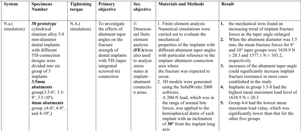
On using Finite Element Analysis (FEM), it was shown that The mean microgap size was
significantly larger for flat-to-flat interface systems compared to conical interface systems.
(Merz et al 2010). For instance, reported the presence of a microgap for external hexagonal
connection systems on the tension side of the implant under oblique or horizontal loading
simulation. (Pessoa et al. 2010) also reported microgap formation on tension sides for internal
hexagonal and external hexagonal connection systems. Conical implant
–
abutment systems
did not seem to develop statistically significant microgaps.
17
2.
Loading / fatigue performance and Torque maintenance:
Eleven in vitro trials assessed the changes in preload, mainly the loss or gain of the Implant –
abutment removal torque.
The change in torque was evaluated after initial tightening and, by how it was influenced by
the following:
(1) Fatigue loading.
(2) The presence of biofilm in the Implant-abutment interface.
(3). Increased/decreased initial tightening torque.
(4) Repeated tightening and removal cycles.
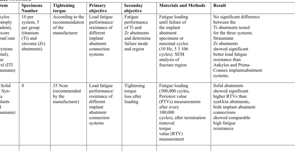
Two researchers; (Pereira, Jorge. et al. 2016, and Ricomini, Filho. et al. 2010), addressed
seal performance in addition to load / fatigue performance, whereas others were mainly
focused on stress/load performance, particularly dealing with load fatigue performance of the
implant
–
abutment connection.(Ricomini Filho et al. 2010;
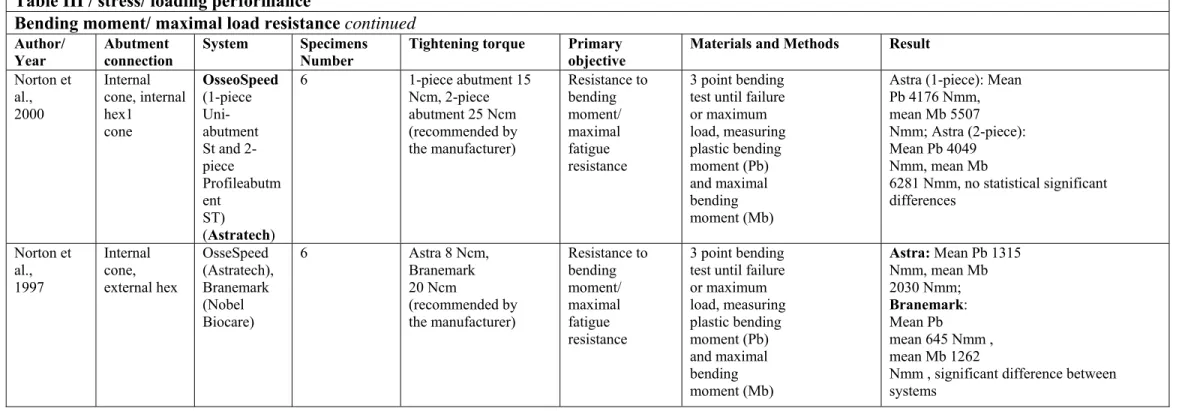
Norton MR 1997;
Koutouzis T, et
al. 2011).
(Pereira Jorge, et al. 2016) conducted an in vitro study to evaluate the influence of fatigue
loading on the removal torque values of Morse taper and external hexagon abutments in a
simulated oral environment, and concluded that the mean values of torque loss, microgap
size, and biofilm density recorded at Morse taper joints were lower in comparison to those
recorded at external hexagon implant-abutment joints after fatigue tests which simulated six
months of normal chewing function.
18
(Shin, HM.; et al in 2014) on the other hand compared the removal torque loss between
external hexagon and two conical implant abutment systems of different diameters after
loading and found out that the reverse torque values for the regular external hexagon is higher
than that of the regular diameter Morse taper implant abutment connections after loading, and
the percentage of preload loss was higher for Morse taper than external hexagon connections,
, the results of this study concluded that the implant-abutment interface design and diameter
affect the screw joint stability.( The outcome results of this study were surprisingly
contradicting with what was reported in the literature as it showed that the external hexagon
is more advantageous than the internal cone in terms of torque maintenance, however the
difference was not statistically significant, and it was mainly attributed to the preload loss
caused by the abutment sinking phenomenon ).
(Feitosa, Pinheiro Paulo Cesar et al. in 2013) for instance carried out an experiment to
compare the initial reverse torque (before fatigue test) and final reverse torque (after fatigue
test which simulated one year of chewing function) of three different implant-abutment
connection systems (External, Internal hexagon and Morse taper) and showed that the
internal connections were more stable than the external connections, and Morse taper
connection showed better stability and lowest torque loss after simulated year of clinical
function.
(Ding et al. 2003) assessed the torque loss of two different implant-abutment internal
connections following the initial tightening and demonstrated that both connections (the
internal conical and octagonal groups) showed loss of torque however, this loss was
significantly lower in the internal conical group than the internal octagonal group.
19
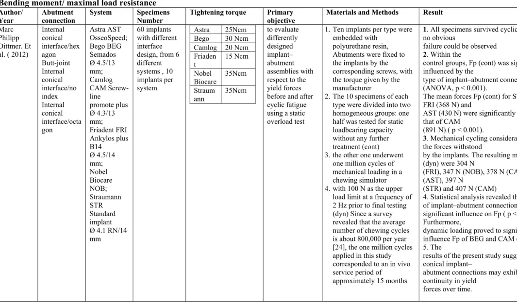
(Norton et al 1999). Compared the torque loss for different conical implant abutment
systems after applying different tightening torque values in wet and dry environment and
showed no cold welding for ITI and Astratech Morse Taper implant
–
abutment connection
systems with tightening torque values between 20 and 40 Ncm. However when Higher
tightening torque values applied (>100 Ncm), the rate of cold welding increased as well as
the rate of fractures, however The environment (dry and wet) did not seem to influence these
outcomes.
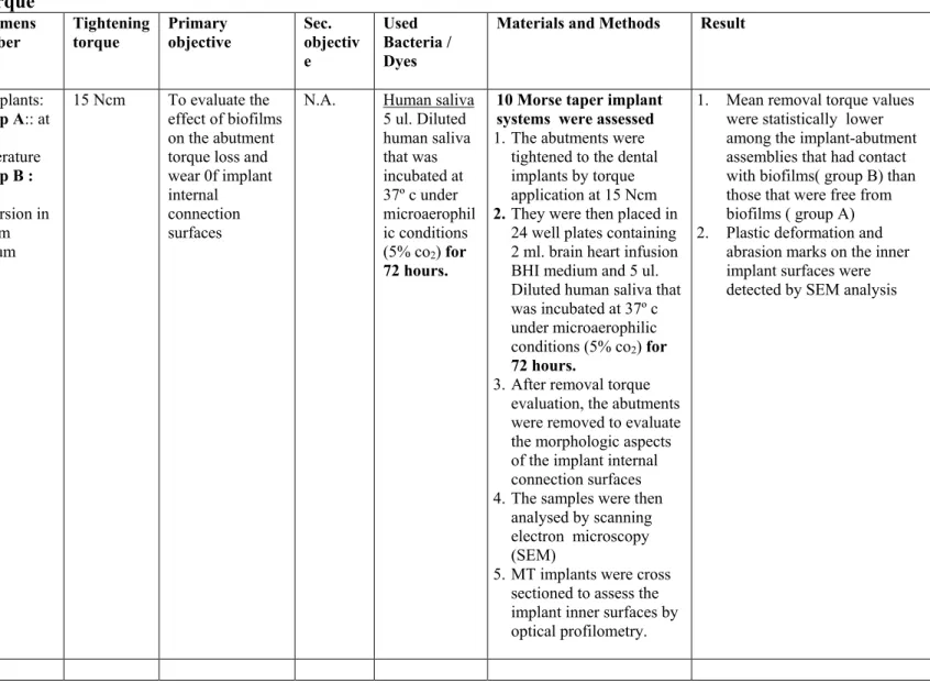
However an in vitro laboratory study conducted by (Prado Abraao and Pereira J. et al in
2016) to evaluate the effect of the presence of biofilms on the abutment torque loss and wear
of the implant internal connection surfaces, and reported a noticeable decrease in the mean
removal torque values on implant-abutment connection after immersion in a biofilm medium,
and concluded that, the presence of biofilm have a lubricating effect that can decrease the
friction between the contacting metal surfaces which can lead to preload loss and
consequently compromise the mechanical integrity of the implant-abutment internal
connections.
A very recent comparative study carried out by (Prado Abraao, et al 2017) to assess the
difference in reverse torque values between Morse taper and external hexagon
implant-abutment connections before and after immersion in biofilm medium, and documented that
the removal torque values for External hexagon after immersion in biofilm were lower in
comparison to those recorded at Morse taper implant-abutment connections, as before
immersion, the removal torque values recorded for both External hexagon and Morse taper
were higher than those recorded after immersion in biofilm medium.
Torque loss was also evident as a result of multiple consecutive closures using different
implant
–
abutment connections. It was shown that when tightening and removal cycles were
20
The influence of Stress/ loading on the removal torque- untightening torque value( the torque
force required to remove the abutment) for different implant-abutment connection designs
was studied it was shown that internal conical implant
–
abutment connection systems had
significantly less torque loss compared to internal octagonal connection systems as well as
external hexagonal connection systems. (Park JK, et al. 2010; Ricomini Filho AP, et al.
2010).
It was also demonstrated that stress/loading can cause cold welding to occur between the
implant and abutment in conical systems.( Ricomini Filho AP, et al. 2010; Koutouzis T,et al.
2011; Ricciardi Coppede A, et al. 2009) Alternatively, (Piermatti J et al. 2006) showed that
there was more loss of torque in the conical connection group compared to the external
hexagonal or internal hexagonal groups after cycling, However, it was suggested that the
screw design was an influencing factor in torque loss rather than the design of the connection
itself ( i.e.: the use of a screw with a thick stem and a journal appeared to provide the least
loss of torque after several cycles or tightening and untightening ).
B.
In vivo studies:
1)
Animal studies:
Four relevant in vivo studies conducted in Animals have been reviewed and included in this
study; (summarised in Table V / Attachment).
(Weng et al. 2011); compared radiographically conical and non-conical implant-abutment
connections in relation to marginal bone level changes for submerged and non-submerged
implant and reported statistically significant differences in marginal bone level changes with
less bone loss around conical connections of submerged and non-submerged implants.
21
2)
Human studies:
Four relevant in vivo comparative studies performed in Human were included in this review;
(summarised in Table VI / Attachment)
Two studies conducted by Crespi et al.,(2009) and Pieri et al., (2011) compared immediately
placed and loaded conical and non-conical internal implant-abutment connection system,
(Crespi et al.,(2009)) reported 100% implant success for both (internal conical and external
hexagon) after 2 years of function, whereas Pieri et al., (2011) reported 94% success for
conical implant system and 100% success for internal hexagon after one year of function,
however both researchers reported less marginal bone loss around conical implant-abutment
connections than non-conical connections.
The other two studies were performed by Kielbassa et al.,(2009), and Bilhan et al., (2010)
followed delayed implant placement protocols with submerged or non-submerged healing
and delayed or immediate loading protocols.
22
III.
Discussion
This focused systematic literature review on the mechanical integrity and performance of
Morse taper implant-abutment connection found some relevant in vitro and in vivo scientific
evidence supporting the hypothesis that conical implant-abutment connections provide better
performance and appear to be superior in terms of abutment fit , torque maintenance and seal
performance than other connections.
In vitro laboratory studies which are summarised in tables ( I, II and III) / Attachment,
reported that most implant-abutment connections systems have a microgap smaller than 10
µm. Astra implants that have conical interface geometric design for instance showed The
smallest microgap among all connections, followed by the Ankylos implants.(
Jansen VK, et
al. 1997; Baixe S, et al. 2010), given these results, the conical interface geometry appeared
to provide a better performance in terms of bacterial seal, however, 100% bacterial seal
couldn’t be achieved. And can only happen after applying much higher torque than the
recommended tightening torque values which most likely could result in cold welding and
damage or distortion at the implant-abutment interface.
Testing the abutment performance under mechanical stress is the most important factor in
determining the abutment stability in the long term, mechanical stress can lead to abutment
micro movement and increase the size of the microgap and lead to bacterial accumulation and
thus compromising the health of the peri implant tissue and consequently the longevity of the
implant system. (Table III) / Attachment.
Under vertical and oblique occlusal loading, no rotational abutment movement or microgap
enlargement for conical connection systems was detected. External and internal hexagonal
connection systems were more prone to abutment micro movements.
23
All tested connection systems showed torque loss following initial tightening. However,
Morse taper connections showed superiority as it gained torque following initial tightening
and moreover showed the lowest torque loss after loading (fatigue tests), see table III.
The Impact of the mechanical stress/loading on the torque values was clearly evidenced on
non-conical implant-abutment connections, as most systems showed significant torque loss
following loading, whereas Morse taper conical connection systems showed either higher
resistance to torque loss or resulted in cold welding between the abutment and the implant,
however no cold welding was addressed for non-conical connection implant systems, see
tables (I, II and III) / Attachment.
The influence of multiple consecutive tightening and re-tightening and untightening was
clearly evidenced for all connection systems, in most cases it resulted in torque loss and
higher microgap size, it was shown that increasing the number of cycles lead to a significant
decrease in the torque value, it was recommended that the number of cycles should be
reduced in order to prevent any further torque loss and abutment loosening after insertion of
the final superstructure.
One of the most important risk factors that promote the formation of microgap and can
further compromise the bacterial seal performance between the abutment and implant inner
surface is the loss of torque and screw loosening, as loose abutment fixtures can encourage
Micro-gaps to form in the abutment/implant interface thus favouring bacterial invasion and
biological and mechanical problems (Gratton DG, et al. 2001)
24
As far as seal performance is concerned, It was recommended that using conical
implant-abutment connection systems is highly favoured than non-conical connection systems as it
was demonstrated that conical connections keep bacterial penetration to the minimum,
furthermore, they have the highest resistance to abutment micro movement.
Concerning the outcome of the included in vivo clinical studies which were performed in
animal and human (summarised in table V and VI,)/ attachment, reported that, marginal bone
loss was observed for all implant systems regardless of whether the implants had been placed
using a submerged or non-submerged placement protocol. Likewise, it has been shown that
the placement of immediate or delayed implants (including early or late loading) had no
influence on the loss of marginal bone. However, in most cases, it was demonstrated that
there was less marginal bone loss around the conical connection systems in comparison to
non-conical connection systems.
In spite of what have been shown and evidenced in the current reviewed literature concerning
the influencing factors for the observed changes in marginal or Cristal bone height; we must
acknowledge, that there are perhaps various other factors that influence marginal bone
heights.
However, according to what have been evidenced in this systematic review, it would seem
that conical implant-abutment connection is more advantageous as far as maintenance of
marginal bone level is concerned.
25
IV.
Conclusion
The outcome of this systematic focused review can be summarised as the following:
A.
In vitro
studies:
The design of the implant-abutment connection system appeared to be an important
influencing factor in relation to the presence of microgap and torque maintenance.
Concerning abutment micro-movement and the resultant formation or generation of
microgaps, it can be concluded that, conical implant-abutment connection systems are more
favourable and more mechanically stable, as they seemed more reluctant to abutment
micro-movement and consequently microgap enlargement than the internal and external hexagon
connections as they appeared less favourable and inferior
Despite the fact that no implant-abutment connection system, currently available, has shown
a 100% bacterial seal. The majority of the reviewed in vitro studies demonstrated that conical
connection systems are more advantageous than, and appear to be superior to non-conical
connections with regard to microgap size and bacterial seal, as they showed the smallest
microgap size.
Concerning the maintenance of torque, it can be concluded that conical implant-abutment
connections are more favourable than other non-conical connection systems, as they showed
the lowest torque loss under loading, compared to other connections
B.
In vivo
studies:
Regarding implant success and survival rates (in vivo studies performed in human and
animal) demonstrated that conical and non-conical systems are almost similar however, most
cases proved that conical implant-abutment connection systems are more favourable and
superior, as they showed less marginal bone loss around them.
V.
References:
Abrahamsson, I.; et al (1998). Soft tissue response to plaque formation at different implant systems. A comparative study in the dog. Clinical Oral Implants Research; 9(2), pp.73–79.
Akca, K.; Cehreli MC.. (2008). A photoelastic and strain-gauge analysis of interface force transmission of internal-cone implants. International Journal of Periodontal Restorative Dentistry; 28(4), pp.391–399. Albrektsson, T..; et al (2012). Working Group 1 Review Implant survival and complications. The Third EAO consensus conference 2012. Clinical Oral Implants Research, 6(10), pp.63-5.
Alkan, I., Sertgoz, A., Ekici, B. (.2004). Influence of occlusal forces on stress distribution in preloaded dental implant screws. Journal of Prosthetic Dentistry; 91(4), pp.319–325.
Aloise, JP.; et al (2010). Microbial leakage through the implant–abutment interface of Morse taper implants in vitro. Clinical Oral Implants Research; 21(3), pp.328-35.
Arvidson, K.; et al (1998). Five year prospective follow-up report of the Astra Tech dental implant system in the treatment of edentulous mandibles. Clinical Oral Implants Research, 9(4),pp.225–234.
Assenza, B.; et al (2011). Bacterial leakage in implants with different implant– abutment connections: An in vitro study. Journal of Periodontology, 83(4), pp. 491-7.
Baixe, S.; et al (2010). Microgap between zirconia abutments and titanium implants. International Journal of Oral and Maxillofacial Implants; 25(3), pp.455–460.
Berglundh, T.; et al (2005). Bone reactions to longstanding functional load at implants: An experimental study in dogs. Journal of Clinical Periodontology; 32(9),pp.925–932.
Bernardes, SR.; et al (2009). Photoelastic analysis of stress patterns from different implant–abutment interfaces. International Journal of Oral and Maxillofacial Implants; 24(5), pp.781–789.
Bilhan, H.; et al (2010). Astra Tech, Branemark, and ITI implants in the rehabilitation of partial edentulism: Two-year results. Implant Dentistry, 19(5), pp.437-446.
Binon, Paul P. (2000).Implants and Components: Entering the New Millennium. International Journal of Oral and Maxillofacial Implants, 15(1), pp.76-94.
Blum, K..; et al (2015). Fatigue induced changes in conical implant-abutment connections. Dental Materials, 31(11), pp.1415-26.
Brånemark, PI.; et al (1977). Osseointegrated implants in the treatment of the edentulous jaw. Experience from a 10-year period. Scandinavian Journal of Plastic and Reconstructive Surgery, 16(4), pp.1-132. Burguete, RL.; et al (1994). Tightening characteristics for screwed joints in osseointegrated dental implants.
Journal of Prosthetic Dentistry, 71(6), pp.592-9.
Carotenuto, G.; et al (1999). Characterization of the interface between prefabricated gold copings and cast dental alloy in implant restorations. Clinical Oral Implants Research, 10(2), pp.131-8.
Cehreli, M.; et al (2004). Implant design and interface force transfer. A photoelastic and strain-gauge analysis.. Clinical Oral Implants Research; 15(2), pp.249–257.
Coppede, AR.; et al (2009). Fracture resistance of the implant–abutment connection in implants with internal hex and internal conical connections under oblique compressive loading: An in vitro study. International Journal of Prosthodontics; 22(3), pp.:283–286.
Crespi, R.; et al (2009). Radiographic evaluation of marginal bone levels around platform-switched and non-platform switched implants used in an immediate loading protocol. International Journal of Oral and Maxillofacial Implants; 24(5), pp.920–926.
DʼErcole, S.; et al (2014). Bacterial leakage in Morse Cone internal connection implants using different torque values: an in vitro study.Implant Dentistry, 23(2), pp.175-9.
Ding, TA.; et al (2003). Evaluation of the ITI Morse taper implant/abutment design with an internal modification. International Journal of Oral and Maxillofacial Implants, 18(6), pp.865–872.
Dittmer, MP.; et al (2012). Influence of the interface design on the yield force of the implant-abutment complex before and after cyclic mechanical loading. Journal of Prosthodontic Research, 56(1), pp. 19-24 Do Nascimento, C.; et al (2012). Leakage of saliva through the implant–abutment interface: In vitro evaluation of three different implant connections under unloaded and loaded conditions. International Journal of Oral and Maxillofacial Implants, 27(3), pp.551–60.
Feitosa, PCP.; et al (2013). Stability of external and internal implant connections after a fatigue test.
European Journal of Dentistry, 7(3),pp. 267–271.
Gehrke, S.A.; et al (2015). Load fatigue performance of conical implant-abutment connection: effect of torque level and interface junction. Minerva stomatologica Journal, 64(1), pp.1-7.
Gehrke, SA.;et. al (.2014). Changes in the abutment - implant interface in Morse taper implant connections after mechanical cycling: a pilot study. The International Journal of Oral & Maxillofacial Implants, 29(4), pp.791-7.
Gross, M.; et al (1999). Microleakage at the Abutment Implant interface of osseointegrated implants: A comparative study. International Journal of Oral and Maxillofacial Implants,14(1), pp.94–100.
Hansson, S. (.2000). Implant–abutment interface: Biomechanical study of flat top versus conical. Clinical Implant Dentistry and Related Research, 2(1), pp.:33–41.
Harder, S.; et al (2010). Molecular leakage at implant–abutment connection—in vitro investigation of tightness of internal conical implant–abutment connections against endotoxin penetration. Clinical Oral Investigations 14(4), pp.427–432.
Jansen, VK; Conrads, G.; Richter, EJ. (1997). Microbial leakage and marginal fit of the implant-abutment interface. International Journal of Oral Maxillofacial Implants, 12(4), pp.527-40.
Jeng, MD.; et al (2017). Load fatigue performance of two internal tapered abutment-implant connection implants under different screw-tightening torques.The Journal of Oral Implantology, 43(2), pp.107-13. Jokstad, A.; et al ( 2003). Quality of dental implants International Dental Journal, 53(6), pp.409-43. Kielbassa, AM.; et al (2009). Randomized controlled trial comparing a variable-thread novel tapered and a standard tapered implant: Interim one-year results. Journal of Prosthetic Dentistry;101(5), pp.293–305. Kitagawa, T.; et al (2005). Influence of implant/abutment joint designs on abutment screw loosening in a dental implant system.. Journal of Biomedical Materials Research Part B , 75(2),pp.457–463.
Levine, RA.; et al (1997). A multicentre retrospective analysis of the ITI implant system used for single-tooth replacements: Preliminary results at 6 or more months of loading. International Journal of Oral Maxillofacial Implants, 12(2), pp.237–242.
Lin, CL.; et al (2007). Factorial analysis of variables influencing mechanical characteristics of a single tooth implant placed in the maxilla using finite element analysis and the statistics-based Taguchi method.
European Journal of Oral Sciences, 115(5), pp.408– 416.
Macedo,J. P.; et al. (2016). Morse taper dental implants and platform switching: The new paradigm in oral implantology. European Journal of Dentistry, 10(1), pp. 148–54.
Maeda, Y ; Satoh, T; Sogo, M.. (2006). In vitro differences of stress concentrations for internal and external hex implant-abutment connections: A short communication. Journal of Oral Rehabilitation, 33(1), pp.75–8. Merz, BR.; et al (2000). Mechanics of the implant–abutment connection: An 8-degree taper compared to a butt joint connection. International Journal of Oral Maxillofacial Implants; 15(4), pp.519–526.
Michalakis, KX; Hirayama, H.; Garefis, PD. (2003). Cement-retained versus screw-retained implant restorations: a critical review. International Journal of Oral Maxillofacial Implants, 18(5), pp. 719-28. Nishioka, RS.; et al (2011). Comparative strain gauge analysis of external and internal hexagon, Morse taper, and influence of straight and offset implant configuration. Implant Dentistry, 20(2), pp.24-32. Norton, MR (2000).. An in vitro evaluation of the strength of a 1-piece and 2-piece conical abutment joint in implant design. Clinical Oral Implants Research;11(5), pp.:458–464.
Norton, MR. (1997). An in vitro evaluation of the strength of an internal conical interface compared to a butt joint interface in implant design. Clinical Oral Implants Research; 8(4), pp.290–298.
Norton, MR. (1999). Assessment of cold welding properties of the internal conical interface of two commercially available implant systems. Journal of Prosthetic Dentistry; 81(2), pp.159–66.
Norton, MR.(2000). In vitro evaluation of the strength of the conical implant-to-abutment joint in two commercially available implant systems. Journal of Prosthetic Dentistry, 83(5), pp.:567–571.
Park, JK.; et al (2010). Effects of abutment screw coating on implant preload. Journal of Prosthodontics, 19(6), pp.458–464.
Pellizzer, EP.; et al (2014). Photoelastic analysis of stress distribution with different implant systems.
Journal of Oral Implantology, .40(2), pp.117-22.
Pereira, J.; et al (2016). Removal Torque and Biofilm Accumulation at Two Dental Implant - Abutment Joints After Fatigue. The International Journal of Oral & Maxillofacial Implants, 31(4), pp.813 - 9.
Pessoa, RS.; et al. (2010). Influence of implant connection type on the biomechanical environment of immediately placed implants—CT-based nonlinear, three-dimensional finite element analysis. Clinical Implant Dentistry and Related Research; 12(3), pp.219–234.
Pieri, F.; et al (2011). Influence of implant– abutment interface design on bone and soft tissue levels around immediately placed and restored single-tooth implants: A randomized controlled clinical trial. International Journal of Oral and Maxillofacial Implants; 26(1), pp.169– 178.
Piermatti, J.; et al (2006). An in vitro analysis of implant screw torque loss with external hex and internal connection implant systems. Implant Dentistry;15(4), pp.427–435.
Prado, AM.; et al (2017). Wear of Morse taper and external hexagon implant joints after abutment removal.
Prado, AM.; Pereira, J.; et al (2016). Biofilm Affecting the Mechanical Integrity of Implant-Abutment Joints. The International Journal of Prosthodontics, 29(4), pp.381-3.
Quaresma, SE.; et al (2008). A Finite element analysis of two different dental implants: Stress distribution in the prosthesis, abutment, implant, and supporting bone. Journal of Oral Implantology, 34(1),pp.1–6. Quirynen, M.; et al (2002). Infectious risks for oral implants: a review of the literature. Clinical Oral Implants Research, 13(1), pp.1-19.
Ricciardi, Abilio.; et al (2009). Fracture Resistance of the Implant-Abutment Connection in Implants with Internal Hex and Internal Conical Connections Under Oblique Compressive Loading: An In Vitro Study.
International Journal of Prosthodontics, 22(3),pp.283-286.
Ricciardi, Coppede. A.; et al (2009). Effect of repeated torque/mechanical loading cycles on two different abutment types in implants with internal tapered connections: An in vitro study. Clinical Oral Implants Research; 20(6), pp.624–632.
Ricomini, Filho AP.; et al (2010). Preload loss and bacterial penetration on different implant–abutment connection systems. Brazilian Dental Journal, 21(2), pp.123– 129.
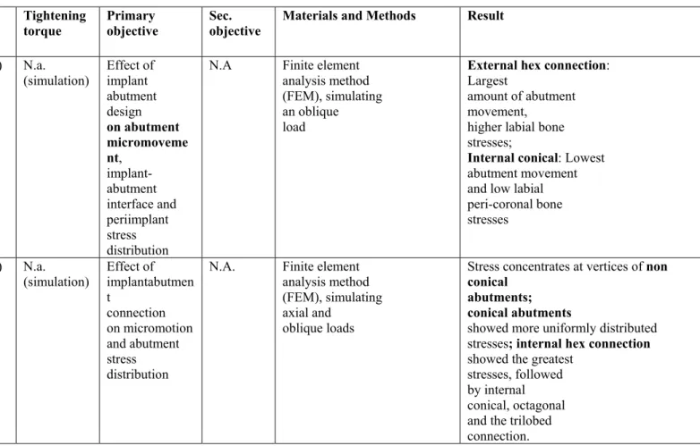
Saidin, S.; et al (2012). Effects of different implant– connections on micromotion and stress distribution: Prediction of microgap formation. Journal of Dentistry; 40(6), pp.:467–474.
Scarano, A. Valbonetti.; et al (2016). Implant-Abutment Contact Surfaces and Microgap Measurements of Different Implant Connections Under 3-Dimensional X-Ray Microtomography. Implant Dentistry,
25(5),pp.656-62.
Schmitt, CM.; et al (2014). Performance of conical abutment (Morse Taper) connection implants: a systematic review. Journal of Biomedical Materials Research Part A, 102(2), pp.552-74.
Seetoh, YL; et al (2011). Load fatigue performance of conical implant–abutment connections. International Journal of Oral Maxillofacial Implants, 26(4), pp.797–806.
Shin, HM.; et al (2014). Influence of the implant-abutment connection design and diameter on the screw joint stability. The Journal of Advanced Prosthodontics, 6(2), pp.126-32.
Tabata, LF.; et al (2011). Platform switching: Biomechanical evaluation using three-dimensional finite element analysis. International Journal of Oral Maxillofacial Implants, 26(3), pp.482–91
Teixeira, W.; et al (2011). Microleakage into and from two-stage implants: An in vitro comparative study.
International Journal of Oral and Maxillofacial Implants; 26(1), pp.56–62.
Tesmer, M.; et al (2009). Bacterial colonization of the dental implant fixture–abutment interface: An in vitro study. Journal of Periodontology; 80(12), pp.1991–1997.
Tripodi, D.; et al (2012). An in vitro investigation concerning the bacterial leakage at implants with internal hexagon and Morse taper implant–abutment connections. Implant Dentistry; 21(4), pp.335–339.
Walton, JN.; MacEntee, MI.. (1997). A prospective study on the maintenance of implant prostheses in private practice. The International Journal of Prosthodontics;10(5),pp.453–458.
Wang, K.; et al (2016). Comparison of the fracture resistance of dental implants with different abutment taper angles. Materials Science and Engineering, 63(6), pp.:164-71.
Weng, D.; et al (2011). Influence of microgap location and configuration on radiographic bone loss in nonsubmerged implants: An experimental study in dogs. International Journal of Prosthodontics; 24(5), pp.445–452.
Xia, Dandan.; et al (2014). Dynamic fatigue performance of implant-abutment assemblies with different tightening torque values. Bio-Medical Materials and Engineering, 24 (4) 2143 –2149.
Yamanishi, Y.; et al (2012). Influences of implant neck design and implant–abutment joint type on periimplant bone stress and abutment micromovement: Three-dimensional finite element analysis. Dental Materials; 28(5), pp.1126–1133.
VI.
Attachment:
23
Table I / Seal Performance
The presence of Microgap and seal performance after fatigue test
Author/ Year Abutment
connection System Specimens Number Tightening torque Primary objective Used Bacteria / Dyes Materials and Methods Result
Pereira Jorge,
et al. 2016 1.Morse Taper 2.External hexagon (4mm diameter) Titamax CM, Neodent 60 implants ; 30 implants per group Morse Taper
group : 15 Ncm
External
Hexagon group :
32 Ncm.
To evaluate the removal torque and in vitro biofilm accumulation at
Morse taper and
external hexagon
implant abutment connections after
fatigue test in a simulated oral environment for 72 hours
Human saliva (10 mL) was collected from four individuals and diluted (1:5) in phosphate-buffered solution (PBS) every day over a period of 4 days then , 5 μL of the initial suspension was inoculated in brain-heart infusion (BHI) medium enriched with 5% sucrose (Sigma-Aldrich) for the biofilm growth. ( to simulate actual conditions)
1. Sixty dental implants were divided into two groups: (1) 30 Morse taper and (2)
30external hexagon
2. The samples were then immersed in 2 mL of BHI growth medium containing human saliva for 72 hours at 25°C
3. Fatigue tests on the implant-abutment assemblies were performed at a normal force (50N) at 1.2 Hz for 500.000 cycles (to simulate fatigue over a period of 6 months of mastication.)
4. Removal torque mean values (n = 10) were measured after fatigue tests.
5. Groups of implant-abutment assemblies (n =8) were cross-sectioned at 90 degrees relative to the plane of the implant-abutment joints for the microgap
measurement by
field-emission guns scanning electron microscopy. (
FEG-SEM)
RT (before fatigue)
RT Ncm
(after fatigue)
MT 24 ± 0.5 22.1 ± 0.5
EH 24.8 ± 0.6 21.1 ± 0.7
1 .Mean values of the removal torque on abutments were significantly lower for both Morse taper (22.1 ± 0.5 Ncm) and external hexagon (21.1 ± 0.7 Ncm) abutments after fatigue tests than those recorded without fatigue tests
(respectively, 24 ± 0.5 Ncm and 24.8 ± 0.6 Ncm) Microgap ( before fatigue) μm Microgap ( after fatigue)
MT 1.7 ± 0.4 3.2 ± 0.8
EH 1.5 ± 0.4 8.1 ± 1.7
2-Mean values of microgap size for the Morse taper joints were statistically signicantly lower without fatigue tests (1.7 ± 0.4 μm) than those recorded after fatigue tests (3.2 ± 0.8 μm).
3-mean values of microgap size for external hexagon joints free of fatigue were statistically signicantly lower (1.5 ± 0.4 μm) than those recorded
after fatigue tests (8.1 ± 1.7 μm) (P < .05).
-Conclusion: The mean values of
removal torque loss, microgap size, and biofilm density recorded at Morse taper joints were lower in comparison to those recorded at external hexagon
24
Table I / Seal Performance
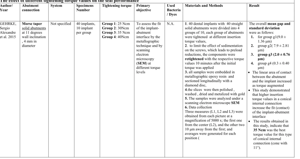
The effect of different tightening torque values on the seal performance
Author/
Year Abutment connection System Specimens Number Tightening torque Primary objective Used Bacteria
/ Dyes
Materials and Methods Result
GEHRKE, Sergio Alexandre et al. 2015
Morse taper
solid abutments at 11 degrees wall inclination , 4 mm in diameter
Not specified 40 implants, 10 implant per group
Group 1: 25 Ncm
Group 2: 30Ncm
Group 3: 35 Ncm
Group 4: 40Ncm
To assess the fit of the implant-abutment interface by the metallographic technique and by scanning electron microscopy (SEM) at different torque levels
N.A. 1. 40 dental implants with 40 straight solid abutments were divided into 4 groups of 10, each group of abutments were tightened at different insertion torque values,
2. to limit the effect of sedimentation on the screws, which leads to preload reductions, the components were
retightened with the respective torque values 10 minutes after the initial torque was applied
3. all samples were embedded in metallographic epoxy resin and sectioned longitudinally with a diamond disc,
4.the slices were then polished , washed , dried and metalized with gold
5. The samples were analyzed under a scanning electron microscope SEM 6. Data collection
Three measures (L1, L2 and L3) were obtained from each picture at a magnification of 5000 x; the first one from the center (L2), and the other two 10 μm away from the first; and averages were generated for each position (
The overall mean gap and standard deviation
were as follows:
1. for group g1(9.0 ± 1.36 μm)
2. group g2( 7.9 ± 2.81 μm)
3. group g3 (2.0 ± 0.76
μm)
4. group g4 (0.3 ± 0.40 μm)
The linear area of contact between the abutment and the implant increased as torque augmented
This study demonstrated that higher insertion torque values in a conical internal connection increase the fit (contact) of the implant-abutment interface
The results obtained in this study, indicate that
35 Ncm was the best
torque value for this type of conical internal connection (cone with 11°).
25
Table I
/Seal Performance
Continued
Author/
Year Abutment connection System Specimens Number Tightening torque Primary objective Used Bacteria
/ Dyes
Materials and Methods Result
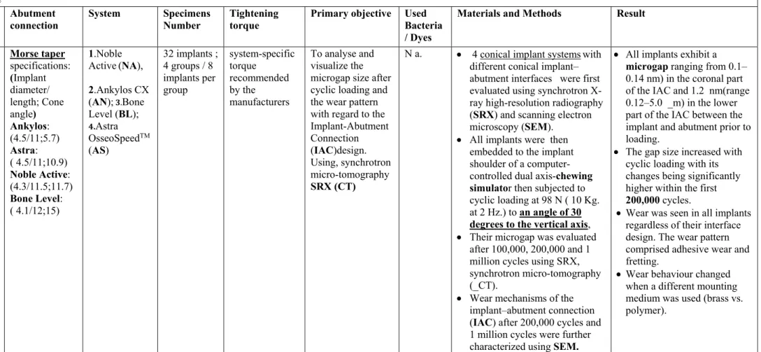
Blum, Kai
et al , 2015 Morse taperspecifications:
(Implant diameter/ length; Cone angle) Ankylos: (4.5/11;5.7) Astra: ( 4.5/11;10.9)
Noble Active: (4.3/11.5;11.7)
Bone Level: ( 4.1/12;15)
1.Noble Active(NA),
2.Ankylos CX (AN);3.Bone
Level (BL); 4.Astra
OsseoSpeedTM
(AS)
32 implants ; 4 groups / 8 implants per group system-specific torque recommended by the manufacturers
To analyse and visualize the microgap size after cyclic loading and the wear pattern with regard to the Implant-Abutment Connection (IAC)design. Using, synchrotron micro-tomography
SRX (CT)
N a. 4 conical implant systemswith different conical implant– abutment interfaces were first evaluated using synchrotron X-ray high-resolution radiography (SRX) and scanning electron microscopy (SEM).
All implants were then embedded to the implant shoulder of a computer-controlled dual axis-chewing simulator then subjected to cyclic loading at 98 N ( 10 Kg. at 2 Hz.) to an angle of 30 degrees to the vertical axis,
Their microgap was evaluated after 100,000, 200,000 and 1 million cycles using SRX, synchrotron micro-tomography (_CT).
Wear mechanisms of the implant–abutment connection (IAC) after 200,000 cycles and 1 million cycles were further characterized using SEM.
All implants exhibit a
microgap ranging from 0.1–
0.14 nm) in the coronal part of the IAC and 1.2 nm(range 0.12–5.0 _m) in the lower part of the IAC between the implant and abutment prior to loading.
The gap size increased with cyclic loading with its changes being significantly higher within the first
200,000 cycles.
Wear was seen in all implants regardless of their interface design. The wear pattern comprised adhesive wear and fretting.
Wear behaviour changed when a different mounting medium was used (brass vs. polymer).
26
Table I
/
Seal Performance
/ Bacterial leakage
continued
Author/ Year Abutment
connection System Specimens Number Tightening torque Primary objective Used Bacteria / Dyes Materials and Methods Result
D’Ercole, Slimonetta et al , 2014
Morse
Taper Oralplant ;
Cordenon s, PD, Italy 30, 10 implants per group
Group1: 20N
Group2: 30N
Group3: 40N
To assess whether there was a
decrease of
Bacterial leakage with increasing
torque values in conical Morse cone
connection implants
Pseudomonas aeruginosa suspension incubated for 24 hours at 37°C ; Aggregatibact er actinomycete mcomitans incubated for 48 hours at 37°C in 5% CO2
A total of 30 Morse taper implants divided in to 3 groups( 10 implants each), G1 abutments were connected to the implants with 20N,
G2 with 30N,
G3 with 40N, each group was later subdivided into two groups of 5 implants which were then inoculated with 2 different
bacterial suspensions and monitored for 14 days
Bacterial contamination
In group 1 (20 N), was found in 2 of the 5 implant-abutment assemblies seeded with the P. aeruginosa, all on the sixth
day
in group 2 , Two assemblies at 30N and inoculated with P. aeruginosa showed the evidence of bacterial leakage after 13 days of incubation
In tgroups 1,2 and3 he assemblies at 20 N ,30 and 40 N seeded with A.
actinomycetemcomitans, no contamination was found.
-In groups 1 and 2, bacterial contamination was found in 2 of the 10 implants, only in the specimens seeded with P, aeruginosa.
-In group 3, no contaminated samples were found.
Nascimento
et al., 2012 Morse taper, internal and external hex SIN, Sistema de Implante Nacional 20 per group, 10 loaded and 10 unloaded
According to the recommendation of the
manufacturer (20 Ncm)
Saliva leakage into the implant– abutment interface under loaded and unloaded conditions
Human saliva Implant abutment connection and incubation in human saliva.
Detecting saliva leakage. Half of the specimens: Cycling with 120 N, 500,000 cycles at 1.8 Hz
Contamination:
External hex: Loaded 10 out of 10, unloaded 3 out of 10
Internal hex: Loaded 10 out of 10,
unloaded 4 out of 10,
Morse taper: Loaded 9 out of 10, unloaded 1 out of 10
27
Table I
/
Seal Performance
continued
Author/ Year Abutment
connection System Specimens Number Tightening torque Primary objective Used Bacteria / Dyes Materials and Methods Result
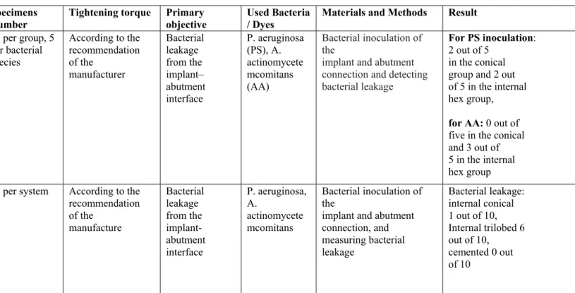
Tripodi et al.,
2012 Internal cone,
internal hex
Universal II HI and CM, (Implacil De Bortoli)
10 per group, 5 per bacterial species
According to the recommendation of the
manufacturer
Bacterial leakage from the implant– abutment interface
P. aeruginosa (PS), A. actinomycete mcomitans (AA)
Bacterial inoculation of the
implant and abutment connection and detecting bacterial leakage
For PS inoculation: 2 out of 5
in the conical group and 2 out of 5 in the internal hex group,
for AA: 0 out of five in the conical and 3 out of 5 in the internal hex group Assenza et al.,
2011 Internal cone,
internal trilobed, cemented
Ankylos (Dentsply Friadent), Replace Select (Nobel Biocare), Bone System
10 per system According to the recommendation of the
manufacture
Bacterial leakage from the implant-abutment interface
P. aeruginosa, A.
actinomycete mcomitans
Bacterial inoculation of the
implant and abutment connection, and measuring bacterial leakage
28
Table I
Seal Performance
continued
Author/
Year Abutment connection System Specimens Number Tightening torque Primary objective Sec. objective Used Bacteria /
Dyes
Materials and
Methods Result
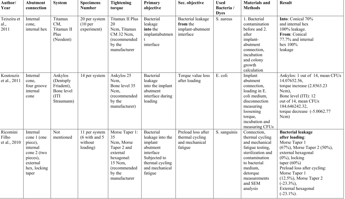
Teixeira et al., 2011 Internal cone, internal hex Titamax CM, Titamax II Plus (Neodent)
20 per system (10 per experiment)
Titamax II Plus 20
Ncm, Titamax CM 32 Ncm, (recommended by the
manufacturer
Bacterial leakage
into the implantabutmen t
interface
Bacterial leakage
from the
implant-abutment interface
S. aureus 1. Bacterial contamination before and 2. after implant-abutment connection, incubation and colony growth calculation
Into: Conical 70% and internal hex 100% leakage.
From: Conical 77.7% and internal hex 100%
leakage
Koutouzis
et al., 2011 Internal cone, four groove internal cone Ankylos (Dentsply Friadent), Bone level (ITI Straumann)
14 per system Ankylos 25 Ncm, Bone level 35 Ncm, (recommended by the manufacturer) Bacterial leakage into the implant abutment interface during loading
Torque value loss
after loading E. coli Implant abutment connection, loading in E. coli medium, disconnection measuring loosening torque, incubation and measuring CFUs
Ankylos: 1 out of 14, mean CFUs 14.07652.56,
torque increase (2.8563.23 Ncm),
Bone level (ITI): 12 out of 14, mean CFUs 184.646242.32,
torque decrease (-5.0062.77 Ncm)
Ricomini Filho et al., 2010
Internal cone 1 (one piece), internal cone 2 (two pieces), external hex, locking taper
Not
mentioned 11 per system (6 with and 5 without loading)
Morse Taper 1: 35
Ncm, Morse Taper 2 and external hexagonal: 15 Ncm, (recommended by the manufacturer Bacterial leakage into the implant abutment interface Subjected to thermal cycling and mechanical fatigue
Preload loss after thermal cycling and mechanical fatigue
S. sanguinis Connection, thermal cycling and mechanical fatigue testing, sterilization and contamination to bacterial medium, detorque measurements and SEM analysis Bacterial leakage after loading: Morse Taper 1
(67%), Morse Taper 2 (50%), external hexagonal
(0%), locking taper (60%)
Preload loss after cycling: Morse Taper 1
(12.5%), Morse Taper 2 (-23.3%),
29
Table I
/
Seal Performance
continued
Author/
Year Abutment connection System Specimens Number Tightening torque Primary objective Sec. objective Used Bacteria /
Dyes
Materials and
Methods Result
Aloise et al., 2010 Internal cone, internal cone Bicon Implant System (Bicon), Ankylos (Dentsply Friadent)
10 per system Ankylos 25 Ncm, Bicon tapped, (recommended by the manufacturer Bacterial leakage from the implant-abutment interface.
N.a. S. sanguinis Inoculation S.
sanguinis, connecting abutment and implant, incubation and proof of bacterial presence or absence Bactarial leakage: Ankylos 20%, Bicon 20%. Harder et al., 2010 Internal cone, internal cone OsseoSpeed (AstraTech) , Ankylos (Dentsply Friadent
8 per system According to the recommendation of the manufacturer Molecular leakage of endotoxin along the implant abutment interface
N.a. LPS of
Salmonella enterica
Inoculation of implant with LPS, connection to abutment and incubation, endotoxin detection and measuring concentration over time (168h)
Endotoxin detection in both
groups after 5 minutes. Significant less endotoxin concentration (mean) for OsseoSpeed units over the whole examination period Baixe et al., 2010 Internal cone (2x), external flat, internal flat Ankylos (Dentsply Friadent), OsseoSpeed (Astratech), Standard ITI (ITI Straumann, Nobel Replace Tapered Groovy (Nobel Biocare)
5 per system Nobel 35 Ncm, ITI
15 Ncm, Astra 25 Ncm, Ankylos 15 Ncm (recommended by the manufacturer Microgap between implant and abutment Microgap comparing titanium and zirconia abutments
N.a. Longitudinal
cutting and scanning electron microscopy ( SEM)
The mean microgap was larger
for flat-to-flat interface systems compared
30
Table I
/
Seal Performance
continued
Author/
Year Abutment connection System Specimens Number tightening torque Primary objective Sec. objective Used Bacteria /
Dyes
Materials and
Methods Result
Tesmer et al., 2009 Internal cone, manipulated internal cone, trichannel internal connection Ankylos and manipulated Ankylos (Dentsply Friadent), Nobel Replace Select (Nobel Biocare)
10 per system Ankylos and manipulated Ankylos 25 Ncm, Nobel Replace Select 35 Ncm, (recommended by the manufacturer) Bacterial invasion into the implant abutment interface
N.a. A.
actinomycete mcomitas, P. gingivalis Implant abutment connection, contamination with bacterial solution (Aa and Pg), disconnection, incubation and detecting bacterial contamination Bacterial contamination Ankylos:
(Aa 3/10, Pg 0/ 10, median CFUs; Aa 0, Pg
0), Nobel Replace select: (Aa 9/10, Pg 9/
10, CFUs; Aa 24.5, Pg 12),
Manipulated Ankylos:
(Aa 10/ 10, Pg 10/10, CFUs; Aa 81, Pg 55) Coelho et al., 2008 Internal cone, trilobed internal, internal hex Standard SLA implant (ITI, Straumann), Replace Select (Nobel Biocare), Intralock short collar implant (Intra-lock Int.)
5 per system According to the recommendation of the manufacture Sealing capability of implant system
N.a. Toluidin
Blue dye Contamination of implant interface, connection to abutment and measuring dye leakage over time with spectrophotometri c analysis
Total release after 144h:
ITI Straumann 55%,