*Corresponding author: Alexander Welker Biondo
Departamento de Medicina Veterinária, Universidade Federal do Paraná – UFPR Rua dos Funcionários 1540, CEP 80035-050 Juveve, Curitiba - PR, Brazil Phone: +55 (41) 3350-5723 / 3350-5623; e-mail: abiondo@illinois.edu
www.cbpv.com.br/rbpv
A review of the occurrence of hemoplasmas
(hemotrophic mycoplasmas) in Brazil
Uma revisão da ocorrência dos hemoplasmas (micoplasmas hemotróficos) no Brasil
Alexander Welker Biondo1,8*; Andrea Pires dos Santos2; Ana Marcia Sá Guimarães2,9; Rafael Felipe da Costa Vieira1,3; Odilon Vidotto3; Daniel de Barros Macieira4; Nádia Regina Pereira Almosny4; Marcelo Beltrão Molento1;
Jorge Timenetsky5; Helio Autran de Morais6; Félix Hilário Diáz González7; Joanne Belle Messick2
1Departamento de Medicina Veterinária, Universidade Federal do Paraná – UFPR
2Department of Veterinary Pathobiology, Purdue University
3Departamento de Medicina Veterinária Preventiva, Universidade Estadual de Londrina – UEL
4Departamento de Patologia Clínica Veterinária, Universidade Federal Fluminense – UFF
5Departamento de Microbiologia, Universidade de São Paulo – USP
6Department of Clinical Sciences, Oregon State University
7Departamento de Patologia Clínica Veterinária, Universidade Federal do Rio Grande do Sul – UFRGS
8Department of Veterinary Pathobiology, University of Illinois
9CAPES – Fulbright Program
Received January 14, 2009 Accepted April 16, 2009
Abstract
Recent studies have been conducted in Brazil using molecular techniques for the detection of hemotrophic mycoplasmas in several mammals. In domestic cats, Mycoplasma haemofelis, ‘Candidatus M. haemominutum’, and ‘Candidatus M. turicensis’ infections have been identified. These species have also been found in free-ranging and captive neotropical felid species. Two canine hemoplasmas, Mycoplasma haemocanis and ‘Candidatus Mycoplasma haematoparvum’, have been identified in dogs. In commercial swine populations, Mycoplasma suis was found to be highly prevalent, especially in sows. Moreover, novel mycoplasma species have been identified in Brazilian commercial pigs and domestic dogs. A hemoplasma infection in a human patient infected with the human immunodeficiency virus (HIV) was also recently documented. In conclusion, hemoplasma species are common and important infectious agents in Brazil. Further studies should be conducted to better understand their impact on pets, production animals, and wildlife fauna, as well as their role as zoonotic agents, particularly in immunocompromised patients.
Keywords: Hemoplasma, Mycoplasma haemofelis, Mycoplasma suis, HIV.
Resumo
Estudos recentes utilizando técnicas moleculares para a detecção de micoplasmas hemotróficos em diferentes mamíferos têm sido conduzidos no Brasil. Em gatos domésticos, infecções por Mycoplasma haemofelis, ‘Candidatus M. haemominutum’ e ‘Candidatus M. turicensis’ foram identificadas. Estas espécies também foram encontradas em felídeos neotropicais de vida livre e de cativeiro. Dois hemoplasmas caninos, Mycoplasma haemocanis e ‘Candidatus Mycoplasma haematoparvum’, foram identificados em cães domésticos. Em populações comerciais de suínos, Mycoplasma suis possui alta prevalência, especialmente em porcas. Além disso, novas espécies de hemoplasmas foram detectadas em suínos comercias e cães. Infecção por um hemoplasma em um paciente humano infectado com o vírus da imunodeficiência humana (HIV) foi recentemente documentada. Em conclusão, espécies de hemoplasmas são comuns e importantes agentes de infecções no Brasil. Estudos futuros devem ser conduzidos para melhor entender seu impacto em cães e gatos, animais de produção e na fauna silvestre, e também para determinar o seu papel como agentes zoonóticos, particularmente em pacientes imunocomprometidos.
Introduction
Hemoplasmas (hemotrophic mycoplasmas) are obligate erythrocyte bacteria that infect a wide range of mammalian species and may induce anemia in an infected host. Initially, hemoplasmas were classified in the order Rickettsiales based on morphology and response to antibiotic therapy (WEISS; MOULDER, 1984). However, molecular studies based on the 16S rRNA gene have shown that the genera Haemobartonella and Eperythrozoon are more closely related to the pneumoniae group of mycoplasmas, leading to the reclassification within the genus Mycoplasma (RIKIHISA et al., 1997; NEIMARK et al., 2001). Mycoplasmas are characterized by the absence of cell wall and diminutive genome that gives strict dependence to the host cell. Accordingly, hemoplasmas range from 0.3 to 1 µm in diameter and can be found in form of cocci, rings, or rods attached to and forming indentations on the surface of red blood cells (MESSICK et al., 2004). In contrast to several mucosal mycoplasmas, these organisms have never been grown successfully in culture.
Species of hemoplasmas (i.e., Mycoplasma haemofelis) have been described in all continents, except Antarctica, and it is thus believed that these organisms have a worldwide distribution. Their main form of transmission is probably through blood-sucking arthropods; there appears to be a high prevalence of hemotrophic mycoplasmas in tropical regions, likely due to environmental conditions favoring transmission of these organisms by vector-borne agents (MACIEIRA, 2008). Nevertheless, studies related to hemoplasma transmission are scarce and mostly not conclusive. Since hemoplasmas cannot be cultured in vitro, the dose of organisms in experimental infections cannot be accurately measured and interferes with the results. Experimental studies suggest fleas (Ctenocephalides felis) as responsible for the transmission of M. haemofelis and ‘Candidatus M. haemominutum’ in cats (WOODS et al., 2005), the tick Rhipicephalus sanguineus as the main vector of M. haemocanis in dogs (SENEVIRATNA et al., 1973), the lice Polyplax serrata and P. spinulosa as vectors for
M. coccoides transmission in mice (BERKENKAMP; WESCOTT, 1988), and Stomoxys calcitrans and Aedes aegypti as vectors of
M. suis in pigs (PRULLAGE et al., 1993).
Animal hemoplasmosis ranges from a severe anemia to a chronic infection without clinical signs. Acutely infected animals may present a mild to life-threatening hemolytic anemia in addition to anorexia, fever, icterus, and hypoglycemia, depending on the hemoplasma species involved. The chronic form has been commonly associated with the absence of clinical signs; however, the long-term consequences of hemoplasma infection in animals are still unclear. Chronically infected animals may become anemic following immunossupression events, and growth retardation in feeder pigs and poor reproductive performance in sows have been described (HOELZLE, 2008). In addition, hemoplasmas vary in pathogenicity and virulence; species-specific identification should always be conducted when performing the diagnosis (MESSICK, 2004).
Diagnosis of the infection has historically relied on the evaluation of Romanowsky stained blood smears. Hemoplasmas are mostly found attached to the erythrocytes; some organisms
may also be found in the plasma because of detachment from the cells. Due to cyclic parasitemia and chronic infection, this technique is neither sensitive nor specific for infection diagnosis. Consequently, molecular diagnostic techniques, such as standard and real-time PCR, have been extensively and successfully used to identify acute and chronically infected animals (MESSICK, 2004; HOELZLE, 2008).
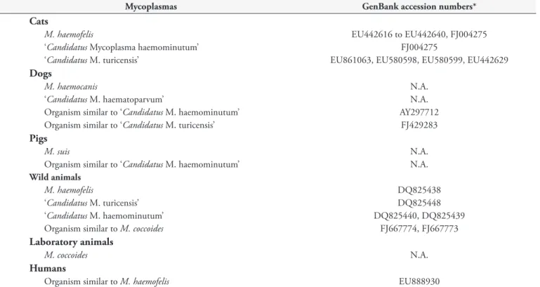
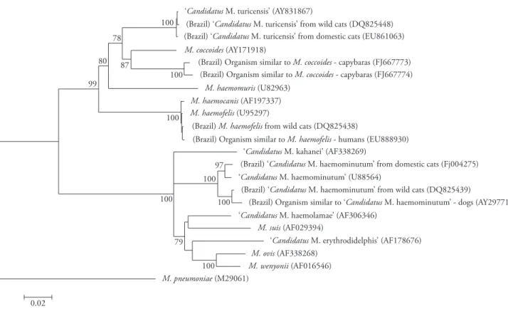
Over the last few years, there has been a growing interest in hemoplasma infection of domestic and wild animals in Brazil. Acute and life-threatening cases of the disease have been observed in domestic pigs and cats (unpublished data). Additionally, new hemoplasma species have been described in pigs (MESSICK et al., 2007) and dogs (SANTOS, 2008). Our goal in this review is to summarize all recent molecular studies that characterize the occurrence of hemoplasmas in Brazil (Table 1, Figure 1).
Companion Animals
Although hemoplasmas have been frequently found on blood smears of cats in Brazil, molecular approaches for detecting hemotrophic mycoplasma species have increased the detection sensitivity in anemic and non-anemic cats from different regions of the country. The three currently known species of feline hemoplasmas(M. haemofelis, ‘Candidatus M. haemominutum’, and ‘Candidatus M. turicensis’) have been molecularly detected in Brazilian domestic and wild cats (DE MORAIS et al., 2007; SANTOS, 2008; WILLI et al., 2007).
Using PCR, 14 of 37 (38%) anemic cats [packed cell volume (PCV) <24%] from a Curitiba veterinary hospital harbored
M. haemofelis, 4 (11%) were positive for ‘Candidatus M. haemominutum’, and 3 (8%) were co-infected with both organisms (BAUMANN et al., 2006). Blood smear evaluations in this study were not reliable for hemoplasma diagnosis. Mycoplasma-like bodies were found in only about 1/3 of PCR positive samples (SANTOS, 2008).
In Porto Alegre, 371 cats were tested by PCR for the three feline hemoplasmas; 21.3% were infected by at least one species. The most prevalent species in this study was ‘Candidatus M. haemominutum’ with 50 positive cats (13.5%), whereas ‘Candidatus M. turicensis’ and M. haemofelis were found in 10 (2.7%) and 8 (2.2%) cats, respectively (SANTOS, 2008). Male cats and cats with outdoor access were more likely to be infected with hemoplasmas in this study. This was also the first report of ‘Candidatus M. turicensis’ in domestic cats in Brazil.
In a study at the Small Animal Hospital, University of São Paulo, Southeast Brazil, M. haemofelis and ‘Candidatus M. turicensis’ were detected by PCR and confirmed by 16S rRNA gene sequencing in 23 (8.5%) and 1 (0.37%) out of 270 anemic cats (PCV < 30%), respectively (HORA, 2008). Another survey compared 80 healthy and 74 sick cats from Botucatu, São Paulo State; 8 (10%) healthy cats and 15 (20%) of the symptomatic cats were positive for
M. haemofelis and/or ‘Candidatus M. haemominutum’ (BATISTA, 2004). Of the 23 positive samples, 11 had M. haemofelis, 4 had
M. haemominutum, and 8 were co-infected with both organisms. Association between clinical signs and infection with M. haemofelis
Table 1. Hemotrophic mycoplasmas described in Brazil.
Mycoplasmas GenBank accession numbers*
Cats
M. haemofelis EU442616 to EU442640, FJ004275
‘Candidatus Mycoplasma haemominutum’ FJ004275
‘Candidatus M. turicensis’ EU861063, EU580598, EU580599, EU442629
Dogs
M. haemocanis N.A.
‘Candidatus M. haematoparvum’ N.A.
Organism similar to ‘Candidatus M. haemominutum’ AY297712
Organism similar to ‘Candidatus M. turicensis’ FJ429283
Pigs
M. suis N.A.
Organism similar to ‘Candidatus M. haemominutum’ N.A.
Wild animals
M. haemofelis DQ825438
‘Candidatus M. turicensis’ DQ825448
‘Candidatus M. haemominutum’ DQ825440, DQ825439
Organism similar to M. coccoides FJ667774, FJ667773
Laboratory animals
M. coccoides N.A.
Humans
Organism similar to M. haemofelis EU888930
*Accession numbers from Brazilian isolates of hemoplasmas (SANTOS, 2008). N.A.: not available in GenBank database.
compared to M. haemominutum. One hundred and forty-nine cats from Rio de Janeiro were also screened for hemoplasmas using PCR and Southern blot; 15 (10%) were positive for ‘Candidatus
M. haemominutum’, 6 (4%) for M. haemofelis, and 3 (2%) were co-infected with both hemoplasmas (MACIEIRA et al., 2006; MACIEIRA et al., 2008); eight positive samples were selected for sequencing of the 16S rRNA gene and showed 100% identity to published sequences on the GenBank.
Hemoplasma frequency comparisons among different studies must be cautiously performed. Differences in animal population characteristics as well as diagnostic techniques are likely to influence the results. In addition, the fact that not all hemoplasmas are evaluated in every study further confounds the ability to compare these results. Nevertheless, the prevalence of cat hemoplasmas seems to vary between cities from Southern and Southeastern Brazil. Hemoplasma infection in anemic cats ranged from 8.5% in São Paulo to 38% in Curitiba. Prevalence in samples including anemic and non-anemic cats showed an overall prevalence of 16% in Rio de Janeiro and 15% in Botucatu. Cats infected with feline immunodeficiency virus (FIV), either alone or in association with feline leukemia virus (FeLV), were at a higher risk of harboring ‘Candidatus M. haemominutum’ than retroviral negative cats in studies in Rio de Janeiro and São Paulo (HORA, 2008; MACIEIRA et al., 2008). It can be concluded that hemoplasma infections are common in cats from Southern and Southeastern Brazil.
Dogs are known to be infected with at least two hemoplasmas,
M. haemocanis and ‘Candidatus M. haematoparvum’. Mycoplasma haemocanis was described in blood smears of dogs from Belo Horizonte, Southeast Brazil (O’DWYER et al.1997), and
later characterized by PCR in four non-splenectomized dogs from a veterinary hospital in Londrina, South Brazil (DE MORAISet al., 2003) as well as in other canine populations (SANTOS, 2008; TRAPP et al., 2006). Recently, a PCR survey was conducted in Belo Horizonte and the surroundings to compare infection rates for
M. haemocanis between urban and rural areas. Twenty (11.3%) of 176 dogs living in rural areas were positive, whereas 6 of 104 (5.8%) dogs from urban areas harbored the organism. Although a survey with 78 healthy blood donors in São Paulo showed no positive sample for M. haemocanis, only blood smear evaluation was made and therefore results lack sensitivity (SANTOS, 2008).
A PCR survey for both M. haemocanis and ‘Candidatus M. haematoparvum’ was conducted in Londrina (SANTOS, 2008). A total of 147 dogs with at least one laboratory abnormality (leucopenia, thrombocytopenia, pancytopenia, or anemia) were tested, resulting in 2 (1.4%) positive samples for M. haemocanis
and 11 (7.5%) for ‘Candidatus M. haematoparvum’. However, the sequenced PCR amplicons of ‘Candidatus M. haematoparvum’ were only 85% similar to the ‘Candidatus M. haematoparvum’ described by Sykes et al. (2005), but showed 96% homology to M. haemofelis
and 96% to M. haemocanis. Results suggested that, although amplified by a standard ‘Candidatus M. haematoparvum’ PCR protocol, this particular species of hemoplasma is phylogenetically closer to M. haemocanis than to ‘Candidatus M. haematoparvum’. It is unknown if this organism is a new species of hemoplasma or a strain variation of M. haemocanis.
99 80 87
78
100
100 100 100 100
100 100
97
79
0.02
‘Candidatus M. turicensis’ (AY831867)
‘Candidatus M. kahanei’ (AF338269)
(Brazil) ‘Candidatus M. haemominutum’ from domestic cats (Fj004275)
(Brazil) ‘Candidatus M. haemominutum’ from wild cats (DQ825439)
(Brazil) Organism similar to ‘Candidatus M. haemominutum’ - dogs (AY297712) ‘Candidatus M. haemominutum’ (U88564)
‘Candidatus M. haemolamae’ (AF306346)
‘Candidatus M. erythrodidelphis’ (AF178676) (Brazil) ‘Candidatus M. turicensis’ from wild cats (DQ825448) (Brazil) ‘Candidatus M. turicensis’ from domestic cats (EU861063)
M.coccoides (AY171918)
(Brazil) Organism similar to M.coccoides - capybaras (FJ667773) (Brazil) Organism similar to M.coccoides - capybaras (FJ667774)
M.haemomuris (U82963)
M.haemofelis (U95297)
(Brazil) M.haemofelis from wild cats (DQ825438)
(Brazil) Organism similar to M.haemofelis - humans (EU888930)
M.haemocanis (AF197337)
M. suis (AF029394)
M. ovis (AF338268)
M. wenyonii (AF016546)
M. pneumoniae (M29061)
Figure 1. Phylogenetic tree of 16S rRNA gene sequences of Brazilian hemoplasma isolates and other hemoplasma sequences held in the GenBank database. Analyses were performed using MEGA software version 4 (TAMURA et al., 2007). Briefly, selected sequences were aligned using the ClustalW tool and manually adjusted. The phylogenetic tree was constructed with the neighbor-joining method based on a distance matrix corrected for nucleotide substitutions by the Kimura two-parameter, and the data set was resampled 1000 times to generate bootstrap percentage values. Mycoplasma pneumoniae 16S rRNA gene sequence was used as outgroup. Bootstrap values >70% are shown. Only sequences >1100 bp long were used.
16S rRNA gene sequence) than to ‘Candidatus M. haematoparvum’ was detected by PCR in a dog from Londrina and a novel hemoplasma with 98-100% identity with the ‘Candidatus M. turicensis’ found in cats was also amplified in 7 of 10 splenectomized dogs from Porto Alegre (SANTOS, 2008). Again, it is unclear if this is a new canine hemoplasma species or a demonstration that ‘Candidatus M. turicensis’ also can infect dogs.
Production Animals
Mycoplasma suis was surveyed by PCR and Southern blot in 186 pigs (121 sows, 61 piglets and 4 boars) from four farms in Southern Brazil, the region responsible for most of the pork production in the country. In this study, 22 sows (18.2%) were positive by PCR, whereas 40 (33.1%) were positive by Southern blot; 1 piglet (2%) and 1 of 4 boars were also positive. Sequence of PCR products from 4 positive sow samples showed over 99% identity to the published 16S rRNA gene sequence of M. suis
(AF029394). There was no association between PCV and the infection, suggesting a subclinical or chronic form of the disease in pigs (GUIMARÃES et al., 2007a). Therefore, although acute infection and high mortality rates have declined due to the addition
of tetracycline to the food, M. suis infection is still present in pig herds and may adversely affect weight gain and meat production. In addition, a novel mycoplasma species was detected in pigs from these farms (MESSICK et al., 2007); 14 (7.5%) of 186 domestic pigs were PCR-positive for an organism with 98-99% identity to ‘Candidatus M. haemominutum’, some of them coinfected with
M. suis. When first described in the 1950s, eperythrozoonosis was a disease known to have two distinct causative agents: E. parvum
and E. suis. However, pigs infected with E. parvum showed mild clinical signs. This parasite has not yet been fully characterized, except for three reports more than 40 years ago and another description more than 25 years ago (NEIMARK et al., 2005). The novel species characterized in this study is likely to be the putative parasite E. parvum.
Wildlife Animals
The three species of feline hemoplasmas have been found in both free-ranging and captive wild-cat species (WILLI et al., 2007). In a study with 110 captive wild felids, including five non-native species, 11 were positive for ‘Candidatus M. haemominutum’ (1 lion, 1 geoffroy’s cat, 1 margay, 4 ocelots, 3 oncillas, and 1 puma), 2 were positive for M. haemofelis (1 margay and 1 ocelot), and 1 was positive for ‘Candidatus M. turicensis’ (1 ocelot) by PCR; results were also confirmed by DNA sequencing (Table 1). In another study, a 12-year-old lion was PCR positive for ‘Candidatus
M. haemominutum’ at the Zoological Garden of Curitiba, Southern Brazil (GUIMARÃES et al., 2007a). Recently, a total of 57 neotropical felids maintained in captivity at Foz do Iguaçu in Southern Brazil were tested for M. haemofelis and ‘Candidatus
M. haemominutum’ infection by PCR. Only a 7-year-old margay showed a positive result for M. haemofelis. It is of particular interest that flea infestation is more common in São Paulo zoos (ADANIAet al., 1998) and uncommon in Refúgio Bela Vista at Foz do Iguaçu (GUIMARÃES, 2008). This may explain the differences in hemoplasma infection reported in these studies.
In Marana and Clara, Northeastern Brazil, another survey in 29 neotropical felids was conducted. Six ocelots and 1 jaguarundi were positive (24.1%) by PCR. Three of them were co-infected with
M. haemofelis and ‘Candidatus M. haemominutum’ (2 ocelots and 1 jaguarundi), and 4 with only ‘Candidatus M. haemominutum’ (SANTOS, 2008).
A search for parasites on blood smears in 374 small wild mammals, including nine rodent (Akodon montensis, A. serrensis,
Delomys dorsalis, D. sublineatus, Nectomys squamypes, O1ligorysomys nigripes, Oxymycterus sp., Thaptomys nigrita, and Trynomis dimediadus) and six marsupial (Didelphis aurita, Gracilynanus sp., Marmosops incanus, Monodelphis scallops, Philander frenata, Thylamis velutinus) species was undertaken in three areas of the Atlantic Forest in Southeastern Brazil (SILVA et al., 2007). A total of 65 specimens (17.4%) belonging to nine different mammals (six rodents and three marsupials) were infected with hemotrophic mycoplasmasor
Babesia sp. Packed cell volume and total plasma protein were similar between infected and non-infected specimens, suggesting that wild mammals may have a sub-clinical or chronic infection.
Recently, a novel hemoplasma species was detected in capybaras (Hydrochaeris hydrochaeris) (VIEIRA et al., 2009). A total of 10 captive and 21 free-ranging animals were surveyed by PCR for hemoplasma infection. M. coccoides and M. haemomuris were not molecularly detected in these animals; however, 17 (80.95%) free-ranging and 3 (30%) captive capybaras were positive for a not-yet-described hemoplasma species. Sequence analysis of the 16S rRNA gene shows that this isolate is closely related to
M. coccoides, but with only 92% identity. Further studies are in progress to better characterize this infectious agent.
Other native Brazilian wild species may also be infected with hemotrophic mycoplasmas. Mycoplasma spp. detection in squirrel monkeys (Saimiri sciureus) in French Guiana, which borders Northern Brazil, raises the possibility of hemoplasma infection in native Brazilian monkeys (CONTAMIN et al., 1999; MICHEL et al., 2000; NEIMARK et al., 2002). Our research
group is currently conducting a survey in Brazilian peccaries (T. pecari and T. tajacu) since M. suis infection was described in the USA (HANNONet al., 1985).
Laboratory Animals
Mycoplasma coccoides (formerly known as Eperythrozoon coccoides), which causesa mild hemolytic anemia in laboratory and wild mice, has been described in the blood smears of laboratory mice in Brazil (DE ANDRADE Jr et al., 1986). No further work has been published to date, and epidemiological, clinical and molecular characterization of M. coccoides in Brazil remains to be determined.
Public Health
Hemotrophic mycoplasmas infect a wide variety of mammals. There are also a few anecdotal reports of human infections. Recently, the first molecularly documented case of a human hemoplasma infection was found in Brazil, where a Mycoplasma haemofelis-like organism (Table 1) was detected in a HIV-positive patient co-infected with Bartonella henselae (SANTOSet al., 2008). Although this finding may represent an isolated case with no epidemiological consequence, it emphasizes the need for further research on the zoonotic potential of hemoplasma species.
Conclusion
In conclusion, hemoplasma species are common in the Brazilian cities where surveys were conducted. Further studies should be conducted in other regions. Also, studies should be conducted in which pathogenicity, host immune defense, disease transmission, and prevention of hemoplasmosis in non-human mammals, as well as the role of these animals as zoonotic agents, are concurrently addressed.
References
ADANIA, C. H. et al. Avaliação das condições veterinárias e de manejo dos pequenos felinos neotropicais em cativeiro do Estado de São Paulo.
Revista de Educação Continuada do CRMV-SP, v. 1, n. 1, p. 44-54, 1998.
BATISTA, T. N. Frequência de infecção do Mycoplasma haemofelis
e ‘Candidatus Mycoplasmahaemominutum’em gatos (Felis catus).
Botucatu, 2004. 44f. Dissertação (Mestrado) – Universidade Estadual Paulista Júlio de Mesquita Filho. Disponível em:<http://servicos.capes. gov.br/capesdw/resumo.html?idtese=200436733004064022P3>.
BAUMANN, A. et al. Mycoplasma haemofelis and ‘Candidatus Mycoplasma haemominutum’ detection by PCR in anemic domestic cats (Felis catus) from Curitiba, Brazil: A Preliminary Study. Veterinary Clinical Pathology, v. 35, n. 3, p. 370, 2006.
CONTAMIN, H.; MICHEL, J. C. Haemobartonellosis in squirrel monkeys (Saimiri sciureus): antagonism between Haemobartonella sp. and experimental Plasmodium falciparum malaria. Experimental Parasitology, v. 91, n. 4, p. 297-305, 1999.
De ANDRADE Jr, H. F. et al. Malaria experimental: contaminação de cepas e animais de biotério por Eperythrozoon coccoides. Revista do Instituto de Medicina Tropical de São Paulo, v. 28, n. 4, p. 246-252, 1986.
De MORAIS, H. S. A. et al. Mycoplasma haemocanis (previously Haemobartonella canis) in non-splenectomized dogs in Brazil: 4 cases (1999-2001). Journal of Veterinary Internal Medicine, v. 17, n. 3, p. 421, 2003.
De MORAIS, H. A. et al. Co-infection with Mycoplasma haemofelis and ‘Candidatus Mycoplasma haemominutum’ in three cats from Brazil. Journal of Feline Medicine and Surgery, v. 9, n. 6, p. 518-520, 2007.
GUIMARÃES, A. M. S. et al. Exploratory study of Mycoplasma suis (Eperythrozoon suis) on four commercial pig farms in Southern Brazil.
Veterinary Record, v. 160, n. 3, p. 50-53, 2007a.
GUIMARÃES, A. M. S. Detecção de micoplasmas, bartonelas e vírus da leucemia felina em pequenos felídeos neotropicais mantidos em cativeiro no Refúgio Bela Vista, Foz do Iguaçu, 2008. São Paulo, 2008. 129f. Dissertação (Mestrado) – Universidade de São Paulo.
HANNON, P. G. et al. Eperythrozoon in Captive Juvenile Collared Peccaries in Texas. Journal of Wildlife Diseases, v. 21, n. 4, p. 439-440, 1985.
HOELZLE, L. E. Haemotrophic mycoplasmas: recent advances in Mycoplasma suis. Veterinary Microbiology, v. 130, n. 3-4, p. 215-226, 2008.
HORA, A. S. Associação da infecção por Mycoplasma haemofelis e os vírus da leucemia e imunodeficiência em felinos anêmicos. São Paulo, 2008. 115f. Dissertação (Mestrado) – Universidade de São Paulo.
MACIEIRA, D. B. et al. Hemoplasmas in domestic cats and possible assoassociation to feline immunodeficiency virus (FIV) and/or feline leukaemia virus (FELV) in naturally infected animals. Veterinary Clinical Pathology, v. 35, n. 3, p.368, 2006.
MACIEIRA, D. B. Hemoplasmas em gatos domésticos: prevalência e sua associação à infecção natural pelos vírus das imunodeficiência e/ou leucemia felinas. Seropédica, 2008. 90f. Tese (Doutorado) – Universidade Federal Rural do Rio de Janeiro.
MACIEIRA, D. B. et al. Prevalence and risk factors for hemoplasmas in domestic cats naturally infected with feline immunodeficiency vírus and/or feline leukemia vírus in Rio de Janeiro, Brazil. Journal of Feline Medicine and Surgery, v. 10, n. 2, p. 120-129, 2008.
MESSICK, J. B. Hemotrophic mycoplasmas (hemoplasmas): a review and new insights into pathogenic potential. Veterinary Clinical Pathology, v. 33, n. 1, p. 2-13, 2004.
MESSICK, J. B. et al. A novel haemoplasma in pigs. Veterinary Pathology, v. 44, n. 5, p. 769, 2007.
MICHEL, J. C.; de THOISY, B.; CONTAMIN, H. Chemotherapy of haemobartonellosis in squirrel monkeys (Saimiri sciureus). Journal of Medical Primatology, v. 29, n. 2, p. 85-87, 2000.
NEIMARK, H. et al. Proposal to transfer some members of the genera Haemobartonella and Eperythrozoon to the genus Mycoplasma with descriptions of ‘Candidatus Mycoplasma haemofelis’, ‘Candidatus
Mycoplasma haemomuris’, ‘Candidatus Mycoplasma haemosuis’ and ‘Candidatus Mycoplasma wenyonii’. International Journal of Systematic and Evolutionary Microbiology, v. 51, p. 891-899, 2001.
NEIMARK, H. et al. The putative haemobartonella that influences Plasmodium falciparum parasitaemia in squirrel monkeys is a haemotrophic mycoplasma. Microbes Infection, v. 4, n. 7, p. 693-698, 2002.
NEIMARK, H.; HOFF, B.; GANTER, M. Mycoplasma ovis comb. nov. (formerly Eperythrozoon ovis), an eperythrocytic agent of haemolytic anaemia in sheep and goats. International Journal of Systematic and Evolutionary Microbiology, v. 54, p. 365-371, 2004.
NEIMARK, H. et al. Phylogenetic analysis and description of Eperythrozoon coccoides, proposal transfer to the genus Mycoplasma as Mycoplasma coccoides comb. nov. and request for an opinion.
International Journal of Systematic and Evolutionary Microbiology, v. 55, p. 1385-1391, 2005.
O’DWYER, L. H.; GUIMARÃES, L.; MASSARD, C. L. Ocorrência de infecção múltipla por Babesia canis, Hepatozoon canis e Haemobartonella canis, em um cão esplenectomizado. Revista Brasileira de Ciência Veterinária, v. 4, n. 2, p. 83-84, 1997.
PRULLAGE, J. B.; WILLIAMS, R. E.; GAAFAR, S. M. On the transmissibility of Eperythrozoon suis by Stomoxys calcitrans and Aedes aegypti. Veterinary Parasitology, v. 50, n. 1-2, p. 125-135, 1993. RIKIHISA, Y. et al. Western immunoblot analysis of Haemobartonella muris and comparison of 16S rRNA gene sequences of H. muris, H. felis and Eperythrozoon suis. Journal of Clinical Microbiology, v. 35, n. 4, p. 823-829, 1997.
SANTOS, A. P. Infecção por hemoplasmas em felinos domésticos da região de Porto Alegre, Rio Grande do Sul, Brasil. Porto Alegre, 2008. 162 f. Dissertação (Doutorado) – Universidade Federal do Rio Grande do Sul.
SANTOS, A. P. et al. Hemoplasma Infection in HIV-positive Patient, Brazil. Emerging Infectious Diseases, v. 18, n. 12, p. 1922-1924, 2008.
SENEVIRATNA, P.; WEERASINGHE, N.; ARYIADASA, S. Transmission of Haemobartonella canis by the dog tick Rhipicephalus sanguineus. Research in Veterinary Science, v. 14, n. 1, p. 112-114, 1973.
SILVA, M. A. M. L. et al. Blood parasites, total plasma protein and packed cell volume of small wild mammals trapped in three mountain ranges of the Atlantic Forest in Southeastern Brazil. Brazilian Journal of Biology, v. 67, n. 3, p. 531-535, 2007.
SYKES, J. E. et al. ‘Candidatus Mycoplasma haematoparvum’, a novel hemotropic mycoplasma from a dog. International Journal of Systematic and Evolutionary Microbiology, v. 55, pt. 1, p. 27-30, 2005.
TAGAWA, M.; MATSUMOTO, K.; INOKUMA, H. Molecular detection of Mycoplasma wenyonii and ‘Candidatus Mycoplasma haemobos’ in cattle in Hokkaido, Japan. Veterinary Microbiology, v. 132, n. 1-2, p. 177-180, 2008.
TAMURA, K. et al. MEGA4: Molecular Evolutionary Genetics Analysis (MEGA) software version 4.0. Molecular Biology and Evolution, v. 24, n. 8, p. 1596-1599, 2007.
TRAPP, S. M.; MESSICK, J. B.; VIDOTTO, O.; JOJIMA, F. S.; DE MORAIS, H. S. A. Babesia gibsoni genotype Asia in dogs from Brazil.
VIEIRA, R. F. C. et al. Detection of a novel hemoplasma based on 16S rRNA gene DNA in captive and free-ranging capybaras (Hydrochaeris hydrochaeris). Veterinary Microbiology, 2009. (in press).
WEISS, E.; MOULDER, J. E. Order I: Rickettsiales. In: KRIEG, N. R.; HOLT, J. G. Bergey’s manual of systematic bacteriology. Baltimore: Williams and Wilkins, 1984.
WILLI, B. et al. Worldwide occurrence of feline hemoplasma infections in wild felid species. Journal of Clinical Microbiology, v. 45, n. 4, p. 1159-1166, 2007.