CASE REPORT
Choreoathetosis after subarachnoid hemorrhage
related to an aneurysm of the posterior fossa
Ju´lio Leonardo Barbosa Pereira,ILucas Alverne Freitas de Albuquerque,IMauro Cruz Machado Borgo,I Gerival Vieira Junior,IPaulo Pereira Christo,IIGerva´sio Teles C. de CarvalhoIII
INeurosurgery, Santa Casa de Belo Horizonte, Belo Horizonte/MG, Brazil. IIResearch Program of the Santa Casa, Belo Horizonte/MG, Brazil. IIINeurosurgery, Santa Casa de Belo Horizonte and Faculdade de Cieˆncias Medicas de Minais Gerais, Belo Horizonte/MG, Brazil.
Email: [email protected] Tel.: 31 3238-8100
INTRODUCTION
Non-traumatic subarachnoid hemorrhage (NSAH) is a neurological emergency with a high rate of death and complications.1,2A ruptured intracranial aneurysm accounts for approximately 80% of NSAH cases.
The classic clinical manifestations of NSAH are headache, nausea and vomiting, focal neurological signs, meningeal irritation, and a reduction in the level of consciousness. In the present work, we describe a case of choreoathetosis that developed in a young patient who presented with a subarachnoid hemorrhage (SAH) related to an aneurysm of the posterior fossa. We also review the corresponding literature.
CASE REPORT
A 17-year-old boy, with no previous co-morbidity or neurological disability, reported the sudden onset of a severe headache associated with an alteration of his level of consciousness and meningeal signs. Upon admission to our department, three days after the acute event, he was confused, dysarthric, exhibited choreoatetoid movements of the distal upper limbs, exhibited postural instability, and exhibited the inability to walk, stand up, or sit up without help. The choreoathetosis began just after the onset of the SAH. The results of the motor, sensitivity , and reflex examinations were normal. The patient’s hyperkinesia ceased with sleep. In addition, there was no family history of movement disorders.
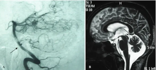
A computed tomography (CT) scan revealed a hemor-rhage in the fourth ventricle but no evidence of a parenchyma lesion. A cerebral angiography (Figure 1A) revealed a small saccular aneurysm of the basilar artery, which was located close to the emergence of the anterior inferior cerebellar artery (AICA). No evidence of vasospasm was observed.
On the fourth day after the ictus, the patient was administered haloperidol, and a progressive reduction in the frequency of choreoatetoid movements was observed. However, the dysarthria and postural instability remained. The patient was administered phenytoin for seven days
prior to the endovascular aneurysm repair, which was completed uneventfully. A control CT scan did not reveal hydrocephalus.
At the one-year follow-up visit, the patient did not exhibit any choreoatetoid movements; however, the cerebellar alterations remained, with an important static and dynamic imbalance and significant dysmetria. Magnetic Resonance Imaging (MRI) at the one year follow-up revealed cerebellar atrophy (Figure 1B).
DISCUSSION
Stroke-related movement disorders are uncommon (3.6%) and are very rare in SAH cases . Chorea, tremor, dystonia, Parkinsonism, and myoclonus have all been associated with cerebral infarcts and hemorrhaging.3 Movement disorders, which represent a portion of the clinical spectrum of acute stroke, may be delayed or progressive.
The first case of a movement disorder (chorea) after a SAH was reported by Sakai et al.4and occurred eight days after the SAH onset. The CT scans revealed a SAH with ventricular dilation and periventricular lucency involving the bilateral caudate nuclei. The chorea was attributed to the vasospasm and hydrocephalus.4
In another case, reported by Morigaki et al.,5 the
involuntary movements began shortly after the SAH onset. There was no acute hydrocephalus, and the authors attributed the symptoms to a hematoma on the corpus callosum that resulted from a rupture of an aneurysm of the distal accessory anterior cerebral artery. The hyperkinetic involuntary movements were suggested to have occurred due to the interruption of the cortico-striato-pallido-tha-lamo-cortical feedback loop.
Alarco´n et al. analyzed 1,500 consecutive stroke patients over a period of ten years to identify patients with a movement disorder, which was observed in only 56 patients (3.6%). Chorea was the most common movement disorder (35.7%). Thirty-nine (69.6%) patients experienced an ischemic stroke, 14 (25%) experienced a parenchymal hemorrhage, and only three (5.3%) experienced a SAH. All of the patients with a movement disorder that was secondary to a SAH in the Alarco´n et al. series presented with a tremor (Table 1) as their principal involuntary movement.3
The time that elapses between a stroke onset and the development of a movement disorder is variable. Alarco´n et al. observed involuntary movements that began on the first day of the stroke in 12.5% of their patients but reported
Copyrightß2011CLINICS– This is an Open Access article distributed under the terms of the Creative Commons Attribution Non-Commercial License (http:// creativecommons.org/licenses/by-nc/3.0/) which permits unrestricted non-commercial use, distribution, and reproduction in any medium, provided the original work is properly cited.
CLINICS 2011;66(9):1655-1657 DOI:10.1590/S1807-59322011000900026
cases in which the abnormal movement occurred much later, including Parkinsonism that began ten months after the stroke.3
While analyzing the literature data on movement dis-orders after a SAH (Table 1), we observed a total of six patients (including this reported case). Of these six patients, five were female and one was male, with a mean age of 58.6 years (SD 21.6, ranging from 17 to 74 years). Chorea or choreoathetosis was observed in three of these cases; tremor was observed in the other three cases. One patient in the tremor group exhibited associated dystonia. Our case is very atypical because he is the youngest patient ever described and was the only male to present with SAH-related involuntary movement.
In our case, no vasospasm, hydrocephalus, or even direct injury to the basal ganglia was observed that could explain the choreoathetosis. We suggest that the hematoma that was
located in the fourth ventricle may have disturbed the dentato-rubro-thalamo-cortical pathways, leading to transi-ent choreoatetoid movemtransi-ents. Another hypothesis, without evidence of a vasospasm, is that the movement disorder was secondary to an ischemic lesion in the thalamogeniculate artery (a branch of the posterior cerebral artery), which disturbed the posterior ventral thalamic areas related to the basal ganglia circuit.
REFERENCES
1. Brisman JL, Song JK, Newell DW. Cerebral aneurysms. N Engl J Med. 2006;355:928-39, doi: 10.1056/NEJMra052760.
2. Suarez JI, Tarr RW, Selman WR. Aneurysmal subarachnoid hemorrhage. N Engl J Med. 2006;354:387-96, doi: 10.1056/NEJMra052732.
3. Alarco´n F, Zijlmans JCM, Duen˜as G, Cevallos N. Post-stroke movement disorders: report of 56 patients. J Neurol Neurosurg Psychiatry. 2004;75:1568–74, doi: 10.1136/jnnp.2003.011874.
A B
Figure 1 -A) Cerebral angiography at admission revealed a small saccular aneurysm of the basilar artery near the emergence of the anterior inferior cerebellar artery (AICA). B) T2-weighted Magnetic Resonance Imaging (MRI) at the one-year follow-up revealed cerebellar atrophy.
Table 1 -Movement disorders after SAH.
Author (Year) Age Sex MD
Time from SAH to MD
Aneurysm
localization MD Recovery Hypothesis
Sakai et al. (1991)4
71 F Chorea 8 days ICA - AchoA Total Vasospasm Hydrocephalus
Alarco´n et al. (2004)3
74 F Tremor * ** ** Intraventricular hemorrhage
Alarco´n et al. (2004)3
55 F Tremor * ** ** Hydrocephalus
Alarco´n et al. (2004)3
63 F Tremor, dystonia, ataxia and dysmetria
* ** ** Hydrocephalus
Morigaki et al. (2008)5
72 F Choreoathetosis Ictus Distal accessory ACA Total Corpus callosum hematoma
Pereira et al. (2011)
17 M Choreoathetosis Ictus Basilar artery at AICA emergence
Total Disturbance of the dentato-rubro-thalamo-cortical
pathways
F = female; M = male; MD = movement disorder; AICA = anterior inferior cerebellar artery; ICA – AchoA = internal carotid artery - anterior choroidal artery; ACA = anterior cerebral artery.
*Only time in days (18.7 days, SD = 12.8 days) reported between the diagnosis of stroke and the onset of abnormal movements in a group of 14 patients with tremor after stroke. Six patients experienced an ischemic stroke, five had parenchymal hemorrhage, and three experienced a subarachnoid hemorrhage.
**not mentioned.
Choreoathetosis after subarachnoid hemorrhage
Pereira JLB et al. CLINICS 2011;66(9):1655-1657
4. Sakai K, Kyoshima K, Ohigashi Y, Unoki T, Kobayashi S, Meguro M. Generalized choreic movement associated with subarachnoid hemor-rhage. No To Shinkei. 1991;43:875–80.
5. Morigaki R, Uno M, Matsubara S, Satoh K, Nagahiro S. Choreoathetosis due to rupture of a distal accessory anterior cerebral artery aneurysm. Cerebrovascular Diseases. 2008;25:285-7, doi: 10.1159/000119640.
CLINICS 2011;66(9):1655-1657 Choreoathetosis after subarachnoid hemorrhage Pereira JLB et al.