einstein CASE REPORT
This content is licensed under a Creative Commons Attribution 4.0 International License. 1 Hospital Israelita Albert Einstein, São Paulo, SP, Brazil.
2 Faculdade de Medicina de Itajubá, Itajubá, MG, Brazil.
Corresponding author: Priscila Mina Falsarella – Hospital Israelita Albert Einstein, Department of Interventional Radiology – Avenida Albert Einstein, 627/701, Morumbi – Zip code: 05652-900 – São Paulo, SP, Brazil – Phone: (55 11) 2151-0195 – E-mail: [email protected]
Received on: Feb 3, 2017 – Accepted on: May 4, 2017 DOI: 10.1590/S1679-45082017RC4014
ABSTRACT
Preservation of the knee joint has enormous advantages in terms of mobility and rehabilitation of an amputee. Any cause of breakdown requiring revision to an above-knee amputation is a major setback because it reduces the patient’s rehabilitative potential. We report a case of intra-arterial thrombolysis use to save a below-knee amputation stump with acute ischemia. A 56-year-old man who sought the emergency department with 1-day history of acute pain on his right below-knee stump. The angiography confirmed popliteal artery occlusion. Pharmacomechanical thrombectomy, with Aspirex (rotational catheter to restore blood flow in occluded vessel, by removing occlusion material from the vessel) and recombinant tissue plasminogen activator, was performed. After 9 years of follow-up the patient remained asymptomatic, capable of independent ambulation with prosthetic limb. Intra-arterial fibrinolysis seems to be a safe and effective treatment for cases of acutely ischemic amputation stump.
Keywords: Fibrinolysis; Ischemia; Amputation stumps; Case reports
RESUMO
A preservação da articulação do joelho tem grandes vantagens para a mobilidade e a reabilitação de um amputado. Qualquer causa que exija revisão para uma amputação acima do joelho é um grande revés, porque reduz o potencial de reabilitação do paciente. O objetivo aqui foi descrever o uso de trombólise intra-arterial para salvar um coto de amputação abaixo do joelho com isquemia aguda. Homem, 56 anos, procurou pronto atendimento de nosso hospital com histórico de 1 dia de dor aguda em seu coto de amputação infrapatelar direito. A angiografia confirmou oclusão
da artéria poplítea. Foi realizada trombectomia farmacomecânica com Aspirex (cateter rotativo para restabelecer o fluxo sanguíneo em vasos ocluídos, removendo material de oclusão do vaso) e ativador do plaminogênio tecidual recombinante. Após 9 anos de seguimento, o paciente permanecia assintomático, capaz de deambulação independente com membro protético. A fibrinólise intra-arterial parece ser um tratamento seguro e eficaz para casos selecionados de coto de amputação com isquêmica aguda.
Descritores: Fibrinólise; Isquemia; Cotos de amputação; Relatos de casos
INTRODUCTION
The principle of improving the inflow to achieve a lower level of amputation is well known. Preservation of the knee joint has enormous advantages in terms of
rehabilitation of an amputee.(1) Any cause of breakdown
requiring revision to an above-knee amputation is a major setback, as it reduces the patient’s rehabilitative potential because of the lower stability and the highest
energetic cost of a gait with above-knee amputees.(2,3)
Acute lower limb ischemia is one of the most challenging conditions in vascular surgical practice and carries a high risk of amputation and death if not treated. Intra-arterial thrombolytic therapy can be considered standard of care for acute peripheral arterial occlusions. It can reclaim arterial perfusion and identify suspicious lesions that can be treated with
endovascular techniques.(4)
Intra-arterial fibrinolysis for the management of acute ischemia
on a below-knee amputation stump. Case report
Fibrinólise intra-arterial para tratamento da isquemia aguda em
coto de amputação infrapatelar. Relato de caso
Breno Boueri Affonso1, Joaquim Maurício da Motta Leal Filho1, Rafael Noronha Cavalcante1,
einstein
2 Affonso BB, Leal Filho JM, Cavalcante RN, Falsarella PM, Galastri FL, Cardoso RS, Nasser F
This paper describes the use of intra-arterial thrombolytic therapy with recombinant tissue plasminogen activator (rTPA) for the revascularization of a below-knee amputation stump with acute ischemia.
CASE REPORT
A 56- year-old man sought our service with 1-day history of acute pain on his right below-knee amputation stump. He had undergone right below-knee amputation for critical limb ischemia secondary to trauma 3 years before. After surgery, he had excellent rehabilitation and independent ambulation with prosthetic limb.
Upon clinical examination he had normal femoral pulse, absent popliteal pulse, decreased temperature and cyanosis in the distal third of the below-knee amputation stump. Doppler scan showed above-knee popliteal artery occlusion.
Heparin was administered (80UI/Kg), and right lower limb angiography and catheter directed thrombolysis,
with Aspirex® (rotational catheter to restore blood flow;
Straub Medical AG, Wangs, Switzerland), were carried out, in the attempt to save the below-knee amputation stump.
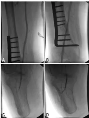
Right limb angiography showed superficial and deep femoral arteries without lesions and confirmed popliteal artery occlusion (Figure 1A). A 0,014” guide wire was
passed through the occluded area, and a thrombectomy catheter was placed intra-thrombus (Figure 1B). An initial bolus dose of 10mg of rTPA was administered and mechanical thrombectomy was performed with
Aspirex® (Figure 1C). Angiography showed partial
popliteal artery recanalization (Figure 1D).
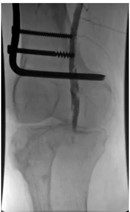
The thrombectomy device was replaced for a multiperforated catheter, and the patient was referred to the intensive care unit to continue rTPA infusion at dose of 3mg/hour and heparinization in full dose (16UI/kg/hour) for 8 hours under close monitoring. A new angiography showed patency of the popliteal artery with residual stenosis and thrombus (Figure 2). Angioplasty was performed with a 4x20 Paseo-35 balloon (Biotronik, Bulach, Switzerland), followed by injection of 10mg rTPA (Figure 3A and 3D). Final angiography demonstrated patency of the popliteal artery, without residual stenosis or thrombus, and excellent inflow to the below-knee amputation stump (Figures 4A and 4D).
The patient had remission of symptoms and was discharged after 5 days. After recovery, he was able for independent ambulation with his prosthetic limb. After 9 years of clinical follow-up the patient remained asymptomatic.
Figure 2. Digital subtraction angiography. Patency of the popliteal artery with residual stenosis and thrombus
Figure 1. Digital subtraction angiography. (A) Popliteal artery occlusion. (B and C) Thrombectomy catheter placed intra-thrombus. (D) Partial popliteal artery recanalization
A
C D
3
Intra-arterial fibrinolysis for the management of acute ischemia on a below-knee amputation stump
einstein
who already have a lower stability due to loss of muscle mass and osteopenia, to maintain as highest as possible number joints is benefic for the rehabilitation process,
which is more difficult among older individuals.(3)
Despite the technical progress in vascular surgery, acute peripheral arterial occlusion is still characterized
by high rates of morbidity and mortality.(5) The
management of this condition includes anticoagulation, surgical revascularization, and pharmacomechanical thrombectomy, depending on the clinical condition of the patient.
So far, there is no standard of care for management of acutely ischemic stump. Bunt described five cases of ascending gangrene followed by amputation that underwent immediate surgical revascularization. The
overall mortality rate of these patients was high (60%).(6)
In order to avoid stump gangrene, inflow revascularization prior to amputation is usually recommended when there is severe vascular disease, particularly when occlusion of the iliac or common femoral artery exists in combination with occlusion or stenosis of the deep femoral artery. In that sense, Wolosker et al., described the use of intraoperative stenting through the open end of the fibular artery to improve the inflow and achieve a lower level of
amputation.(7)
To our best knowledge, there are only two previous reports on below-knee ischemic stump revascularization for preservation of the amputation level, none of
them with thrombolysis.(2) Karkos et al., described
a femoropopliteal subintimal recanalization of an ulcerated stump in a previously rehabilitated patient. The authors described that the patient was pain-free
and able to mobilize at 6-month follow-up.(2) Warner
et al., described a deep femoral artery angioplasty in a patient with bellow-knee amputation not rehabilitated, presenting pain at rest and non-healing stump wound. After surgery, pain symptoms and wound healing
improved significantly.(8)
In our case, we decided to intervene because patient had a severely ischemic stump, pain at rest and cyanosis. If revascularization were not achieved, the only alternative would be an above-knee amputation. In our service, we usually decide for endovascular approach first and then, if there is failure, we choose open surgical revascularization. In literature, catheter based
thrombolysis achieves success in 75 to 92% of cases.(9)
There is no consensus in literature on the fibrinolytic dose and infusion duration but it is well established that best results are seen in cases with less than 2-week
duration.(10) In our practice, infusion rates of 0.5 to
DISCUSSION
The importance of maintaining the highest number of joints in an amputee patient is mainly due to the complex mechanism needed to keep stability. In elderly patients Figure 3. Digital subtraction angiography. (A and B) Popliteal artery angioplasty performed with a 4x20 Paseo-35 balloon. (C and D) Partial popliteal recanalization
A
C
B
D
Figure 4. Final angiography demonstrated patency of the superficial femoral and popliteal arteries, without residual stenosis or thrombus
A B
D
einstein
4 Affonso BB, Leal Filho JM, Cavalcante RN, Falsarella PM, Galastri FL, Cardoso RS, Nasser F
5mg/hour are usually chosen for arterial thrombolysis, usually with a control angiography being performed after 6 to 12 hours. It is known that catheter-directed infusion achieves higher limb salvage rates (about 80%) than intravenous systemic therapy (45%) and that the method of fibrinolytic agent delivery (infusion, pulse spray, and dose rate) does not influence limb salvage rates.(10)
Van den Berg in a review article concluded that there is no difference between surgery and thrombolysis, in terms of mortality and amputation, for acute peripheral
arterial occlusion of the lower limb.(4) Although the use
of catheter-directed thrombolysis is reasonably safe and effective, the risk of bleeding remains. Complications reported include major hemorrhage (5.1%), minor
hemorrhage (14.8%), and distal embolization (<1%).(4)
CONCLUSION
In selected cases, intra-arterial fibrinolysis seems to be a safe and effective treatment for acutely ischemic amputation stump.
REFERENCES
1. Tisi PV, Than MM. Type of incision for below knee amputation. Cochrane Database Syst Rev. 2014;8(4):CD003749. Review.
2. Karkos CD, Bright E, Bolia A, London NJ. Subintimal recanalization of the femoropopliteal segment to promote healing of an ulcered below-knee amputation stump. J Endovasc Ther. 2006;13(3):420-3.
3. Kamali M, Karimi MT, Eshraghi A, Omar H. Influential factors in stability of lower-limb amputees. Am J Phys Med Rehabil. 2013;92(12):1110-8. Review. 4. Van den Berg JC. Thrombolysis for acute arterial occlusion. J Vasc Surg.
2010;52(2):512-5. Review.
5. Earnshaw JJ. Demography and etiology of acute leg ischemia. Semin Vasc Surg. 2001;14(2):86-92. Review.
6. Bunt TJ. Gangrene of the immediate postoperative above-knee amputation stump: role of emergency revascularization in preventing death. J Vasc Surg. 1985;2(6):874-7.
7. Wolosker N, Nakano L, Duarte FH, De Lucia N, Leao PP. Peroneal artery approach for angioplasty of the superficial femoral artery: a case report. Vasc Endovascular Surg. 2003;37(2):129-33.
8. Warner BE, Richards AJ, Biswas M, Chick C, Lewis P, Harding KG. Chronic wound and postamputation claudication pain in a diabetic patient. Ann R Coll Surg Engl. 2013;95(7):115-7.
9. da Motta Leal Filho JM, Santos AC, Carnevale FC, de Oliveira Sousa W Jr, Grillo LS Jr, Cerri GG. Infusion of recombinant human tissue plasminogen activator through the superior mesenteric artery in the treatment of acute mesenteric venous thrombosis. Ann Vasc Surg. 2011;25(6):840.e1-4. 10. Kessel DO, Berridge DC, Robertson I. Infusion techniques for peripheral arterial