503
Correspondence to: Srdjan BABIĆ
Institute for Cardiovascular Disease “Dedinje” Heroja Milana Tepića 1 11000 Belgrade Serbia
[email protected] [email protected]
Srp Arh Celok Lek. 2016 Sep-Oct;144(9-10):503-506 DOI: 10.2298/SARH1610503B
ОРИГИНАЛНИ РАД / ORIGINAL ARTICLE UDC: 617.582-089.873-085.83
SUMMARY
Introduction The stump wound complications after above-knee amputation lead to other problems, such as prolonged rehabilitation, delayed prostheticrestoration, the increase in total treatment cost and high mortality rates.
Objective To evaluate the safety and outcomes of negative-pressure wound therapy (NPWT) using Vacuum-Assisted Closure (VAC®) therapy in patients with stump complication after above-knee ampu-tation (AKA).
Methods From January 2011 to July 2014, AKA was performed in 137 patients at the University Cardio-vascular Clinic. Nineteen (12.4%) of these patients (mean age 69.3 ± 9.2 years) were treated with NPWT. The following variables were recorded: wound healing and hospitalization time, rate of NPWT treatment failure, and mortality.
Results AKA was performed in 17 (89.5%) patients afterthe vascular or endovascular procedures had been exhausted, while urgent AKA was performed in two (10.5%) patients due to uncontrolled infection. The time before NPWT application was 3.1 ± 1.9 days and the duration of the NPWT use ranged from 15 to 54 days (mean 27.95 ± 12.1 days). During NPWT treatment, operative debridement was performed in 12 patients. All the patients were kept on culture-directed intravenous antibiotics. The average hospital length of stay was 34.7 days (range 21–77 days). There were four (20.9%) failures during the treatment which required secondary amputation. During the treatment, one (5.3%) patient died due to multi-organ failure after 27 days.
Conclusions The use of NPWT therapy in the treatment of AKA stump complication is a safe and effective procedure associated with low risk and positive outcome in terms of wound healing time and further complications.
keywords: above-knee amputation; wound complication; negative-pressure wound therapy (NPWT)
Treatment of stump complications after above-knee
amputation using negative-pressure wound therapy
Srdjan Babić1, Slobodan Tanasković1, Branko Lozuk1, Dražen Samardžić1, Petar Popov1,2, Predrag
Gajin1,2, Predrag Matić1,2, Vesna Marić3, Djordje Radak1,2,4
1Institute for Cardiovascular Disease “Dedinje”, Belgrade, Serbia; 2University of Belgrade, School of Medicine, Belgrade, Serbia; 3Clinical Center of Serbia, Clinic of Eye Diseases, Belgrade, Serbia; 4Serbian Academy of Sciences and Arts, Belgrade, Serbia
INTRODUCTION
Lower extremity amputation definitely remains the therapeutic option for the management of ischemic disease that fails revascularization, extensive traumatic tissue loss, infection and ischemic disease of the leg where attempts of revascularization failed [1]. Major lower ex-tremity amputations, especially above-knee amputation (AKA), are associated with high morbidity and mortality rates [2, 3]. Previ-ous study reports 11% rate of stump compli-cations [2]. Also, patients after AKA have the worst functional outcomes and survival rates of all other levels of amputation. In addition, the stump wound complications lead to other problems, such as the increase in total treat-ment cost, prolonged rehabilitation, delayed prosthetic restoration, and finally the reduction in the quality of life of patients [4–7].
Majority of the published papers are focused on the treatment of diabetic foot ulcers, venous leg ulcers and postoperative groin wound in-fection [8–11]. However, reports of the use of negative-pressure wound therapy (NPWT) af-ter AKA and stump complications are limited to a few patients [12, 13].
To further elucidate this issue, we report of 19 patients with stump complications treated with NPWT after AKA.
OBjECTIvE
The purpose of this study was to evaluate the safety and outcomes of NPWT using Vacuum-Assisted Closure (VAC®) therapy in patients with stump complication after AKA.
METHODS
Patients involved in the study
504
doi: 10.2298/SARH1610503B
Babić S. et al. Treatment of stump complications after above-knee amputation using negative-pressure wound therapy
criterion was massive tissue necrosis, which required early re-amputation.
Preoperative evaluation consisted of the following: clinical examination, ankle-brachial index, multi-slice computed tomography angiography (Lightspeed VCT, GE Healthcare, Milwaukee, WI, USA), and appropriate laboratory tests. The indication for AKA was made by at-tending vascular surgeon, only if all other options for vas-cular and endovasvas-cular procedures were either exhausted or contraindicated. All stump complications in the series were recorded. Initially, ceftriaxone (2 g / 24 hours) and/ or amikacin (1.5 g / 24 hours) were administered to all the patients, who signed the informed consent form for use of their data for the analysis. The study was approved by our hospital’s Ethical Committee. Standard descriptive statistics methods were used (number and means), me-dian, minimum, and maximum values. Cox univariate and multivariate analyses were carried out to assess predictors of NPWT treatment failure. SPSS version 17.0 (SPSS Inc., Chicago, IL, USA) was used for all statistical calculations.
Negative-pressure wound therapy
If any signs of stump (primary closed) complication ap-peared (infection, secretion, or skin necrosis), the wound would be opened and assessed daily for at least three days. If there was no sign of massive tissue necrosis, the indication for NPWT would be considered set. In case of an uncon-trolled infection (e.g. necrotizing fasciitis or gas gangrene), guillotine amputation was performed to sustain the infec-tion. The wound was assessed daily by the attending surgeon. In all the patients wound swabsweretaken routinely before the application of the negative pressure wound device.
The NPWT system used in this study was Vacuum-Assisted Closure (VAC®, KCI Medical, San Antonio, TX, USA) and it consisted of three components: 1) a negative pressure-generating unit with a disposable canister, 2) a pad with evacuation tube, and 3) a reticulated, open cell sterile polyurethane or a dense open-pore polyvinyl alco-hol foam dressing, cut to fit the open amputation stump wound. Non-occlusive wound dressings were used as protection for exposed vessels. VAC® was applied to the wound as specified by the manufacturer’s guidelines [14].
Negative pressure used to treat these patients ranged from 75 mmHg to 125 mmHg. The treatment was contin-ued until several goals were achieved: 1) the wound swabs were negative; 2) sufficient granulation tissue formation for possible secondary suture; 3) absence of necrotic tissue, and 4) absence of secretion from the wound three days after VAC® system removal.
However, the treatment was terminated if NPWT caused progressive worsening of the local ischemic pro-cess and necrosis. The polyurethane foam was changed regularly in three-day intervals. When necessary, surgical debridement was performed to healthy muscle and fat. Antibiotics were administered according to the results of antibiogram, generated from intra- or post-operatively obtained wound swab.
RESULTS
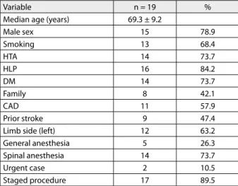
During the observation period, 19 (12.4%) of 137 patients met study criteria and were treated with NPWT. Baseline demographic characteristics of enrolled patients, indica-tion for treatment, and type of anesthesia administered are shown in Table 1.
The mean patient population age was 69.2 ± 9.2 years (range 51–82 years) and the patients were predominantly male (78.9%). Staged procedure was performed in 17 (89.5%) patients, while urgent AKA was performed in two (10.5%) patients due to uncontrolled infection. In 17 patients (staged procedure), 49 open vascular (2.9 per patient) and 24 en-dovascular (1.4 per patient) procedures were performed before AKA. Remaining two patients with uncontrolled in-fection were admitted to our (tertiary) institution; however, after our assessment and patient status, the revascularization procedures were contraindicated. The time before NPWT application was 3.1 ± 1.9 days and the duration of therapy ranged from 15 to 54 days (mean 27.95 ± 12.1 days). During the therapy, operative debridement was performed in 12 pa-tients, after a mean of 19.2 days. All the patients were kept on cultudirected intravenous antibiotics. The swab culture re-sults are shown in Table 2. The majority of the infections were polymicrobial and two patients had negative swab cultures.
When the conditions were met (sterile swab culture, suf-ficient granulation tissue formation and absence of necrotic debris), the secondary suturing was done. All the patients were followed in our vascular surgery wound clinic until the
Table 2. Culture results from wounds
Culture result N
Polymicrobial 7
Klebsiella 2
Methicillin-resistant Staphylococcus aureus 2
Serratia 2
Enterococcus 2
Proteus vulgaris 1
Escherichia coli 1
Negative 2
Table 1. Demographic characteristics of enrolled patients
Variable n = 19 %
Median age (years) 69.3 ± 9.2
Male sex 15 78.9
Smoking 13 68.4
HTA 14 73.7
HLP 16 84.2
DM 14 73.7
Family 8 42.1
CAD 11 57.9
Prior stroke 9 47.4
Limb side (left) 12 63.2
General anesthesia 5 26.3
Spinal anesthesia 14 73.7
Urgent case 2 10.5
Staged procedure 17 89.5
505
Srp Arh Celok Lek. 2016 Sep-Oct;144(9-10):503-506
www.srpskiarhiv.rs wounds completely healed. Our practice is to use NPWT
only in the institutional setting because of the increased risk of morbidity and hemorrhaging in these patients. The aver-age hospital length of stay was 34.7 days (range 21–77 days). There were four (20.9%) failures during the treatment. Re-amputation was required in three of these four patients, and coxofemoral disarticulation in the remaining one, all caused by massive stump tissue necrosis. During the treat-ment, one patient died due to multi-organ failure 27 days after the amputation.
Univariate analysis evaluating the factors such as age, sex, risk factors for vascular disease, presence of cardiac artery diseases, previous vascular/endovascular intervention, uni-lateral iliac or deep femoral artery occlusion and limb side, failed to identify any variable predictive of NPWT failure.
DISCUSSION
The main result of this study is that NPWT can have a significant role in the treatment of AKA stump complica-tions. Our study shows that the NPWT reduces morbidity, total treatment cost, the rate of secondary amputations, and the mortality rate. Treatment with NPWT has been used for over two decades. The first paper was published by Fleischman et al. [15] in 1993, reporting the treatment of soft tissue damage caused by open fracture.
In recent years, the advance in technology has extend-ed the use of NPTW to more extensive and complicatextend-ed wounds, such as diabetic foot wounds, post-surgery diabetic foot wounds, inflammatory ulcers, dehisced sternal wounds, open abdominal wounds, and traumatic wounds [16].
Additional indications for NPWT in vascular surgery are
ulcus cruris treatment, MESH-grafting, secondary healing,
and infected wounds after several vascular operations/inter-vention, amputations, lymphatic fistulas, soft tissue and skin infections, and infections of vascular grafts [12].
However, majority of these papers reported their results using NPWT in the treatment of diabetic foot ulcers, ve-nous leg ulcer, and vascular groin infection [8–11].
On the other hand, reports on NPWT use in patients with stump complication are limited to a few patients [12, 13].
Unfortunately, major lower extremity amputation is still a commonly performed operation that is indicated in pa-tients with failed attempts of revascularization, extensive traumatic tissue loss, and infection [1]. Further problem after major lower extremity amputation is a significant risk of perioperative morbidity and mortality. Aulivola et al. [2] found significantly higher 30-day mortality rate for AKA patients (16.5%) than for below-knee amputation (BKA) patients (5.7%). A consistent result of 13.3% 30-day mortality rate for AKA was shown in series published by Feinglass et al. [3]. During the study period in our series, 30-day mortality in all of the patients (137 AKA) was 5.1%, and only one (out of 19 patients) died, which occurred 27 days after the index intervention. Also, it has been shown that stump complications increase mortality risk from car-diac complications, pneumonia, renal failure, and stroke [1, 2, 3, 17, 18]. Regarding lower mortality rate than that
in previous series [2, 3], we could hypothesize that the use of NPWT in these patients reduces mortality risk.
Regarding the morbidity after AKA, Aulivola et al. [2] reported an 11% rate of the stump complication, which is similar to our series. Patients requiring guillotine ampu-tation as a consequence of sepsis are a special problem. Reported mortality rate in these patients is 14.3% (includ-ing AKA and BKA) [2]. In our study, both patients that underwent guillotine amputation had extended postopera-tive periods (47 and 36 days), and after secondary suture the wounds were completely healed.
We had four (20.9%) treatment failures. Re-amputation was required in three of these four patients, and coxo-femoral disarticulation in the remaining one, all caused by massive stump tissue necrosis. However, even Cox uni-variate analysis failed to identify any variable predictive of VAC® failure. These four patients had unilateral iliac or deep femoral artery occlusion verified with preoperative multi-slice computed tomography (MSCT) angiography. We can conclude that patients like these should not be candidates for NPWT and that local findings (absence of the necrotic mass during the first several days after the amputation) can be misleading.
There are several advantages of NPWT which could be ap-plied in the treatment of AKA wound complications. NPWT decreases the healing time of the wound, reduces the bacterial counts in the wound, possibly decreases the need for future amputations, and signifi cantly improves the results in the subjective pain scores at the follow-up [9, 11, 17–20].
In a recently published case report by Richter and Knudson [13], NPWT was used in a AKA wound with extremely large tissue deficit. After multiple surgical pro-cedures and three months of NPWT, the residual limb improved to a prosthetic-ready state.
In case of wound complications after AKA, general prac-tice in our hospital is to use NPWT until the following goals are achieved: two or more negative swab cultures, presence of sufficient granulation tissue formation, absence of secretion from the wound for three days after NPWT system removal, and absence of necrotic tissue. When these criteria are met, we perform secondary suturing for several reasons: AKA wound defect is too large for second intention healing, and it reduces the length of hospitalization and hospital related complication rate. Also, we avoid the use of skin grafts in this scenario. In addition, this method allows early rehabilitation and prepares the patient to begin prosthetic restoration.
There are some limitations of this study – a relatively small number of patients included (n = 19), and the lack of control group (i.e. patients treated without NPWT or any other therapeutic option).
CONCLUSION
506
doi: 10.2298/SARH1610503B REFERENCES
1. Tunis SR, Bass EB, Steinberg EP. The use of angioplasty, bypass surgery, and amputation in the management of peripheral vascular disease. N Engl J Med. 1991; 325:556–62.
[DOI: 10.1056/NEJM199108223250806] [PMID: 1857391] 2. Aulivola B, Hile CN, Hamdan AD, Sheahan MG, Veraldi JR, Skillman
JJ, et al. Major lower extremity amputation: outcome of a modern series. Arch Surg. 2004; 139(4):395–9.
[DOI: 10.1001/archsurg.139.4.395] [PMID: 15078707]
3. Feinglass J, Pearce WH, Martin GJ, Gibbs J, Cowper D, Sorensen M, et al. Postoperative and late survival outcomes after major amputation: findings from the Department of Veterans Affairs National Surgical Quality Improvement Program. Surgery. 2001; 130:21–9. [DOI: 10.1067/msy.2001.115359] [PMID: 11436008] 4. Apelqvist J, Armstrong DG, Lavery LA, Boulton AJ. Resource
utilization and economic costs of care based on a randomized trial of vacuum-assisted closure therapy in the treatment of diabetic foot wounds. Am J Surg. 2008; 195(6):782–8.
[DOI: 10.1016/j.amjsurg.2007.06.023] [PMID: 18355797] 5. Francis W, Renton CJ. Mobility after major limb amputation for
arterial occlusive disease. Prosthet Orthot Int. 1987; 11:85–9. [DOI: 10.3109/03093648709078184] [PMID: 3658652] 6. Sandnes DK, Sobel M, Flum DR. Survival after lower-extremity
amputation. J Am Coll Surg. 2004; 199:394–402. [DOI: 10.1016/j.jamcollsurg.2004.05.270] [PMID: 15325609] 7. Harker J. Wound healing complications associated with lower limb
amputation. World Wide Wounds; September 2006. Available from URL: http://www.worldwidewounds.com.
8. Dosluoglu HH, Loghmanee C, Lall P, Cherr GS, Harris LM, Dryjski ML. Management of early (<30 day) vascular groin infections using vacuum-assisted closure alone without muscle flap coverage in a consecutive patient series. J Vasc Surg. 2010; 51(5):1160–6. [DOI: 10.1016/j.jvs.2009.11.053] [PMID: 20356703] 9. Armstrong DG, Lavery LA. Diabetic Foot Study Consortium.
Negative Pressure Wound Therapy after partial diabetic foot amputation; a multicentre, randomized controlled trial. Lancet. 2005; 366:1704–10.
[DOI: 10.1016/S0140-6736(05)67695-7] [PMID: 16291063]
10. Eginton MT, Brown KR, Seabrook GR, Towne JB, Cambria RA. A prospective randomized evaluation of negative-pressure wound dressings for diabetic foot wounds. Annals of Vascular Surgery. 2003; 17(6):645–49.
[DOI: 10.1007/s10016-003-0065-3] [PMID: 14534844]
11. Vuerstaek, JD Vainas T. State-of-the art treatment of chronic leg ulcers: Assessing the role of Vacuum Assisted Closure (V.A.C.®) in wound healing. J Vasc Surg. 2006; 44(5):1029–37.
[DOI: 10.1016/j.jvs.2006.07.030] [PMID: 17000077]
12. Beno M, Martin J, Sager P. Vacuum assisted closure in vascular surgery. Bratisl Lek Listy. 2011; 112(5):249–52.
[DOI: 10.4149/BLL_2011_051] [PMID: 21682077]
13. Richter K, Knudson B. Vacuum-assisted closure therapy for a complicated, open, above-the-knee amputation wound. J Am Osteopath Assoc. 2013; 113(2):174–6. [PMID: 23412679]
14. V.A.C. Therapy Clinical Guidelines. A Reference Source for Clinicians. KCI, San Antonio, TX; 2004
15. Fleischmann W, Strecker W, Bombelli M, Kinzl L. Vacuum sealing as treatment of soft tissue damage in open fractures. Unfallchirurg. 1993; 96(9):488–92.
[DOI: 10.9738/INTSURG-D-14-00018.1] [PMID: 8235687] 16. World Union of Wound Healing Societies (WUWHS). Principles of
best practice: Vacuum assisted closure: recommendations for use. A consensus document. London: MEP Ltd; 2008.
17. Porter JM, Baur GM, Taylor LM. Lower extremity amputations for ischemia. Arch Surg. 1981; 116:89–92.
[DOI: 10.1001/archsurg.1981.01380130065015] [PMID: 7469738] 18. Keagy BA, Schwartz JA, Kotb M, Burnham SJ, Johnson G Jr. Lower
extremity amputation: the control series. J Vasc Surg. 1986; 4:321– 26. [DOI: 10.1016/0741-5214(86)90223-5] [PMID: 3761472] 19. Frykberg RG, Williams DV. Negative-pressure wound therapy
and diabetic foot amputations. J Am Podiatr Med Assoc. 2007; 97(5):351–59. [PMID: 17901338]
20. Morykwas MJ, Argenta LC, Shelton-Brown EL, McGuirt W. Vacuum-assisted closure: a new method for wound control and treatment: animal studies and basic foundation. Ann Plast Surg. 1997; 38(6):553–62.
[DOI: 10.1097/SAP.0b013e3181a72f77] [PMID: 9188970]
КРАТАК САДРжАЈ
Увод Компликације ампутационог патрљка након натко-лене ампутације представљају комплексан проблем који води ка продуженом лечењу пацијената, порасту трошкова лечења и повећаноj стопи смртности.
Циљ рада Циљ овог истраживања је процена безбедности и ефикасности терапије негативним притиском код пације-ната са компликацијама ампутационог патрљка након нат-колене ампутације.
Методе рада У периоду од јануара 2011. до јула 2014. годи-не натколена ампутација је урађена код 137 пацијената. Код 19 (12,4%) пацијената (просечне старости 69,3 ± 9,2 година) због компликација ампутационе ране примењена је тера-пија негативним притиском. Праћене су следеће варијабле: зарастање ране и време хоспитализације, неуспех терапије негативним притиском и смртност.
Резултати Натколена ампутација је урађена код 17 (89,5%) пацијената након што су васкуларне или ендоваскуларне
могућности реваскуларизације исцрпљене, а у два (10,5%) пацијента због прогресивне и неконтролисане инфекције. Просечно време трајања терапије негативним притиском било је 27,95 ± 12,1 дана (распон 15 до 54 дана). Током те-рапије хируршка обрада ране урађена је код 12 пацијената. Просечно време хоспитализације је било 34,7 дана (распон 21–77 дана). Неуспех терапије је забележен код четири па-цијента (20,9%), што је резултирало реампутацијом. Током третмана, код једног (5,3%) пацијента дошло је до смртног исхода након 27 дана као последица мултиорганског по-пуштања.
Закључак Терапија негативним притиском у лечењу ком-пликација ране након натколене ампутације је безбедан и ефикасан терапијски избор, праћен ниским ризиком и до-брим исходом у погледу зарастања ране.
Кључне речи: натколена ампутација; компликације ране; терапија негативним притиском
Терапија негативним притиском у лечењу компликација ампутационог
патрљка након натколене ампутације
Срђан Бабић1, Слободан Танасковић1, Бранко Лозук1, Дражен Самарџић1, Петар Попов1,2, Предраг Гајин1,2, Предраг
Матић1,2, Весна Марић3, Ђорђе Радак1,2,4
1Институт за кардиоваскуларне болести „Дедиње“, Београд, Србија; 2Универзитет у Београду, Медицински факултет, Београд, Србија; 3Клинички центар Србије, Клиника за очне болести, Београд, Србија; 4Српска академија наука и уметности, Београд, Србија
Примљен • Received: 08/07/2015 Ревизија • Revision: 11/02/2016 Прихваћен • Accepted:18/02/2016