Issues in solid-organ transplantation in children:
translational research from bench to bedside
Steven E. Lipshultz,I,II,VI* Jayanthi J. Chandar,IIIPaolo G. Rusconi,IIAlessia Fornoni,IIICarolyn L. Abitbol,III George W. Burke III,IIIGaston E. Zilleruelo,IIISi M. Pham,IVElena E. Perez,VRuchika Karnik,IIJuanita A. Hunter,IIDanielle D. Dauphin,VIJames D. WilkinsonVI
IWayne State University School of Medicine and Children’s Hospital of Michigan, Department of Pediatrics, Detroit/MI, United States.IIUniversity of
Miami Miller School of Medicine, Department of Pediatrics, Division of Pediatric Cardiology, Miami/FL, United States.IIIUniversity of Miami Miller School
of Medicine, Department of Pediatrics, Division of Pediatric Nephrology, Miami/FL, United States.IVUniversity of Miami Miller School of Medicine/Jackson
Memorial Division of Heart/Lung Transplant and Artificial Heart Programs, Transplant Institute, Miami/FL, United States.VUniversity of Miami Miller
School of Medicine, Department of Pediatrics, Division of Pediatric Immunology and Allergy, Miami/FL, United States.VIUniversity of Miami Miller School
of Medicine, Department of Pediatrics, Division of Pediatric Clinical Research, Miami/FL, United States.
In this review, we identify important challenges facing physicians responsible for renal and cardiac transplantation in children based on a review of the contemporary medical literature. Regarding pediatric renal transplantation, we discuss the challenge of antibody-mediated rejection, focusing on both acute and chronic antibody-mediated rejection. We review new diagnostic approaches to antibody-mediated rejection, such as panel-reactive antibodies, donor-specific cross-matching, antibody assays, risk assessment and diagnosis of antibody-mediated rejection, the pathology of antibody-mediated rejection, the issue of ABO incompat-ibility in renal transplantation, new therapies for antibody-mediated rejection, inhibiting of residual antibodies, the suppression or depletion of B-cells, genetic approaches to treating acute antibody-mediated rejection, and identifying future translational research directions in kidney transplantation in children. Regarding pediatric cardiac transplantation, we discuss the mechanisms of cardiac transplant rejection, including the role of endomyocardial biopsy in detecting graft rejection and the role of biomarkers in detecting cardiac graft rejection, including biomarkers of inflammation, cardiomyocyte injury, or stress. We review cardiac allograft vasculopathy. We also address the role of genetic analyses, including genome-wide association studies, gene expression profiling using entities such as AlloMapH, and adenosine triphosphate release as a measure of immune function using the CylexH ImmuKnowTM cell function assay. Finally, we identify future
translational research directions in heart transplantation in children.
KEYWORDS: Child; Translational Medical Research; Transplantation; Kidney Transplantation; Renal Transplantation; Heart Transplantation; Cardiac.
Lipshultz SE, Chandar JJ, Rusconi PG, Fornoni A, Abitbol CL, Burke III GW, et al. Issues in solid-organ transplantation in children: translational research from bench to bedside. Clinics. 2014;69(S1):55-72.
E-mail: [email protected] *corresponding author
Tel.: (313) 745-5870
& INTRODUCTION
Solid-organ transplantation is an accepted treatment for end-stage renal and cardiac diseases in children. Over the past few decades, better methods of matching donor-recipient pairs and newer immunosuppressive drugs have substantially improved the overall survival of transplant recipients. Consequently, the number of children receiv-ing solid-organ transplants has increased tremendously.
However, improving the long-term management and quality of life of recipients continues to be a challenge. The main challenges are allograft rejection, the deleterious effects of the immunosuppressive drugs, infections, malignancies, nephro-toxicity, post-transplant lymphoproliferative disorders, and, in some cases, recurrence of the primary disease.
The University of Miami’s Miller School of Medicine is deeply involved in caring for a large number of children receiving solid-organ transplants. In this article, we review many of the challenges in caring for these children, which we accomplish with an interdisciplinary team of pediatric cardiologists, pediatric nephrologists, pediatric immunolo-gists, cell bioloimmunolo-gists, molecular bioloimmunolo-gists, transplant surgeons, pathologists, epidemiologists, and computational scientists.
We focus on antibody-mediated rejection (AMR) and the recurrence of focal segmental glomerulosclerosis (FSGS) in the renal section and on biomarkers for rejection and Copyrightß2014CLINICS– This is an Open Access article distributed under
the terms of the Creative Commons Attribution Non-Commercial License (http:// creativecommons.org/licenses/by-nc/3.0/) which permits unrestricted non-commercial use, distribution, and reproduction in any medium, provided the original work is properly cited.
No potential conflict of interest was reported.
immunotolerance in the cardiology section. At the end of this review, we propose future research directions to identify the most appropriate children to list for transplan-tation and to improve the post-transplant care of these children.
& TRANSLATIONAL RESEARCH IN PEDIATRIC RENAL TRANSPLANTATION
The Challenge of Antibody-Mediated Rejection
Kidney transplantation has had a long and successful history since the human leukocyte antigen (HLA) was discovered in the 1960s. The realization that graft rejection was an immunological phenomenon resulted in the devel-opment of immunosuppressive drugs, which allowed for organ transplantation from genetically different donors (1). Although the primary consideration in tissue and organ transplantation is to ensure ABO blood group compatibility, large national databases suggest that graft survival improves with better HLA antigen matching and that this matching is an important factor in long-term graft survival (2,3). The establishment of the International Histocompatibility Workshop in 1965 set the stage for histocompatibility testing in transplantation. In the ensuing years, the techniques and standardization of HLA typing evolved, and new antigens were characterized. In recent years, molecular technology has improved the accuracy and reproducibility of tissue typing, cross-matching, and detection of anti-HLA antibodies (4,5).
In the 1960s, immediate allograft failure was found to decrease markedly with negative cross-matching between donor lymphocytes and recipient serum. This finding eventually resulted in the development of the comple-ment-dependent cytotoxicity assay (6,7). Although immu-nosuppressive therapy regimens and the short-term survival of kidney allografts have improved substantially since then, acute rejection in the first year and chronic allograft nephropathy continue to be major determinants of long-term graft survival (8). Traditionally, transplant rejec-tion has been considered to be predominantly mediated by T-cells. However, increasing evidence suggests that inade-quate control of the humoral arm of the immune system contributes to chronic allograft nephropathy (9).
Halloran et al. first described an atypical form of acute rejection occurring a few days to weeks after transplantation that was characterized by a rapid deterioration in renal function and a high incidence of failure in a previously functioning graft. Pathologic features were similar to hyperacute rejection and were associated with donor-specific HLA antibodies (10,11). This phenomenon has since been termed AMR (12,13).
Traditionally, the complement-dependent cytotoxicity cell-based assay was used to detect donor-specific anti-HLA antibodies and was useful in predicting hyperacute rejection. However, this assay is not sensitive enough to detect low or marginal titers of antibodies, which are relevant to the early outcomes of the transplant. The advent of solid-phase assays (SPA), which have greater sensitivity and specificity, has resulted in increased identification of AMR (14). The incidence of AMR is less than 5% in unsensitized patients but between 40% and 90% in sensitized patients (15). The strength of the antibody response appears to be strongly associated with the risk of rejection (14-17). Occurring from preformed or new anti-HLA antibodies, AMR generally has a worse prognosis and
requires different management than does T-cell-related rejection (18).
A higher degree of HLA mismatches, acute rejection episodes, patient nonadherence to treatment, inadequate immune suppression, previous organ transplantation, blood transfusion, and pregnancy result in sensitization and increased risk for AMR. The first six causes are important in children (14,18,19). Currently, 17% of patients with end-stage kidney disease on the waiting list for kidney transplants have had previous transplants. Given that 50% of all childhood kidney transplant recipients will receive a second kidney transplant by the age of 25 years, a substantial proportion of children will be sensitized as young adults (20). The true incidence and prevalence of AMR in children is not known.
Clinical Features of Acute and Chronic Antibody-Mediated Rejection
In acute AMR,patients present with an acute loss of graft function, most often in the first few weeks after transplanta-tion. The clinical presentation is indistinguishable from acute cellular rejection. It can also occur years after transplantation when immune suppression is decreased or stopped, either iatrogenically or because of nonadherence by the patient (21). It can occur in both sensitized patients and in those with a negative pre-transplant cross-match.
Chronic AMR is an insidious process associated with fluctuating levels of donor-specific antibodies (DSAs) and results in irreversible structural damage. Clinically, it manifests as proteinuria, hypertension, and declining graft function over time.
New Diagnostic Approaches to Antibody-Mediated Rejection
In the past decade, there have been major technologic advances in assays that detect anti-HLA antibodies. Solid-phase assays and the use of single HLA antigen beads have increased the sensitivity and specificity of detection. Knowing the presence and specificity of HLA anti-bodies in organ transplant candidates is important for identifying compatible donors, interpreting cross-match results, and assessing the risk of post-transplant rejection. After transplantation, knowledge of anti-HLA antibodies aids in diagnosing AMR and in monitoring alloreactive antibodies (4,22). Therefore, these assays are useful for the preemptive management of sensitized patients, who are at high risk for AMR, and for managing AMR when it occurs.
Panel Reactive Antibodies
efficiency of organ allocation, increase the identification of compatible donors, and increase the likelihood of successful transplantation in sensitized patients (23,24). Knowledge of the HLA antibody specificity of the recipient and the HLA type of the potential donor can predict compatibility, a process called the ‘‘virtual cross-match.’’
Donor-Specific Cross-matching
Donor-specific cross-matching directly measures the reactivity of the patient’s serum to the donor cells. The development of flow cytometry has increased the sensitivity of cross-match testing.
N
The complement-dependent cytotoxicity assay is atraditional cell-based assay that determines whether the donor and recipient are compatible and helps predict immediate graft loss from hyperacute or accelerated rejection. The limitations are that it may detect non-HLA antibodies that are not necessarily harmful, such as autoantibodies and IgM antibodies. The sensitivity is low, so there are false negative reactions, and distin-guishing class I and class II antibody specificities is difficult (4,22).
N
Flow cytometry cross-matching is an antibody-binding assay that detects antibodies to HLA antigens on the surface of target cells. It is more sensitive than the complement-dependent cytotoxicity assay in detecting low-titer complement-fixing and non-fixing antibodies and IgG sub-types associated with an increased risk of allograft rejection. Some centers have abandoned the complement-dependent cytotoxicity cross-match and instead use flow cytometry and solid-phase binding assays exclusively.N
The ELISA cross-match test with donor antigen usespurified HLA molecules from the donor, which are bound to a well in a microtiter plate. HLA antibody is then detected by an enzyme-linked immunosorbent assay.
Antibody Assays
Solid-phase assays (SPAs), such as flow PRA and flow-specific beads that use purified HLA antigens attached to microparticles, can detect anti-HLA antibodies missed by the complement-dependent cytotoxicity assay. They allow for better definition of B-cell cross-matches, which have been attributed to non-HLA-specific autoantibodies in the past. A positive B-cell cross-match from class I and II HLA antibodies can result in acute AMR. Antibodies to HLA DP antigens have been associated with acute rejection. SPAs help identify these antigens because donors are not routinely typed for HLA-DP antigens.
In summary, techniques more sensitive to anti-HLA antibodies allow for early recognition of the risk of allograft injury from AMR and for preemptive management. A concern with SPAs is the clinical relevance of the low-level anti-HLA antibodies that they detect, which may not always be harmful. Therapy with monoclonal antibodies can also interfere with the assays. These tests have to be interpreted in the context of the clinical presentation (4).
The HLA Matchmaker program was developed to review each HLA antigen as a string of epitopes. Because antibodies are induced only against a small proportion of immunogenic epitopes, this information is useful in
determining HLA compatibility at a molecular level and can identify acceptable mismatches (25,26).
Risk Assessment and Diagnosis of Antibody-Mediated Rejection
Donor-specific antibodies can occur in the sensitized individual (before transplant) or in the unsensitized individual after transplant. In living donors, preformed antibodies can be detected before transplant, whereas in deceased donors, the target antigens may not be known in advance, although they can be detected retrospectively. Kidney transplant recipients can also develop new (de novo) HLA and non-HLA antibodies after transplantation, even when they were at low immunological risk before transplant (9,14,27,28).
The development ofde novoantibodies increases the risk of acute and chronic graft injury, which occurs at a median of 2 years after transplant in children (14,28). The frequency of occurrence is variable and depends on the sensitivity of the assay, the type of immune suppression, and the patient. Anti-HLA antibodies often develop before allograft injury (28). Patients withde novoDSAs have a higher risk of acute rejection, higher creatinine concentrations, proteinuria, and a higher incidence of graft loss (14). De novo DSAs are usually class II antibodies and are associated with a worse prognosis than are class I HLA antibodies (9,28).
Studies in animals and humans have found that T-cell recognition of the processed antigen through the indirect pathway activates the humoral response (29). However, not all patients with anti-HLA antibodies have acute rejection or graft loss. Sutherland et al. developed a C1q assay that detects complement binding DSAs, and they hypothesized that complement activation by DSAs may be important in initiating tissue injury (30). Patients with C1q-binding DSAs were more likely to have allograft injury and loss than were patients with non-C1q-binding DSAs (30). Antibody-mediated rejection can be caused by antibodies to major histocompatibility complex (MHC) class I chain-related gene A and gene B (MICA and MICB), angiotensin type I receptors, endothelial antigens, and vimentin, which is a cytosolic protein (Table 1) (31).
Table 1 -Target Antigens in Antibody-mediated Rejection of Renal Transplants in Children.
Target Antigen Antigen Subgroup
Major HLA1Antigens
Class I Class II Minor HLA1Antigens
MICA2
MICB3
Non-HLA1Antigens
Angiotensin II type I receptor Endothelial and monocyte antigens Vimentin
Agrin Percalan
Collagen types 4 and 6 Myosin
ABO Blood Group Antigens
1Human leukocyte antigen
Pathology of Antibody-Mediated Rejection
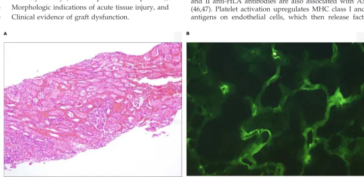
In AMR, alloantibodies preferentially attack the peritub-ular capillaries and glomerperitub-ular capillaries; by contrast, T cell-mediated rejection involves tubular, interstitial, and intimal infiltration of inflammatory cells (32-34). Acute cellular rejection can coexist with acute AMR. In many circumstances, AMR is mediated by activation of the classical complement pathway. The C4d biomarker is a degradation product of activated C4b, which is a classical component of complement. It is covalently bound to tissues and deposited in peritubular capillaries in AMR. C4d is diagnosed by immunohistologic staining. It is strongly associated with DSAs, helps confirm the diagnosis, and is the best marker of complement-fixing circulating antibodies [Figure 1A and 1B (34-36)].
Antibodies to class I and II HLA antigens are found in 88% to 95% of patients with C4d deposition and acute graft dysfunction (36). The deposition of C4d without circulating antibodies can be the result of absorption by the graft, as was proven by eluting anti-HLA antibodies from rejected grafts (21). Additional staining with C3d, a cleavage product of the complement component C3, may be useful in some cases (37). C4d deposition can occur beginning several years after transplantation, even though previous biopsies were C4d negative (34,38). C4d deposition is found in 2% to 26% of ABO-compatible, histologically normal renal allografts. The long-term importance of this deposition is unknown (39). Antibody-mediated rejection has been detected in C4d-negative grafts, and in such cases, evidence of microcircu-latory injury and the presence of class II DSAs portend a bad outcome (40).
The Banff criteria for diagnosing AMR are (41):
1) Circulating antibodies to donor MHC antigens, 2) Diffuse deposition of the complement split product
C4d in peritubular capillaries as an indicator of antibody activity (.50% of peritubular capillaries), 3) Morphologic indications of acute tissue injury, and 4) Clinical evidence of graft dysfunction.
Morphologic evidence of acute tissue injury includes 1) acute tubular injury, 2) neutrophils or monocytes in the peritubular capillaries or glomeruli, and 3) intimal arteritis, intramural or transmural inflammation, or fibrinoid necro-sis of the arteries. Antibody-mediated rejection is diagnosed in 1% to 6% of protocol renal biopsies in unsensitized patients and in 60% to 70% of patients with suspected acute rejection (34,38). Protocol biopsies have revealed diffuse peritubular capillary staining with C4d and DSAs with no histological evidence of injury. The importance of this finding is not clear, but some authors postulate that there may be accommodation to the graft (32,35). Accommodation is the acquired resistance of the allograft to immune-mediated injury (42).
Chronic AMR is characterized by injury to the glomerular and tubular basement membrane. Capillary injury is considered the initiating event, as evidenced by an up-regulation of the endothelial antigen, PV1 (plasmalemmal vesicle associated protein 1) (43). The glomerular lesion is termed ‘‘transplant glomerulopathy’’ and is characterized by thickened capillary loops and double contours. On electron microscopy, the glomerular basement membrane (GBM) shows reduplication and lamination. There are peri-tubular basement membrane multilayers with margination of mononuclear leukocytes. Ultimately, the peritubular capillaries are destroyed, resulting in tubular atrophy and interstitial fibrosis (44,45).
Transplant glomerulopathy has been described as the ‘‘ABCD’’ tetrad: Anti-donor antibodies, capillary Basement membrane multilayering, C4d deposition, and GBM Duplication (44). C4d deposition may or may not be present in chronic AMR, and donor-specific antibody concentrations usually fluctuate.
Other than these findings, complement-independent mechanisms caused by antibodies with specificities to MHC class II (expressed constitutively in the endothelial cells of capillaries in human kidneys) or a mixture of class I and II anti-HLA antibodies are also associated with AMR (46,47). Platelet activation upregulates MHC class I and II antigens on endothelial cells, which then release factors
Figure 1 - A)A 4-year-old child who had good allograft function initially and then developed acute antibody-mediated rejection 2 weeks after deceased donor kidney transplantation. Renal biopsy reveals marked acute tubular necrosis and interstitial hemorrhage. There is evidence of glomerulitis and tubulitis (H&E, 406).B)Immunofluorescence in this patient reveals diffuse C4d staining of the
that activate T-cells (48). Elevated endothelial- and adhe-sion-molecule-associated gene transcripts and antibodies against the GBM are implicated in the morphologic lesion of transplant glomerulopathy (49,50).
Small animal models of cardiac transplantation are frequently used because of the ease of surgery and vigor of rejection, although the concept of AMR is controversial in cardiac transplantation. Animal models suggest that the lesions occur because of chronic endothelial cell injury, and B-cell-deficient mice do not develop fibrous chronic allograft nephropathy (51). However, these models are limited by antigenic differences between murine and human organs (29,52).
ABO-Incompatible Renal Transplant
Historically, one of the main barriers to living kidney donation has been ABO incompatibility. In Japan, because of the lack of deceased donors, ABO-incompatible donors have been used in kidney transplantation, spurring the development of aggressive immunosuppressive protocols (53). The three-year survival in ABO-incompatible trans-plants in adults is similar to that of ABO-compatible kidney transplants. According to the 2010 report of the North American Pediatric Renal Trials and Collaborative Studies, 0.6% of childhood transplants are across ABO barriers (54). The main problem with ABO-incompatible transplants is the development of hemagglutinin antibodies to blood groups not present in the recipient (53,55,56).
Decreasing hemagglutinin antibody titers, caused by B-cell depletion therapies before transplantation, has improved the short- and mid-term survival of children with kidney transplants (53). Strategies to decrease these titers include treatment with rituximab, splenectomy, immunoadsorption of hemagglutinin antibodies, and intravenous immunoglobulin (IVIG) before transplant. However, acute rejection rates are higher than with ABO-compatible transplants, and long-term outcomes remain to be established (57).
New Therapies for Antibody-Mediated Rejection
Sensitization to HLA antigens limits access to and the success of transplantation. Pre-transplant desensitization protocols have made it possible to convert positive anti-HLA cross-matches to negative, thus enabling transplanta-tion in patients who otherwise could not have undergone transplantation. These patients can be managed with desensitization before transplant and treatment of AMR after transplant. Apheresis and IVIG-based protocols can convert a positive lymphocytotoxic cross-match to a negative one before transplant. These protocols have led to higher transplantation rates and improved short-term graft survival. Despite such protocols, however, many patients continue to experience clinical and subclinical AMR after transplant (14).
The protocols used for pre-transplant desensitization and post-transplant treatment of acute AMR are similar and are based on four concepts.
1. Eliminating or reducing circulating antibodies 2. Inhibiting residual antibodies
3. Suppressing or depleting B-cells 4. Suppressing T-cell response
Plasmapheresis can reduce the total IgG HLA antibodies. In one case series, some patients required 15 to 30 sessions
of plasmapheresis, alternating with IVIG, to substantially lower antibody titers (58). However, these treatments also remove clotting factors and require replacement with fresh frozen plasma and albumin. Anemia requiring packed red cell transfusion, bleeding diathesis, allergic reactions, and blood-borne infections are some of the complications associated with plasmapheresis. A Sepharose-bound sta-phylococcal protein A column with a high affinity for binding IgG is used in immunoadsorption. This technique has higher specificity compared with plasmapheresis and does not require replacing large volumes of plasma. Most columns used in Europe and Japan are not approved by the FDA (14). However, anti-HLA antibodies may rebound to baseline or higher levels a few weeks after both plasma-pheresis and immunoadsorption.
Inhibiting Residual Antibodies
Intravenous immunoglobulin inhibits the immune res-foponse in several ways, including neutralizing anti-HLA antibodies and inhibiting complement (14). Various proto-cols have been used to inhibit antibodies. Montgomery et al. used low-dose IVIG (100 mg/kg/day), alternating with plasmapheresis, as part of their desensitization and treat-ment protocol (58). The University of Maryland protocol uses 6 sessions of plasmapheresis, triple immune suppres-sion, and low-dose IVIG after each plasmapheresis in living kidney donors (59).
Jordan et al. initially used high-dose IVIG at 2 g/kg/ month until cross-matching was negative (60,61). However, they subsequently modified the protocol to 2 doses of IVIG and 1 dose of rituximab (62). The advantage of this method is its applicability to patients on the deceased donor list. A report of the use of high-dose IVIG in 2 highly sensitized patients showed both to be rejection-free with excellent renal function at 15 and 19 months, respectively (25).
Despite these desensitization protocols, the incidence of AMR remains high in the first year after transplant because these protocols have no effect on memory B-cells.
Suppressing or Depleting B-cells
Rituximab is a chimeric humanized monoclonal antibody against the cell surface marker, CD20, which is expressed in pre-B and mature B-cells. Rituximab destroys CD20-positive cells in several ways, including antibody-dependent cell-mediated cytotoxicity, complement-cell-mediated cytotoxicity, and apoptosis. Rituximab is generally combined with plasmapheresis, IVIG, or both because it is not effective against antibody-producing plasma cells if used alone (13,14,18,21). The response to rituximab in chronic anti-body-mediated-rejection is variable, and there is currently no way to distinguish responders from nonresponders (63). Infectious complications can increase when rituximab is used with other powerful immunosuppressive agents, such as anti-thymocyte globulin.
Splenectomy immediately reduces the B-cell and plasma cell pool and has been used as a last resort for salvaging transplanted kidneys (64,65). It has also been used in highly sensitized patients in whom desensitization therapy has failed.
Other Therapies
rituximab to treat children undergoing heart transplant (66). Plasmapheresis removes only some of the alloantibodies, and re-equilibration with extra vascular antibodies occurs in 48 to 72 hours. Rituximab blocks CD20-positive cells, and plasma cells do not express this marker. Therefore, bortezomib is useful in blocking anti-HLA antibody production. However, bortezomib alone may not be sufficient to reduce anti-HLA antibody levels because it requires activated plasma cells, such as those present in acute AMR (67). Moreover, it may not sufficiently target memory B-cells.
Eculizumab is an anti-complement C5 humanized mouse monoclonal antibody that prevents the formation of the membrane attack complex. It is based on evidence that activation of the terminal component of complement is necessary for the development of acute antibody-mediated rejection. It has been used as a rescue therapy in patients not responding to other treatments for AMR (68,69). Infectious complications, such as polyoma virus type BK nephritis, have been reported after its use. Stegall et al. reported decreased rates of acute and chronic AMR after treatment with eculizumab (69). Eculizumab does not affect DSA or C4d deposition, but it does decrease tissue injury and graft dysfunction (70). Experience with this drug is currently limited.
C1 inhibitor therapy may eventually be an option for treating refractory AMR. Its use is based on the theory that C1q-binding DSAs harm the allograft (71).
Secondary Immunodeficiency
While several reports have concluded that immunomo-dulatory therapies, such as IVIG–rituximab desensitization, do not significantly hinder cell-mediated or humoral immunity (72,73), the potential immunologic consequences of these and newer therapies used in desensitization or ‘‘antibody reduction therapies’’ (74) should not be over-looked, especially with long-term immunosuppression. Secondary immunodeficiency can be clinically significant, and it presents with recurrent fungal, bacterial, or viral infections, usually of the respiratory tract; however, it may include gastrointestinal infections and increased autoim-mune complications, depending on the extent of the immune suppression. Infections, including CMV, BK virus, and parvovirus B19, have been treated with IVIG in the post-transplant setting (74). The role of IVIG is both immunomodulatory at high doses and immune replacing at lower doses and may be required in the case of secondary hypogammaglobulinemia. Immune replacement with IVIG or subcutaneous immunoglobulin plays an important role in the post-transplant period for patients with recurrent infection because of secondary immunodeficiency. A consultation with an immunologist to coordinate replace-ment therapy may be an important part of the multi-disciplinary approach. With regard to IVIG protocols to prepare ‘‘sensitized’’ patients for transplant, a consensus approach does not yet exist. However, IVIG has certainly become a keystone of many empirically derived protocols, and a recent review of IVIG use in solid organ transplanta-tion endorsed its use in high-risk groups (75). An earlier review published in the US also recognized the use of IVIG in solid organ transplantation but cautioned that further studies are necessary to optimize use for this indication (76).
General Approach to Treating Acute Antibody-Mediated Rejection
Data from children treated with AMR are scarce (19,77,78). In highly sensitized patients, graft survival is better after desensitization and transplantation. The KDIGO (Kidney disease: improving global outcomes) guidelines (strength of evidence 2C) suggest treating acute AMR with some combination of plasma exchange, IVIG, and anti-CD20 antibody, with or without corticosteroids (79). Depending on the clinical response, a tiered approach to management has been suggested by various authors (Figure 2).
During treatment, antibody strength is monitored using an SPA. Threshold values for antibody levels depend on technical and immunosuppression protocols and are estab-lished by individual transplant centers (14-17). Antibody strength is measured by Luminex and is expressed as the mean fluorescence intensity or molecules of equivalent soluble fluorochrome. Antibody strengths are expressed as medium channel shifts in flow cytometry. In acute AMR, the mean fluorescence intensity values were greater than 5200 in most patients (14,17). Treatment is aimed at lowering the strength of the antibody.
Treating chronic AMR is difficult, and the optimal approach has yet to be identified because of its indolent course. High-dose IVIG and rituximab given to children with chronic AMR slowed the rate of decline in renal function in a single-center study (19).
Other Pre-emptive Measures
Paired organ sharing, such as kidney-paired donation, is a viable option when two donor-recipient pairs are blood type or cross-match incompatible with their intended recipients. The exchange of donor kidneys allows each donor to do-nate a kidney and each recipient to receive a compatible transplant. If this exchange works for specific donor-recipient pairs, less-intense immune suppression and better graft outcomes are expected (80).
Combined liver-kidney transplantation with a liver allograft decreases rejection rates and promotes rejection-free survival of the transplanted organs, even if the cross-match is positive before surgery (81). Combined organ transplantation has been extended to include partial auxiliary liver transplantation to make kidney transplanta-tion possible (82). However, AMR has been reported in the renal allograft in highly sensitized patients (83).
Summary and Future Translational Research Directions in Kidney Transplantation
enzyme inhibitors, statins, and aspirin could potentially modulate endothelial cell function (85). We also need to develop more sensitive, specific, and noninvasive biomarkers of the onset of AMR.
Bench to Bedside in Recurrent FSGS
The role of translational science in bridging the gap between ‘‘bench and bedside’’ is best demonstrated by the progress in unraveling the mechanisms of recurrent disease in renal transplantation. Perhaps the most ominous mechan-ism has been recurrent FSGS, a non-immune podocytopathy that is characterized by nephrotic-range proteinuria and variable progression to end-stage renal disease. Approximately 10% of children undergoing renal transplantation have FSGS as the primary diagnosis (86,87). Since FSGS was first recognized in 1972, the incidence in various ethnic popula-tions has remained approximately 30% (range, 20% to 67%) (86,88,89). Although ‘‘late’’ recurrence may occur years after transplantation, the most frequent and marked recurrence of FSGS is with massive proteinuria when the graft is reperfused at the time of surgery. If remission cannot be achieved by medical intervention, the rate of graft loss increases to more than 50% within the first year (90).
Early speculation that a ‘‘circulating permeability factor’’ (cPF) caused recurrent FSGS (R-FSGS) led to plasmapheresis as a primary treatment with mixed success (91-93). Plasmapheresis continues to be a component of both pre-emptive and maintenance treatment for R-FSGS and has led to the quest to identify circulating factors as new targets for more effective treatments (92-95).
Efforts to identify cPF(s) have been arduous and have involved the complex collaboration of basic and clinical physician scientists (96,97). Evidence for circulating factor(s) include:
1) The immediate recurrence of proteinuria when the graft is reperfused (97,98).
2) The improvement in proteinuria when the presumed cPF is removed with plasmapheresis or immunoad-sorption (99,100).
3) The regression of FSGS when a kidney with recurrent FSGS is re-transplanted into a patient without FSGS (101).
4) The induction of proteinuria in animals injected with a patient’s sera or with specific cPF (102-104).
5) The destruction of the podocyte foot process observed in transplanted kidneys biopsied within 1 to 2 hours after reperfusion in patients with recurrent proteinuria (105). 6) The disruption of the actin cytoskeleton in normal
human podocytes exposed to sera from patients with R-FSGS or to currently suspected cPFs (106,107).
Savin et al. pioneered the development of an assay to identify the character and function of the cPF by studying the sera from patients with R-FSGS (96). They developed an in vitro biological assay of glomerular permeability (91,92,96). Isolated rat glomeruli are incubated with a patient’s serum, plasma, or plasma fraction. The perme-ability of albumin to the glomerular membrane is deter-mined and expressed as the ‘‘albumin permeability index’’ (Palb), which ranges from zero in normal control serum to 1.0 for maximal induced injury. A Palb greater than 0.5 indicates marked injury to the glomerular protein barrier and is not specific to FSGS (92). For example, circulating cytokines and inflammatory markers, such as TNFa and b-1-integrin, may also generate a Palbgreater than 0.5. In patients at high risk for recurrence and in those with rapid progression to end-stage renal disease, a Palb greater than 0.5 has been highly predictive of outcomes (92,96). The cPF was further characterized by concentrating patient samples and applying protein isolation and fractionation techniques to obtain a molecular weight between 30 and 50 kD (108). More recently, the Savin group has identified cardiotrophin-like cytokine-1 (CLC-1) as a potential candidate cPF protein, but further studies are required (109).
A parallel effort using research in molecular biology, genetics, and clinically applicable mechanistic studies has been directed to understand the pathophysiology of proteinuria and is grounded in the work of Mundel and of
Reiser and colleagues (106,107,110,111). Much of this research has taken place at the University of Miami Transplant Institute during the past decade and has led to an intense global collaboration with physician scientists caring for patients with both primary and recurrent FSGS (110-115).
The integrity of the podocyte is paramount in maintaining the ‘‘barrier’’ against proteinuria and in the evolution of many progressive kidney diseases (110,111). This complex genetic, anatomic, and physiologic pathogenesis has become better understood during the past decade. The fundamental research began with Reiser et al. (110), who discovered the relationship between the stimulation of a receptor on the podocyte, termed the ‘‘B7-1’’ (also known as CD80), that, when stimulated in various pathologic settings, disrupted the integrity of the podocyte cytoskeleton and led to pathologic proteinuria and nephrotic syndrome (110).
In a summary of recent discoveries, Mundel and Reiser emphasize the basic physiology involved in the proteinuria and identify potential therapeutic targets (111). They hypothesize that there may be a common pathologic pathway involving the enzymatic cleavage of regulators of the podocyte actin cytoskeleton by cytosolic cathepsin L (111). This cleavage disrupts the podocyte actin cytoskeleton and causes the clinical syndrome of nephrotic proteinuria. In the early stages of nephrotic proteinuria, as in R-FSGS, these changes are potentially reversible. In essence, this work forms the basis of new discoveries in the pathogenesis and development of therapeutic targets for early interven-tion (116).
With this active translational research, the microanatomy of the podocyte has been elaborated, and a number of genetic, immune, and nonimmune diseases of the podocyte have been described (117,118). In concert with these discoveries has been the rapid and exciting evolution of potential therapies (109-115). New insights into the cause-effect relationship between cPF and podocyte injury in recurrent FSGS have come from identifying the soluble urokinase receptor (suPAR) as a cPF that can cause podocyte injury in recurrent FSGS (106,107) and from the discovery that sphingolipid-related enzymes are markedly affected in podocytes in R-FSGS (115). Other discoveries include the nonimmune target mechanism of action of cyclosporine (111,112) and rituximab (115) in preventing and modulating FSGS recurrence. Most recently, in our institution, four patients with severe R-FSGS who were nonresponsive to multiple interventions with plasmapher-esis, rituximab, and calcineurin inhibitors responded to the specific B7-1 receptor inhibitor, abatacept, with resolution of their proteinuria (119).
Despite recent advances in research related to primary and recurrent FSGS, observational and therapeutic trials are needed to better understand and treat this complex disease. Among the current studies are the NEPTUNE and FONT2 trials. The NEPTUNE (Nephrotic Syndrome Study Network) is a multicenter, observational trial whose purpose is to characterize the complex disease known as ‘‘steroid-resistant nephrotic syndrome.’’ The National Institutes of Health and private foundations, including the NephCure Foundation, fund it. FONT2 is a randomized, phase II trial comparing the efficacy of galactose with that of adalimumab for treating steroid-resistant FSGS (120).
In summary, scientific advances in understanding the pathogenesis of FSGS and its recurrence after kidney
transplant during the past decade have enabled the introduction of targeted therapies designed to improve patient outcomes.
& TRANSLATIONAL RESEARCH IN PEDIATRIC CARDIAC TRANSPLANTATION
Allograft rejection and complications related to the immunosuppression necessary for the survival of the transplanted organ remain the major causes of morbidity and mortality in cardiac transplant recipients. The mechan-isms of cardiac transplant rejection are similar to those of other solid-organ rejections and can be manifested through three modalities (121).
1) Acute cellular rejection mediated through T-cells invading and destroying allograft tissue.
2) AMR mediated through complement activation and triggered by antibodies directed against the HLA antigens of the donor.
3) Cardiac allograft vasculopathy (CAV) characterized by intimal thickening and remodeling of the coronary arteries. This type of rejection usually occurs many years after transplantation and is a major limiting factor in long-term allograft survival.
Currently, endomyocardial biopsy is the reference stan-dard for detecting graft rejection. However, it is invasive and limited by sampling error and interobserver variability. Additionally, Nakhleh et al. have established that histolo-gical expression of rejection is patchy in autopsied heart allografts; the foci of rejection were surrounded by large areas of intact myocardium (122). Other drawbacks of endomyocardial biopsy include the need for multiple samples to increase sensitivity, the inability to detect graft events in the period between biopsies, and its high cost (121). Endomyocardial biopsy also has complications, albeit rare, such as damage to the vessels, arrhythmias, conduction abnormalities, biopsy-induced tricuspid regurgitation, and even cardiac perforation (123-127).
Over the past few years, clinicians have looked into other noninvasive diagnostic tools, such as Doppler echocardio-graphy (128,129), high-resolution electrocardioechocardio-graphy (130), intramyocardial electrography (131), scintigraphy to detect antimyosin antibodies (132), and gene expression profiling in peripheral blood lymphocytes (133). Narula et al. studied the imaging of technetium-labeled annexin V using gamma cameras. Annexin V is an endogenous protein with a high affinity to phosphatidylserine, which is a phospholipid released during apoptotic cell death. The uptake of annexin V by the myocardium was associated with moderate grades of allograft rejection, suggesting the use of this imaging technique to detect rejection (134). However, although they may be able to detect rejection, these methods are not as effective as endomyocardial biopsy.
Biomarkers are anatomic, biochemical, or molecular characteristics of body fluids or tissues that indicate or are associated with clinically meaningful changes in physiology (135). To be clinically useful, they should be easily acquired and measured, valid, and have a sensitivity and specificity greater than other relevant technologies. They should also be relatively inexpensive and directly related to treatment decisions. Their validity should be established in prospec-tive multicenter studies (136).
In the field of transplantation, the use of biomarkers is rapidly evolving in two areas: 1) predicting and detecting allograft rejection and 2) detecting allograft tolerance for directing the weaning and proper adjustment of immuno-suppression (135).
Overview of Rejection and Cardiac Biomarkers
Rejection of the transplanted heart starts as a proinflam-matory state that ultimately leads to graft failure marked by the signs and symptoms of heart failure. The immunological mechanism of rejection begins when recipient T-cells recognize the graft antigens by non-self MHC type I and type II antigens and by the recruitment of cytotoxic T-lymphocytes. The CD4+T-cells then help the B-lymphocytes to produce antibodies against the MCH antigens. This step is followed by the release of chemical mediators and cytokines and the further proliferation and differentiation of T- and B-lymphocytes, which creates a vicious cycle of inflammation and alloreactivity. The combined actions of the T-cells, antibodies, and natural killer cells cause inflammation, necrosis, and fibrosis in the myocardium (137).
The current knowledge about biomarkers for detecting allograft rejection is derived from markers of different pathophysiologic processes, such as cardiac failure. We review below some of the biomarkers for inflammation, cardiomyocyte injury, and cardiomyocyte stress that have some potential to detect early rejection episodes in cardiac transplantation.
Inflammation
The first biomarker of inflammation was C-reactive protein (CRP), discovered in 1954 (6). A member of the pentraxin superfamily, CRP is a prototypical ‘‘acute phase’’ protein that is synthesized at a low rate under physiologic conditions but that is markedly induced and secreted after tissue injury and inflammation (138). It is produced predominantly by hepatocytes under the influence of cytokines, such as IL-6 or TNF-alpha.
In 1956, Elster et al. found elevated concentrations of CRP in patients with heart failure, and those with higher concentrations had more severe disease (139). Since then, many studies have verified that CRP is an important biomarker of inflammation. It became widely used when Ridker developed a low-cost and high-sensitivity assay in 2001 (140).
Venugopal et al. determined that CRP directly affects vascular endothelium by reducing nitric oxide release, increasing endothelin-1 production, and inducing the expression of endothelial adhesion molecule (141).
More recently, in a prospective observational study of 79 cardiac transplant recipients, Dolz et al. measured various inflammatory markers, such as fibrinogen, IL-6, TNF-alpha, sialic acid determinants, and CRP in endomyocardial biopsies performed during the first year after transplant. They concluded that CRP had the largest area under the
ROC curve, with concentrations less than 0.87 mg/dL being 90% specific for rejection and concentrations greater than 7.3 mg/dL being 100% sensitive for rejection. They identi-fied CRP as the most useful biomarker for the noninvasive screening of acute cellular rejection in the first year after heart transplantation (142). However, because its level is elevated in many inflammatory states, CRP is a nonspecific marker and does not always indicate cardiac rejection alone.
Cardiomyocyte Injury
Cardiomyocyte injury results from a variety of factors, such as ischemia, inflammation, neurohormonal activation, and oxidative stress. Over the past two decades, myofi-brillar proteins —the cardiac troponins I and T—have emerged as sensitive and specific markers of cardiomyocyte injury and have proven useful in stratifying the risk of coronary syndromes.
In a recent pilot study, Dyer et al. evaluated the use of a high-sensitivity assay for cardiac troponin to detect acute graft rejection (AR) in children with heart transplants. Plasma samples for measuring cardiac troponin T were drawn at the same time as endomyocardial biopsy. Children with AR had much higher cardiac troponin T levels than those without AR. On follow-up, troponin concentrations decreased as the rejection was resolving. These researchers concluded that cardiac troponin could be a useful biomarker for monitoring rejection in transplant recipients (143).
Cardiomyocyte Stress
Several other biomarkers of myocardial stress have been studied. Brain natriuretic peptide (BNP) is a 134-amino acid molecule synthesized by cardiomyocytes in response to ventricular dilatation and increased wall tension. It is then cleaved into pro-hormone BNP, which consists of 108 amino acids. A circulating endoprotease, termed ‘‘corin,’’ further cleaves the pro-hormone into two polypeptides: the inactive N-terminal proBNP (NT-proBNP) and the bioactive peptide BNP (144).
The physiologic effects of BNP include arterial vasodila-tation, diuresis, natriuresis, inhibition of the renin-angio-tensin-aldosterone system, and inhibition of sympathetic nervous activity (144). Today, BNP and NT-proBNP are the most widely used biomarkers for heart failure and are being evaluated for their ability to detect graft rejection in patients with cardiac transplants.
Dyer et al. were the first to study NT-proBNP as a biomarker of graft rejection in children receiving trans-plants. In this prospective observational study of 42 children (mean age, 11 years), high-sensitive cardiac troponin T (hscTnT) and NT-proBNP were assayed at the time of endomyocardial biopsy. Acute rejection was defined as an International Society for Heart & Lung Transplantation (ISHLT) grade of 2 or higher. Median (25thto 75thpercentile) hscTnT and NT-proBNP concentrations were higher in children with acute rejection than in those without: hscTnT, 66 (45 to 139) vs. 7 (2 to 13) pg/mL; p= 0.001 and NT-proBNP, 11,169 (280 to 23,317)vs. 334 (160 to 650) pg/mL; p,0.01). They concluded that elevations in NT-proBNP concentrations are associated with episodes of rejection in these children (143).
Chronic Rejection and Cardiac Allograft Vasculopathy
Allograft vasculopathy is a form of chronic rejection that is associated with graft failure and re-transplantation in both adults and children after heart transplantation (146,147). There is clearly a need to understand and detect this process as early as possible.
Allograft vasculopathy is characterized by remodeling of the coronary arteries through diffuse fibromuscular intimal hyperplasia and focal atherosclerosis. Endothelial damage is caused by a combination of alloimmune and non-alloim-mune responses and other factors that induce endothelial activation and death. Endothelial damage is concomitant with endothelial repair. During this process of damage and repair, apoptotic circulating endothelial cells and endothe-lial microparticles are released into the circulation. During the regeneration process, endothelial progenitor cells are released. All these circulating endothelial components may become useful biomarkers of CAV (148).
Singh et al. studied 52 cardiac transplant patients under-going coronary angiography between 5 and 15 years after transplant. In a logistic regression model, apoptotic circulat-ing endothelial cells (p= 0.011) and apoptotic endothelial microparticle concentrations (p= 0.014) were independent predictors of vasculopathy (C statistic, 0.86; 95% confidence interval, 0.76 to 0.95). This research is an initial step in predicting CAV and in determining the need for coronary angiography and intravascular ultrasound when it is suspected.
Biomarkers of rejection have been evaluated in several studies. However, the need for larger, multicenter trials remains. Validated biomarkers will detect allograft rejection before acute clinical decompensation and graft failure. However, few biomarkers have yet become commercially available.
In addition to detecting early rejection, clinicians must also manage the severe immunosuppression required for these transplant recipients. In the next sections, we review the present and future efforts being made to address this challenge.
Tolerance in Transplantation
Rejection of a transplanted allograft is an immunologi-cally mediated process that occurs because of differences between donor and recipient concentrations in HLA. Allorecognition is the term for the immunological recogni-tion of histo-incompatible antigens between genetically disparate individuals from the same species. Alloresponse
in transplantation is the beginning of an adaptive immune response in which allospecific T-cells are recruited. This alloresponse rejects the transplanted organ in nontolerant or inadequately immunosuppressed individuals (149).
To protect against alloresponse, cardiac transplant reci-pients must take immunosuppressive medications, which in the long run increase their susceptibility to infection, malignancy, and accelerated cardiovascular disease, thereby increasing their morbidity and mortality.
A new direction in preventing alloresponse has been to induce ‘‘transplant tolerance,’’ which would avoid the need for immunosuppression and its associated adverse effects. Transplant tolerance is defined as a well-functioning graft that lacks histological signs of rejection in an immunocom-petent host in the absence of any immunosuppressive drugs (150).
‘‘Operational transplant tolerance’’ is a clinical situation defined as stable graft function without clinical features of chronic rejection and in the absence of any immunosup-pressive drugs, usually for longer than 1 year (151). However, current tests and biomarkers cannot indicate tolerance to the graft. Validated indicators or biomarkers of immunologic tolerance and biomarkers able to predict and diagnose graft dysfunction, acute and chronic rejection, and the level of immunosuppression potentially allow for individualized therapy and the safe minimization or with-drawal of immunosuppressive therapy in certain transplant recipients (150).
Experimental models of the immune system’s response to transplantation have identified two key events that lead to tolerance in the recipient: the deletion of a considerable fraction of alloreactive cells and the development of T-regulatory cells that protect the graft from being attacked by the immune system (150). Some evidence indicates that the terminal pathway in destroying the transplanted organ, as occurs in acute rejection, is the same pathway that destroys tissues in many other inflammatory processes, such as immune-mediated tumor rejection, autoimmune disease, cardiovascular events, chronic obstructive pulmonary dis-ease, and infection clearance. This common pathway has been called the ‘‘immunologic constant of rejection’’ (152).
Microarrays that quickly analyze thousands of genes have been used to study the mechanisms and pathways of acute rejection in organ transplantation. In an interesting meta-analysis, Spivey et al. reviewed the literature for all the most common pathways of acute allograft rejection in humans, as determined by microarray technology. The pathways reported in the immunologic constant of rejection hypoth-esis are the same pathways as those occurring in acute allograft rejection. Microarray analysis may help to deter-mine the mechanism that controls the balance between tolerance and rejection (153). Because of the large number of genes that can be analyzed, the microarray technique can be used to study the mechanism of activation of the immuno-system directly in the human body, bypassing experiments in animal models.
Currently, new techniques that can rapidly measure and analyze large amounts of different biological substances and that will help identify new biomarkers include the following:
1) Genetic analysis with real-time PCR, microarrays, and genome-wide association.
3) Mass spectrometry for measuring and identifying proteins.
Genetic Analysis
Real-time PCR can test a small number of genes with high sensitivity and specificity. Microarrays can quickly analyze thousands of genes and determine whether they are activated or suppressed. Microarray analysis is more cost-effective then real-time PCR for studying a large number of genes, but its sensitivity and specificity are lower. Since 2001, microarrays have been used in various solid-organ transplant studies to identify specific patterns of gene expression that can predict and characterize acute and chronic rejection and transplant tolerance (154). Chen et al. used microarray analysis of the biopsies of transplanted hearts in acute rejection to identify 45 upregulated genes that may be correlated with rejection (155).
Genome-wide association studies can be described as whole-genome scans that can identify single nucleotide polymorphisms in a small amount of DNA (156). The theory is that the association between a polymorphism and a disease phenotype can be used as a genetic marker to aid in diagnosis and prognosis. Transplant centers in the UK and Ireland are currently using this type of analysis to identify genes for kidney transplant failure (157). If this technique is applied to a cardiac transplantation population, it may also identify clinically useful biomarkers in that population.
Gene Expression Profiling
Applying genetic testing to cardiac transplantation has brought about a new, noninvasive test for detecting acute cellular rejection. Gene expression profiling (GEP) is being investigated as a potential adjunct to, or even substitute for, endomyocardial biopsy for monitoring acute cellular rejec-tion of cardiac allografts in certain clinical situarejec-tions. Among the more than 15,000 biomarkers studied so far in relation to solid-organ transplantation, only 2 are approved by the FDA and are commercially available: the AlloMapH and CylexHImmuKnowTMtests (121).
The AlloMapH test uses quantitative real-time PCR to measure the expression of 20 genes (11 informative, 9 control and normalization) in peripheral blood mono-nuclear cells (133,158). The test was developed on the premise that peripheral blood mononuclear cells may reflect host responses to the allograft. It has been available for clinical use through the Clinical Laboratory Improvement Amendment certified XDx, Inc. reference laboratory since January 2005 (159).
The AlloMapHtest was developed using DNA microarray technology and validated with quantitative real-time PCR. The 11 informative genes represent several biologic path-ways, including T-cell activation (PDCD1), T-cell migration (ITGA4), mobilization of hematopoietic precursors (WDR40A andcMIR), and steroid-responsive genes (ILIR2, FLT3, and ITGAM). The remaining nine AlloMapH genes are controls for test accuracy and reproducibility (133,158). A score from 0 to 40 is generated using a multigene algorithm. The test was developed and validated through the Cardiac Allograft Rejection Gene Expression Observational (CARGO) study, with the aim to distinguish between a quiescent state (grade 0R by the revised ISHLT grading) and moderate-to-severe rejection (a grade of 3A/2R or higher by the original and revised ISHLT grading of rejection, respectively).
Importantly, GEP could detect the absence of moderate-to-severe rejection and thus identify a state of quiescence, with score thresholds varying with the time after transplant (.2 to
#6 months,.6 to#12 months, or.12 months).
The test has a high negative predictive value, which ranges from approximately 98% to 99% for scores consid-ered to be appropriate thresholds in each of the three time periods after transplant (133,158,159). One year after transplantation, scores of less than 34 were associated with a negative predictive value of more than 99% for grade 3A/ 2R rejection, suggesting the clinical utility of this test is its ability to rule out acute cellular rejection (158,159). The positive predictive values for GEP of 34 or more were low, however, with values decreasing from 20% to 40% during the first 6 months after transplant to approximately 7.8% at 1 year after transplantation. This decline in PPV is thought to be related to the decrease in the incidence of acute rejection with time from transplantation (133,158,159). These findings indicate that GEP has a high sensitivity and low PPV for detecting acute cellular rejection over time.
AlloMapH scores and the histologic results of cardiac biopsies sometimes differ. High GEP scores in the setting of negative biopsy results is the more common phenomenon and may be caused by early or focal rejection that may be missed on biopsy as a result of sampling error. Additionally, immune processes related to conditions other than acute cellular rejection (e.g., AMR, infection, cardiac allograft vasculopathy, or chronic rejection) may be associated with higher AlloMapH scores. AlloMapH scores also tend to increase with time after transplantation, even with quies-cence, and values must be interpreted in this context. Higher GEP scores in the setting of quiescence are thought to be related to down-titration of corticosteroids and other immunosuppression. However, the clinical importance of an AlloMapH score above a threshold in the setting of quiescence is as yet unknown (133,158,159).
AlloMapH testing is currently being used in clinically stable cardiac transplant recipients aged 15 years or older at 6 months or more after transplantation to identify those at low-risk for moderate or severe (Grade $3A/2R) cellular rejection. The test has also been used as an adjunct to clinical evaluation and endomyocardial biopsy in monitoring for rejection and in lieu of biopsy in patients 6 months or more after transplant who are at low risk of rejection (159,160). Its application should be individualized and the results interpreted in the context of the patient’s overall clinical status and risk of acute rejection. The frequency of surveillance should be individualized and a thorough clinical evaluation performed at the time of testing, including echocardiographic assessment of allograft func-tion (159). Thresholds should also be individualized and decisions regarding biopsy or changes in immunosuppres-sion made according to each patient’s risk for acute rejection.
The AlloMapHtest has distinct advantages over biopsy in that it is a less-invasive method for monitoring acute cellular rejection in cardiac allograft recipients. Its genetic basis also has the advantage of having the potential to predict episodes of acute rejection, which may help to guide risk stratification, monitoring, and treatment (161,162). The clinical usefulness also extends to its applicability in managing immunosuppressive regimes to minimize the risk and complications of toxicity while also minimizing the risk of rejection.
The importance of GEP scores in cardiac allograft vasculopathy and in AMR are also areas of investigation (163). These studies are, however, limited by small patient numbers, and their usefulness in children is limited by the fact that most results are from data in adults. AlloMapH scores in patients less than age 15 years remain to be validated.
Adenosine Triphosphate Release as a Measure of Immune Function
The CylexH ImmuKnowTM cell function assay (CICFA) has been promoted as a way to monitor the integrity of cell-mediated immunity. The assay measures the adenosine triphosphate (ATP) concentrations in CD4+ T-cell
lympho-cytes after stimulation with phytohemagglutinin-L. High CICFA concentrations correspond with immune compe-tence and low concentrations with immune suppression (164). In adult solid-organ transplant patients, low CICFA concentrations were associated with a risk of infection, and high CICFA concentrations were associated with a risk of acute organ rejection (165). In an observational study of 296 heart transplant patients, Kobashigawa et al. found that patients with assay results below normal were at higher risk for infectious complication related to elevated immunosup-pression, but attempts to predict rejection were inconclusive (166).
There have, however, been conflicting results. A retro-spective study of 111 adult heart transplant patients found that CICFA did not predict either infection or rejection (167). In this study, two patients had three episodes of cellular rejection, and the CylexH response did not correlate with these periods of rejection. Few studies have evaluated the use of CICFA in children. Hooper et al. established the normal ranges of immune assay concentrations in healthy children, both those less than and greater than 12 years old, and in stable renal transplant recipients greater than 12 years old (168). These ranges are the basis for studying CylexH ImmuKnowTM assays in children. Gautam et al. used the ImmuKnowTM assay to adjust the dosing of immunosuppressive medications in a child with lympho-proliferative disorder after kidney transplantation (169). The assay can thus be used to predict the degree of immuno-suppression and to adjust medications in young transplant recipients.
However, the use of the ImmuKnowTMassay in pediatric heart transplantation is very limited. We found only one study that assessed its use in children with heart trans-plants. The retrospective study by Rossano et al. of 83 children with heart transplants reported 20 episodes of cellular rejection, but ATP concentrations measured by the ImmuKnowTMassay did not differ between children with and without rejection. The authors concluded that CICFA concentrations did not predict acute rejection or clinically important infections (170). Thus, given these conflicting
results, larger multicenter studies are needed to determine whether ATP concentrations can aid in titrating immuno-suppressive therapy and in reducing adverse effects.
Genome Transplant Dynamic
Because rejection is associated with apoptosis and cell death and because free DNA is released into plasma, Snyder et al. used single nucleotide polymorphism (SNP) technol-ogy to measure the presence of free donor DNA as a percent of recipient DNA in the plasma of transplant recipients at the time of endomyocardial biopsy. The correlation between the percentage of donor DNA and rejection increased with the severity of the rejection. Furthermore, these investiga-tors also found that an elevation in donor DNA was detected even before biopsy-proven rejection and that it returned to normal after treatment. On the ROC curve, a threshold of 1.70% donor DNA identified grade 2R rejection with an 83% true positive rate and a 16% false positive rate (171). A test that directly measures the extent of cardiac damage, along with the AlloMapHtest, which measures the signal of the host immune system indicating rejection, may further increase the sensitivity and specificity of detecting rejection.
Mass Spectrometry
Mass spectrometry can analyze proteins in different body fluids and now includes tandem spectrometry to obtain new protein sequence information (172). Mass spectrometry of bronchoalveolar lavage fluid has proved useful in diagnos-ing bronchiolitis obliterans syndrome in lung transplant recipients with 94% specificity and 74% sensitivity (173).
Flow Cytometry
Flow cytometry and Luminex-based techniques measure specific cells and their subsets along with the production of cytokines and other cell functions. These techniques are useful in transplantation (174). Measuring the concentra-tions of T- and B-lymphocytes and their subsets can help determine the level of immunosuppression and the response to immunosuppressive treatment. The diagnostic specificity and the amount of antibodies can also be determined. Limitations include inter-laboratory variability and the inability to cover all the alleles in the population. Antibodies against HLA in the transplanted organ are associated with chronic rejection and a lower rate of organ survival. In 243 cardiac transplant patients with no DSAs before transplantation, Smith et al. found that the develop-ment of DSAs was associated with a marked decrease in survival (hazard ratio, 4.35), independent of the ability to fix complement. More patients with DSAs died of CAV or acute rejection than did patients with non-DSAs. Although the authors were not able to show a cause–effect relation-ship between DSAs and CAV, having both DSAs and CAV was associated with a worse prognosis. The results of this study also emphasized the importance of regular monitor-ing for the presence of DSAs, startmonitor-ing in the first year after transplantation (175).
Concentrations of alloreactive T-cells derived from the memory pool can also be used to predict outcomes in transplant patients. Ongoing multicenter studies in heart and kidney transplant recipients are assessing the feasibility and standardization of measuring memory T-cells to guide the management of immunosuppressants. However, the high cost and complexity of this technique are major obstacles to its widespread implementation (135).
Summary and Future Translational Research Directions in Heart Transplantation
In this review, we have presented current challenges and approaches to the care of children who have received a heart transplant. Specifically, we have reviewed the mechanisms of cardiac transplant rejection and the roles of endomyo-cardial biopsy and biomarkers, such as biomarkers of inflammation, cardiomyocyte injury, or stress, in detecting cardiac graft rejection. We have also reviewed CAV. Lastly, we addressed the role of genetic analyses, including genome-wide association studies, gene expression profiling using entities like AlloMapH, and adenosine triphosphate release as a measure of immune function using the CylexH ImmuKnowTMcell function assay.
Clearly, there has been significant progress in the care of post-transplant pediatric patients using the approaches described above. One theme that has emerged is the importance of further investigations of existing cardiac biomarkers, whether circulating proteins or genetic biomar-kers, and identifying novel cardiac biomarkers to better predict the clinical course after heart transplantation in children. The goal is to minimize the need for resource-intense and potentially risky myocardial biopsies and to better guide therapeutic interventions. Beyond 1 year of life, most pediatric heart transplants occur in children with cardiomyopathy. Recent reports show the knowledge gap regarding cardiac biomarkers in pediatric cardiomyopathy, heart failure, or congenital cardiovascular malformations, which results from a lack of large multisite clinical trials (177). If noninvasive biomarkers could reliably improve clinical monitoring and prognosis, both before and after heart transplantation, they could serve as important surrogate endpoints in both observational studies and clinical trials. New investigations into mechanistic path-ways, as identified by existing and novel cardiac biomarkers and recipient genotypes as biomarkers (including the expression of genetic polymorphisms), could result in important advances in the monitoring of these children and result in improved outcomes for children receiving a heart transplant (178,179).
While continued translational research regarding mechanistic and genetic studies in children who have received a heart transplant is vital to improved patient outcomes, there is a complementary research approach that also needs to be vigorously pursued to result in the best evidence-based approach to the evaluation, monitoring, and decision making regarding the listing of these children for heart transplant. Many risk factors and outcome predictors in children eligible for cardiac transplantation can be identified early in these children’s clinical course. Since the mid-1990s, the National Heart, Lung, and Blood Institute-funded Pediatric Cardiomyopathy Registry (PCMR) has conducted epidemiological and longitudinal follow-up studies of thousands of children with cardiomyo-pathy, which is the most common indication for heart
transplantation after infancy (180,181). During this period, the PCMR has reported risk factors for either death or heart transplant using their pre-transplant dataset. Additionally, the PCMR has combined their data with the peri- and post-transplant data from the Pediatric Heart Transplant Study (PHTS) (182-184) to better characterize the clinical course and risk factors for the outcomes of these children from cardiomyopathy diagnosis to post-cardiac transplant follow-up.
With regard to pediatric cardiomyopathy, the PCMR and the PCMR-PHTS collaborations have identified the follow-ing predictors of the risk of transplant or death, which have not previously been reported. These results could result in better evidence-based decisions regarding listing for heart transplantation by pediatric cardiologists. In children with dilated cardiomyopathy, those with a familial or myocardi-tis etiology had a very good survival experienceversusother etiologies, particularly idiopathic dilated cardiomyo-pathy (185). In a competing risk analysis of children with idiopathic dilated cardiomyopathy, age .6 years at diag-nosis, heart failure, left ventricular dilation, decreased left ventricular function, and decreased height-for age predicted worse clinical outcomes (186). However, in this same group, increased left ventricular dilation was predictive of an increased risk of transplantation but not death. The conclusion was that left ventricular dilation may be over emphasized in listing decisions in idiopathic cardiomyo-pathy patients by pediatric cardiologists, whereas linear growth retardation may not be considered in listing decisions, despite its risk for increased mortality.