Hypothalamic transcriptional expression of the
kis-speptin system and sex steroid receptors differs among
polycystic ovary syndrome rat models with different
endocrine phenotypes
Rodrigo Rodrigues Marcondes,I,*Ka´tia Caˆndido Carvalho,IGisele Giannocco,II Daniele Coelho Duarte,I Nata´lia Garcia,IJose´ Maria Soares-Junior,I Ismael Dale Cotrim Guerreiro da Silva,III Manuel Maliqueo,IV Edmund Chada Baracat,IGustavo Arantes Rosa MacielI,*
ILaboratorio de Ginecologia Estrutural e Molecular (LIM 58), Disciplina de Ginecologia, Faculdade de Medicina FMUSP, Universidade de Sao Paulo,
Sao Paulo, SP, BR.IILaboratorio de Endocrinologia Molecular e Translacional, Departamento de Medicina, Universidade Federal de Sao Paulo, Sao Paulo,
SP, BR.IIILaboratorio de Ginecologia Molecular e Proteomica, Departamento de Ginecologia, Escola Paulista de Medicina, Universidade Federal de Sao
Paulo, Sao Paulo, SP, BR.IVEndocrinology and Metabolism Laboratory, Department of Medicine, West Division, University of Chile, Santiago, Chile.
OBJECTIVES: Polycystic ovary syndrome is a heterogeneous endocrine disorder that affects reproductive-age women. The mechanisms underlying the endocrine heterogeneity and neuroendocrinology of polycystic ovary syndrome are still unclear. In this study, we investigated the expression of the kisspeptin system and gonadotropin-releasing hormone pulse regulators in the hypothalamus as well as factors related to luteinizing hormone secretion in the pituitary of polycystic ovary syndrome rat models induced by testosterone or estradiol. METHODS:A single injection of testosterone propionate (1.25 mg) (n=10) or estradiol benzoate (0.5 mg) (n=10) was administered to female rats at 2 days of age to induce experimental polycystic ovary syndrome. Controls were injected with a vehicle (n=10). Animals were euthanized at 90-94 days of age, and the hypothalamus and pituitary gland were used for gene expression analysis.
RESULTS:Rats exposed to testosterone exhibited increased transcriptional expression of the androgen receptor and estrogen receptor-band reduced expression of kisspeptin in the hypothalamus. However, rats exposed to estradiol did not show any significant changes in hormone levels relative to controls but exhibited hypothalamic downregulation of kisspeptin, tachykinin 3 and estrogen receptor-agenes and upregulation of the gene that encodes the kisspeptin receptor.
CONCLUSIONS: Testosterone- and estradiol-exposed rats with different endocrine phenotypes showed dif-ferential transcriptional expression of members of the kisspeptin system and sex steroid receptors in the hypothalamus. These differences might account for the different endocrine phenotypes found in testosterone-and estradiol-induced polycystic ovary syndrome rats.
KEYWORDS: Polycystic Ovary Syndrome; Hypothalamus; Animal Models; Kisspeptin; Testosterone.
Marcondes RR, Carvalho KC, Giannocco G, Duarte DC, Garcia N, Soares-Junior, et al. Hypothalamic transcriptional expression of the kisspeptin system and sex steroid receptors differs among polycystic ovary syndrome rat models with different endocrine phenotypes. Clinics. 2017;72(8):510-514
Received for publication onNovember 3, 2016;First review completed onJanuary 16, 2017;Accepted for publication onMarch 14, 2017
*Corresponding authors. E-mail: [email protected]/[email protected]
’ INTRODUCTION
Polycystic ovary syndrome (PCOS) is a highly prevalent heterogeneous condition mainly characterized by hyperan-drogenism, chronic anovulation and polycystic ovaries (1).
Another abnormality that is common in women with PCOS is aberrant gonadotropin-releasing hormone (GnRH) secre-tion, which favors higher production of luteinizing hormone (LH) and an increase in androgen production by the ovaries (2). However, the precise mechanisms underlying LH hyper-secretion in PCOS are not well known.
The kisspeptin system, which includes kisspeptin, dynor-phin A and neurokinin B/tachykinin 3, is essential for GnRH/LH pulse control, and it is believed that impairments in this system might contribute to endocrine dysfunction in PCOS (3). Furthermore, evidence suggests that neuroendocrine dysfunction in insulin signaling alters GnRH/LH secretion and impair reproductive cyclicity (4).
DOI:10.6061/clinics/2017(08)09
Copyright&2017CLINICS–This is an Open Access article distributed under the terms of the Creative Commons License (http://creativecommons.org/licenses/by/ 4.0/) which permits unrestricted use, distribution, and reproduction in any medium or format, provided the original work is properly cited.
Exposure to either estrogen or androgens in neonatal life can induce chronic anovulation and polycystic ovaries resembling human PCOS during rat adulthood (5). However, different exposures may result in various endocrine pheno-types (5). For instance, animals exposed to androgens dur-ing the neonatal period present increased levels of LH and testosterone. This phenotype is not observed in the estrogen model (6). The aim of this study was to investigate tran-scriptional changes in the kisspeptin system and GnRH pulse regulators in the hypothalamus as well as factors related to LH secretion in the pituitary in PCOS rat models induced by testosterone or estradiol.
’ MATERIALS AND METHODS
Animals
The experimental procedures were described previously (6). In brief, thirty female Wistar rats at 2 days of age were allocated into the following groups for the induction of experimental PCOS: subcutaneous injection of 1.25 mg of testosterone (TG; n=10) (7) or 0.5 mg of estradiol (EG; n=10) (8). Control animals received a single injection of olive oil (vehicle) (CG; n=10). At 90-94 days of age, under anesthesia, blood was obtained from the abdominal aortic artery, ani-mals were decapitated, and the hypothalamus and pitui-tary gland were removed and stored in RNAlater solution (Ambion; Thermo Fisher Scientific, USA) for 24 h and frozen until use. The measurement of serum levels of LH, FSH and testosterone is described in our previous study (6). This study was approved by the Institutional Ethics Committee of Faculdade de Medicina da Universidade de São Paulo under protocol number 151/10.
Quantitative real-time polymerase chain reaction (qRT-PCR)
RNA extraction, cDNA synthesis and qRT-PCR conditions were performed as described elsewhere (9). Taqmans
assays manufactured by Life Technologies were selected to analyze the gene expression ofGnrh,Gnrhr,Kiss1,Kiss1r,Tac3,Tacr3,
Esr1,Esr2,Ar,Cyp19a1,PdynandOprk1in the hypothalamus andGnrhr,Kiss1,Kiss1r,InsrandArin the pituitary (Table 1).
The data were analyzed using Sequence Detection Software (version 2.0.6), and Ct values were transformed to quantities using the comparative Ct method (DDCt).
Statistical analysis
Statistical analyses were performed with GraphPad Prism (version 6.05; GraphPad Software, USA). ANOVA followed by the Bonferronipost hoctest was performed to analyze data with a normal distribution. For skewed data, the Kruskal-Wallis test followed by Dunn’spost hoctest was performed. A value ofpo0.05 was considered to indicate statistical
sig-nificance. F-ratios (F) are described for significant differences. The degrees of freedom was equal to 29 for all analyses.
’ RESULTS
Treated rats had closed vaginas, and all rats in the CG had normal estrous cycles. In the hypothalamus, kisspeptin (Kiss1), neurokinin B (Tac3), and estrogen receptor (ER)-a (Esr1) were downregulated in the EG relative to the CG (p=0.0001, F=35.92;p=0.01, F=5.193; and p=0.0015, F=8.342, respectively). The expression levels of Kiss1 and GnRH receptor (Gnrhr) were lower in the EG than in the TG (Kiss1:
p=0.01, F=35.92;Gnrhr:p=0.01, F=6.748), and the expression of the gene encoding the KISS1 receptor (Kiss1r) was higher in the EG than in the CG (p=0.007, F=5.142).Kiss1was also expressed at a lower level in the TG than in the CG (p=0.001, F=35.92). TG rats exhibited upregulation of the androgen receptor (Ar) and ER-b(Esr2) genes compared to CG animals (p=0.04, F=3.522; and P=0.02, F=4.497, respectively). The expression of Gnrh, Oprk1, Pdyn, Tacr3 and Cyp19a1 did not differ significantly among the groups (Figure 1A). No significant difference was observed in pituitary gene expres-sion (Figure 1B), and Kiss1 and Kiss1r mRNA levels were undetectable.
’ DISCUSSION
Our group showed in a previous study that testosterone or estradiol exposure in neonatal life leads to different PCOS-like phenotypes in adult female rats. Testosterone-induced PCOS rat models exhibit increased LH and testosterone
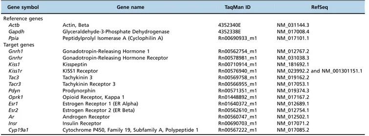
Table 1-Description of the probes and primers used in the study.
Gene symbol Gene name TaqMan ID RefSeq
Reference genes
Actb Actin, Beta 4352340E NM_031144.3
Gapdh Glyceraldehyde-3-Phosphate Dehydrogenase 4352338E NM_017008.4
Ppia Peptidylprolyl Isomerase A (Cyclophilin A) Rn00690933_m1 NM_017101.1 Target genes
Gnrh1 Gonadotropin-Releasing Hormone 1 Rn00562754_m1 NM_012767.2
Gnrhr Gonadotropin-Releasing Hormone Receptor Rn00578981_m1 NM_031038.3
Kiss1 Kisspeptin Rn00710914_m1 NM_181692.1
Kiss1r KISS1 Receptor Rn00576940_m1 NM_023992.2 and NM_001301151.1
Tac3 Tachykinin 3 Rn00569758_m1 NM_019162.2
Tacr3 Tachykinin Receptor 3 Rn00566955_m1 NM_017053.1
Pdyn Prodynorphin Rn00571351_m1 NM_019374.3
Oprk1 Opioid Receptor, Kappa 1 Rn01448892_m1 NM_017167.2
Esr1 Estrogen Receptor 1 (ER Alpha) Rn01640372_m1 NM_012689.1
Esr2 Estrogen Receptor 2 (ER Beta) Rn00562610_m1 NM_012754.1
Ar Androgen Receptor Rn00560747_m1 NM_012502.1
Insr Insulin Receptor Rn00690703_m1 NM_017071.2
levels, anovulation, and polycystic ovaries, while estradiol-induced PCOS rat models exhibit anovulation and polycystic ovaries but no alterations in the serum levels of gonado-tropins or testosterone (6). In the current study, we aimed to further understand the heterogeneity of PCOS endocrine phenotypes by studying the hypothalamic neuroendocrine transcriptional profile of the kisspeptin system and GnRH pulse regulators as well as factors related to LH secretion in the pituitary of these two PCOS rat models.
It has been hypothesized that androgenized rat models have increased GnRH expression (10), and this event, in part, seems to be mediated by the interaction of androgens with androgen receptors (ARs) in the hypothalamus (11). Feng et al. (10) have shown that the AR is co-expressed in GnRH-neurons of adult female rats. In rats exposed to androgens in this study, an upregulation of Aroccurred in the hypotha-lamus. In vitro, AR activation has been shown to increase GnRH-neuron firing activity and GnRH secretion (12, 13).
Kisspeptin is an important factor involved in the stimula-tion of GnRH/LH secrestimula-tion as well as the GnRH/LH surge that induces ovulation (3). Androgen and estrogen exposure downregulate kisspeptin gene expression, with this effect being more pronounced in estrogen-exposed rats. The down-regulation of kisspeptin might be related to anovulation, because those animals seem to be unable to generate the GnRH/LH surge (6). In contrast, the KISS1 receptor gene
was upregulated in rats exposed to estrogen, which is possibly a compensatory mechanism for low levels of kisspeptin.
Despite downregulation of the kisspeptin gene, androgen-exposed rats presented increased levels of LH. However, GnRH pulsatility, which stimulates gonadotropin secretion in a GnRH pulse-dependent manner, as well as LH secretion, is modulated by many factors (14). Other factors might be involved in LH hypersecretion in androgen-exposed rats.
reproduction is not completely known, but this receptor also interacts with an androgen metabolite called 3b-diol (18). However, the role of this metabolite in the GnRH or kiss-peptin system is still unknown.
Neurokinin B is considered an important factor in the modulation of GnRH secretion because loss-of-function mutations in the genes encoding both neurokinin B and its receptor induce hypogonadotropic hypogonadism (19). Fur-thermore, the central administration of a neurokinin B receptor agonist increases LH secretion in female rats (20). Downregulation of the neurokinin B gene (Tac3) may be related to the state of anovulation in estradiol-exposed rats and the lower levels of LH in the estradiol-exposed rats relative to the androgen-exposed rats. Additionally, testos-terone- and estradiol-induced PCOS rat models differ regard-ing GnRH receptor mRNA expression in the hypothalamus, which might contribute to the neuroendocrine differences in those rat models.
To the best of our knowledge, this is the first study to compare the transcriptional expression in neuroendocrine organs in PCOS rat models with different endocrine pheno-types. However, this is a preliminary study, and one limi-tation is the absence of protein expression analysis.
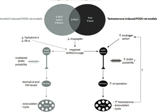
In summary, we found that testosterone- and estradiol-induced PCOS rats with different endocrine phenotypes exhibit differential transcriptional expression of members of the kisspeptin system and sex steroids in the hypothala-mus. It is possible that different insults during development
activate various neuronal circuitries, which might be related to the heterogeneity of PCOS. The hypothesized effects of altered hypothalamic transcriptional expression on endo-crine phenotypes are summarized in Figure 2. These diffe-rences seem to be caused by various mechanisms and might account for the different neuroendocrine and endocrine phenotypes found in androgen- and estrogen-induced PCOS rats and women with PCOS.
’ ACKNOWLEDGMENTS
We thank Thiago H. Gonc¸alves, Luiz F. P. Fuchs, Marinalva de Almeida and Fernanda Condi for their help with animal care and technical support. This research was supported by research grants 2010/17417-3 and 2013/ 12830-8 from Fundac¸ão de Amparo à Pesquisa do Estado de São Paulo (FAPESP) - Brazil, and a Master’s degree scholarship given to R.R.M. (134694/2010-4) by the Conselho Nacional de Desenvolvimento Científico e Tecnológico (CNPq)–Brazil. The funders played no role in the study design, data collection and analysis, decision to publish, or preparation of the manuscript.
’ AUTHOR CONTRIBUTIONS
Maciel GA and Marcondes RR conceived and designed the study. Marcondes RR, Carvalho KC, Giannocco G, Duarte DC and Garcia N performed the experiments. Marcondes RR, Maciel GA, Baracat EC, Maliqueo M, Soares-Junior JM, Silva ID and Carvalho KC analyzed and interpreted the data. Marcondes RR and Maciel GA wrote the manuscript. All authors read and approved thefinal version of the manuscript.
’ REFERENCES
1. Goodarzi MO, Dumesic DA, Chazenbalk G, Azziz R. Polycystic ovary syndrome: etiology, pathogenesis and diagnosis. Nat Rev Endocrinol. 2011;7(4):219-31, http://dx.doi.org/10.1038/nrendo.2010.217.
2. Dumesic DA, Oberfield SE, Stener-Victorin E, Marshall JC, Laven JS, Legro RS. Scientific Statement on the Diagnostic Criteria, Epidemiology, Pathophysiology, and Molecular Genetics of Polycystic Ovary Syndrome. Endocr Rev. 2015;36(5):487-525, http://dx.doi.org/10.1210/er.2015-1018. 3. Lehman MN, Coolen LM, Goodman RL. Minireview: kisspeptin/neuro-kinin B/dynorphin (KNDy) cells of the arcuate nucleus: a central node in the control of gonadotropin-releasing hormone secretion. Endocrinology. 2010;151(8):3479-89, http://dx.doi.org/10.1210/en.2010-0022.
4. DiVall SA, Herrera D, Sklar B, Wu S, Wondisford F, Radovick S, et al. Insulin receptor signaling in the GnRH neuron plays a role in the abnormal GnRH pulsatility of obese female mice. PLoS One. 2015;10(3): e0119995, http://dx.doi.org/10.1371/journal.pone.0119995.
5. Walters KA, Allan CM, Handelsman DJ. Rodent models for human polycystic ovary syndrome. Biol Reprod. 2012;86(5):149, 1-12, http://dx. doi.org/10.1095/biolreprod.111.097808.
6. Marcondes RR, Carvalho KC, Duarte DC, Garcia N, Amaral VC, Simoes MJ, et al. Differences in neonatal exposure to estradiol or testosterone on ovarian function and hormonal levels. Gen Comp Endocrinol. 2015;212: 28-33, http://dx.doi.org/10.1016/j.ygcen.2015.01.006.
7. Mahamed RR, Maganhin CC, Simoes RS, de Jesus Simoes M, Baracat EC, Soares JM, Jr. Effects of metformin on the reproductive system of andro-genized female rats. Fertil Steril. 2011;95(4):1507-9, http://dx.doi.org/ 10.1016/j.fertnstert.2010.07.1093.
8. Alexanderson C, Eriksson E, Stener-Victorin E, Lystig T, Gabrielsson B, Lonn M, et al. Postnatal testosterone exposure results in insulin resistance, enlarged mesenteric adipocytes, and an atherogenic lipid profile in adult female rats: comparisons with estradiol and dihydrotestosterone. Endo-crinology. 2007;148(11):5369-76, http://dx.doi.org/10.1210/en.2007-0305. 9. Marcondes RR, Carvalho KC, Duarte DC, Garcia N, Amaral VC, Simões MJ, et al. Differences in neonatal exposure to estradiol or testosterone on ovarian function and hormonal levels. Gen Comp Endocrinol. 2015;212: 28-33, http://dx.doi.org/10.1016/j.ygcen.2015.01.006.
10. Feng Y, Johansson J, Shao R, Manneras L, Fernandez-Rodriguez J, Billig H, et al. Hypothalamic neuroendocrine functions in rats with dihy-drotestosterone-induced polycystic ovary syndrome: effects of low-frequency electro-acupuncture. PLoS One. 2009;4(8):e6638, http://dx.doi. org/10.1371/journal.pone.0006638.
11. Foecking EM, Szabo M, Schwartz NB, Levine JE. Neuroendocrine con-sequences of prenatal androgen exposure in the female rat: absence of luteinizing hormone surges, suppression of progesterone receptor gene expression, and acceleration of the gonadotropin-releasing hormone pulse generator. Biol Reprod. 2005;72(6):1475-83, http://dx.doi.org/10.1095/ biolreprod.105.039800.
12. Sun J, Moenter SM. Progesterone treatment inhibits and dihydrotest-osterone (DHT) treatment potentiates voltage-gated calcium currents in gonadotropin-releasing hormone (GnRH) neurons. Endocrinology. 2010; 151(11):5349-58, http://dx.doi.org/10.1210/en.2010-0385.
13. Pielecka J, Quaynor SD, Moenter SM. Androgens increase gonadotropin-releasing hormone neuron firing activity in females and interfere with progesterone negative feedback. Endocrinology. 2006;147(3):1474-9, http:// dx.doi.org/10.1210/en.2005-1029.
14. Herbison AE. Control of puberty onset and fertility by gonadotropin-releasing hormone neurons. Nat Rev Endocrinol. 2016;12(8):452-66, http://dx.doi.org/10.1038/nrendo.2016.70.
15. Roa J, Vigo E, Castellano JM, Gaytan F, Navarro VM, Aguilar E, et al. Opposite roles of estrogen receptor (ER)-alpha and ERbeta in the mod-ulation of luteinizing hormone responses to kisspeptin in the female rat: implications for the generation of the preovulatory surge. Endocrinology. 2008;149(4):1627-37, http://dx.doi.org/10.1210/en.2007-1540.
16. Wintermantel TM, Campbell RE, Porteous R, Bock D, Grone HJ, Todman MG, et al. Definition of estrogen receptor pathway critical for estrogen positive feedback to gonadotropin-releasing hormone neurons and fertility. Neuron. 2006;52(2):271-80, http://dx.doi.org/10.1016/j.neuron. 2006.07.023.
17. Radovick S, Levine JE, Wolfe A. Estrogenic regulation of the GnRH neuron. Front Endocrinol. 2012;3:52, http://dx.doi.org/10.3389/fendo. 2012.00052.
18. Hiroi R, Lacagnina AF, Hinds LR, Carbone DG, Uht RM, Handa RJ. The androgen metabolite, 5alpha-androstane-3beta,17beta-diol (3beta-diol), activates the oxytocin promoter through an estrogen receptor-beta path-way. Endocrinology. 2013;154(5):1802-12, http://dx.doi.org/10.1210/en. 2012-2253.
19. Topaloglu AK, Reimann F, Guclu M, Yalin AS, Kotan LD, Porter KM, et al. TAC3 and TACR3 mutations in familial hypogonadotropic hypogonad-ism reveal a key role for Neurokinin B in the central control of repro-duction. Nat Genet. 2009;41(3):354-8, http://dx.doi.org/10.1038/ng.306. 20. Yan X, Yuan C, Zhao N, Cui Y, Liu J. Prenatal androgen excess enhances