Total 3,3
9
,5-Triiodo-
L
-Thyronine and Alters the
Expression of Genes Associated with the Thyroidal Axis
in Female Goldfish (
Carassius auratus
)
Xiaona Zhang, Hua Tian, Wei Wang, Shaoguo Ru*
College of Marine Life Sciences, Ocean University of China, Qingdao, China
Abstract
Our recent study showed that monocrotophos (MCP) pesticide disrupted the hypothalamic-pituitary-thyroid (HPT) axis in male goldfish (Carassius auratus); however, the effects of MCP on the thyroid system in female goldfish are remain unclear.
In the present study, plasma thyroid hormone (TH) and thyroid-stimulating hormone (TSH) levels were evaluated in female goldfish exposed to 0.01, 0.10, and 1.00 mg/L of 40% MCP-based pesticide for 21 days in a semi-static exposure system. Expression profiles of HPT axis-responsive genes, including transthyretin (ttr), deiodinases (d1,d2, andd3),tshb,
thyrotropin-releasing hormone (trh), and corticotrophin-releasing hormone (crh), were determined. The results indicated that MCP
decreased the plasma levels of total 3,39,5-triiodo-L-thyronine (TT3) and the ratio of TT3to total 3,39,5,59-L-thyroxine (TT4), and induced alternative expression of TH-related genes. Exposure to 0.01 and 0.10 mg/L MCP pesticide resulted in the up-regulation ofttrmRNA. The reduction of plasma TT3levels was partly attributed to an increase in the metabolism of T3in
the liver, as revealed by the highly elevated hepatic d1and d3 mRNA levels in the MCP treatment groups, and the expression of hepaticd3showed a negative correlation with the plasma TT3/TT4levels in females. Moreover, the plasma TSH
levels were lower in females exposed to 0.01 and 0.10 mg/L MCP pesticide, whereas the up-regulation oftshbmRNA levels was compensated by the decreased plasma TT3levels. These results indicated that MCP had the potential to influence
several pathways of HPT axis homeostasis in female goldfish.
Citation:Zhang X, Tian H, Wang W, Ru S (2014) Monocrotophos Pesticide Decreases the Plasma Levels of Total 3,39,5-Triiodo-L-Thyronine and Alters the Expression of Genes Associated with the Thyroidal Axis in Female Goldfish (Carassius auratus). PLoS ONE 9(9): e108972. doi:10.1371/journal.pone.0108972
Editor:Vincent Laudet, Ecole Normale Supe´rieure de Lyon, France
ReceivedMarch 10, 2014;AcceptedSeptember 5, 2014;PublishedSeptember 30, 2014
Copyright:ß2014 Zhang et al. This is an open-access article distributed under the terms of the Creative Commons Attribution License, which permits unrestricted use, distribution, and reproduction in any medium, provided the original author and source are credited.
Data Availability:The authors confirm that all data underlying the findings are fully available without restriction. All data are included within the paper.
Funding:This work was supported by the National Natural Science Foundation of China (31202001), www.nsfc.gov.cn, the Natural Science Foundation of Shandong Province (ZR2012CQ010), www.sdnsf.gov.cn, and the Innovation and Research Foundation of Ocean University of China, http://www.moe.edu.cn/. The funders had no role in study design, data collection and analysis, decision to publish, or preparation of the manuscript.
Competing Interests:The authors have declared that no competing interests exist.
* Email: rusg@ouc.edu.cn
Introduction
In fish, growth and reproduction are, at least partly, under the control of thyroid hormones (THs), 3,39,5,59-L-thyroxine (T4) and 3,39,5-triiodo-L-thyronine (T3) [1,2]. Considering that reproduc-tion in females from the early development of follicles to oocyte maturation and ovulation involves a significant investment of energy to support the related physiological functions, the reproductive-related allocation of energy is particularly important during ovarian development in oviparous species, including fish. Since THs have profound effects on energy metabolism, for example, they are the major regulators of oxidative energy metabolism at the level of the mitochondria in teleost fish [3], they should invariably be involved in the multifactorial regulation of metabolism associated with reproduction. They might also act as direct modulators of the reproductive cycle. A previous study on goldfish (Carassius auratus) has shown that T4 could act synergistically with gonadotropin to influence ovarian develop-ment by increasing ovarian sensitivity to gonadotropic stimulation [4]. Moreover, the expression of steroidogenic enzymes and steroid receptors in goldfish could be modulated by THs [5].
in plasma 17b-estradiol (E2) levels and decreases in testosterone (T) levels in both male and female goldfish [11,12]. Balanced plasma TH levels are known to be crucial for normal reproductive function; in mammals, both hyperthyroidism and hypothyroidism were found to result in reproductive impairment and lower fertility [13]. Considering gender differences in the response of the thyroid endocrine system in fish exposed to environmental pollutants, the thyroid disruption effects of MCP in females might be speculated to be similar to those found in males. The effects of MCP on the thyroid endocrine system in females, however, remain unclear.
THs are synthesized and secreted by the thyroid follicles under the control of the HPT axis. In teleosts, the thyrotropin-releasing hormone (TRH) and/or corticotrophin-releasing hormone (CRH), released from the hypothalamus, coordinate the HPT axis function by controlling the release of thyrotropin (TSH) from the pituitary, which could stimulate TH synthesis and release [1,14]. Most of the plasma THs in fish are bound to transthyretin (TTR), a specific TH transport protein in teleosts [15], and only free hormones can enter target cells to elicit a response. In the liver and some other peripheral tissues, three types of deiodinases (type I, D1; type II, D2; and type III, D3) are known to control the conversion of T4 to the more physiologically active T3 or the production of metabolically inactive counterparts [16]. Such complex regulatory pathways are involved in thyroid homeostasis. Therefore, environmental chemicals can act at multiple stages in the HPT axis.
In the present study, following administration of 40% MCP pesticide to female goldfish, plasma TH levels, including TT3, total T4(TT4), and the physiologically relevant free T3(FT3) and free T4(FT4) levels, were determined, and the changes in endocrine-mediated responses along the HPT axis were evaluated, including the regulation of TRH and CRH, synthesis and secretion of TSH, and expression of deiodinases (d1,d2, andd3) and transthyretin (ttr) genes.
Materials and Methods
Pesticide
MCP pesticide (3-hydroxyl-N-methyl-cis-crotonamidedimethyl phosphate, 40% water-soluble preparation) was purchased from the Qingdao pesticide factory in China. The concentration on the label was 40%, which was consistent with the actual concentration determined by gas chromatography (4060.1%) [17]. The half-life of MCP is approximately 66 days at pH 7.0 and 20uC [18].
Fish exposure and sample protocols
Sexually mature goldfish (C. auratus) with mature and fully developed gonads (8.760.8 cm standard length; 21.264.1 g wet weight; 6.860.3% gonadal somatic index) were obtained from a local dealer in Qingdao, PR China, and sampled in late spring. Fish were handled according to the National Institute of Health Guidelines for Handling and Care of Experimental Animals. The animal utilization protocol was approved by the Institutional Animal Care and Use Committee of the Ocean University of China. Fish were maintained in a 70-L aquarium containing 50-L dechlorinated tap water at ambient temperature (1862uC) with dissolved oxygen content of 7.060.1 mg/L and were fed small shrimp daily.
After the female goldfish were acclimated to laboratory conditions for 2 weeks, they were exposed to nominal 0.01, 0.10, and 1.00 mg/L 40% MCP pesticide (4, 40, and 400mg/L equivalent levels of pure MCP), the concentrations of which were 1/10,000; 1/1,000; and 1/100 of the 96-h LC50[11], respectively. The experiments were conducted in the same 70-L aquarium
containing 50-L dechlorinated tap water by using a semi-static toxicity test (20-L water renewal daily to constantly maintain the MCP concentration). Each group of fish was exposed in three aquaria (seven fish/tank) and a control (dechlorinated tap water) was included in the exposure design. Less than 10% mortality was observed in all the treatments during the experiment, and abnormal behaviors such as unusual swimming pattern or bumping against the tank were observed in the group exposed to the highest dose.
After 21 days of exposure, all fish were anesthetized with 75 mg/L MS-222 (Sigma, St. Louis, MO, USA) and sampled between 9:00 and 11:00 h to avoid the possible influences of diurnal fluctuations in hormone levels on the results [19]. Fish from each group were divided into two subgroups at the time of sampling. One subgroup of 9–10 fish was sampled to collect 0.6– 0.8 mL blood for TH measurements, whereas the remaining fish were used to collect blood with the same volume for TSH and cortisol measurements. Blood was collected from the caudal vein by using chilled heparinized syringes and maintained on ice. Plasma samples were obtained after centrifugation at 10006gfor 10 min and stored at280uC until the hormone assay could be performed. The liver, pituitary, and hypothalamus tissues were dissected (n = 9), frozen in liquid nitrogen, and then stored at2
80uC for the quantification of gene expression by real-time polymerase chain reaction (PCR).
Hormone assay
The plasma levels of TT3, TT4, FT3, FT4, TSH, and cortisol were measured using radioimmunoassay (RIA) by using commer-cially available kits following the protocols provided by the manufacturers (Beijing North Institute of Biological Technology, Beijing, China). The RIA kits for human TT3, TT4, FT3, FT4, TSH, and cortisol were validated for use with goldfish samples by showing parallelism between a series of diluted and spiked samples in relation to the standard curve included with the assay kits. Standards and samples were added to test tubes in duplicate. The assay detection limits were 0.05 ng/mL for T3, 2 ng/mL for T4, 0.06 pg/mL for FT3, 0.23 pg/mL for FT4, 0.1mIU/mL for TSH, and 1 ng/mL for cortisol. The inter- and intra-assay coefficients of variation for all the above-mentioned hormones were,10% and
,15%, respectively. Gene expression analysis
Total RNA from each tissue was isolated using the phenolic reagent TRIzol (Invitrogen, Carlsbad, CA, USA) according to the manufacturer’s protocol. The extracted RNA was measured by spectrometry at OD260/280 before treatment with DNase I (Promega, Madison, WI, USA). Equal amounts of RNA (1mg) were reverse-transcribed into cDNA in 20-mL reactions containing 10 pmol oligo(dT)20, 4mL 56RT Buffer, 2mL dNTP mixture, 10 U RNase inhibitor, and 1mL ReverTra Ace (Toyobo, Tokyo, Japan). Reverse transcription reactions were conducted in a Bio-Rad DNA Thermal Cycler (Hercules, CA, USA) at 42uC for 20 min and terminated for 5 min at 85uC.
0.4mL of ROX reference Dye (Takara Bio Inc., Shiga, Japan), and 4mL of first-strand cDNA (template). The thermal profile was 95uC for 30 s, followed by 40 cycles of 95uC for 5 s and 60uC for 30 s. Amplification of a single product was ensured by performing a melting curve analysis by using the PCR products obtained at the end of each PCR run. In addition, 2% agarose gel electrophoresis of the PCR products was performed to confirm the presence of single amplicons having the predicted sizes (data not shown). Theb-actin and 18S rRNA transcripts were used as housekeeping genes to standardize the results and eliminate variations in mRNA and cDNA quantity and quality. Neither the
b-actin nor 18S rRNA levels were affected by any of the experimental conditions used in the study. For each reaction, the relative target gene mRNA expression levels were normalized to the geometric mean ofb-actin and 18S rRNA expression levels by using the formula 22DDCt and plotted on a logarithmic scale [20].
Statistical analysis
The experimental data were presented as the means6standard deviations, and the differences between the control and each exposure group were evaluated using one-way analysis of variance (ANOVA) followed by Tukey’s test. Before parametric analysis, assumptions of normality and homogeneity of variances were assessed using probability plots and normality tests. Pearson’s correlation coefficient was used to calculate the relationship between the expression of certain genes involved in the HPT axis and the plasma TH levels. Values were considered significant when 0.01,P,0.05 and highly significant whenP,0.01.
Results
Effects of MCP pesticide on plasma TT3and TT4levels and the ratio of TT3to TT4
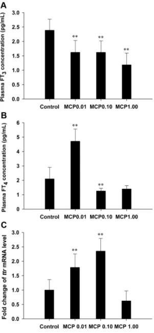
The TT3content in the plasma of the control female goldfish was 1.3160.29 ng/mL, whereas that in the fish exposed to 0.10 and 1.00 mg/L MCP pesticide was significantly decreased by 30% to 0.9260.23 ng/mL (0.01,P,0.05) and 51% to 0.6460.21 ng/ mL (P,0.01), respectively (Fig. 1A). At the end of the MCP treatment, the TT4content was 7.2161.55 ng/mL in the control group, and there was no significant change in the TT4content of any of the MCP pesticide-treated groups (Fig. 1B). There was a dose-related decrease in the ratio of TT3 to TT4 after MCP treatment (P,0.01; Fig. 1C).
Effects of MCP pesticide on plasma FT3and FT4levels and hepaticttrmRNA expression
The basal FT3levels in the plasma of the control female goldfish were 2.3960.39 pg/mL; they were significantly reduced by 32%, 32%, and 50% in the 0.01, 0.10, and 1.00 mg/L MCP pesticide-treated groups, respectively (P,0.01; Fig. 2A). Plasma FT4levels in the control females were 2.1060.80 pg/mL; they were significantly increased to 4.7160.85 pg/mL in the 0.01 mg/L MCP pesticide-treated group (P,0.01) but decreased to 1.26 pg/ mL in the 1.00 mg/L MCP pesticide-treated group (P,0.01; Fig. 2B). Furthermore, the mRNA expression of hepaticttr was significantly higher after treatment with 0.01 and 0.10 mg/L of MCP pesticide (P,0.01), whereas there was no significant difference in the hepaticttrmRNA expression between the group treated with the highest dose and the control group (Fig. 2C).
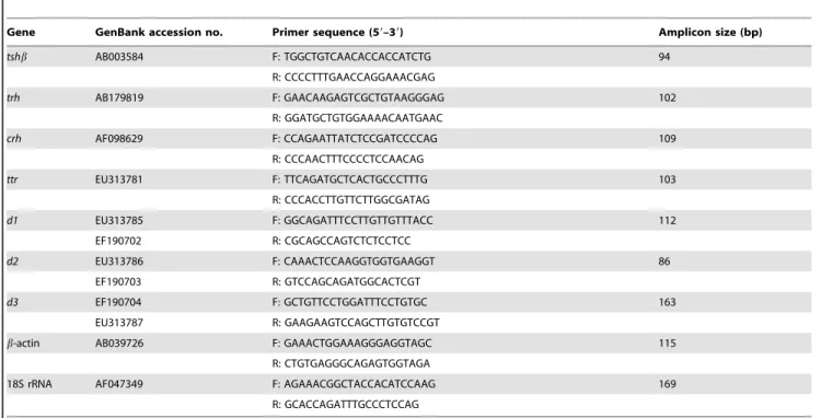
Table 1.Nucleotide sequences of the primers used for real-time polymerase chain reaction and product sizes.
Gene GenBank accession no. Primer sequence (59–39) Amplicon size (bp)
tshb AB003584 F: TGGCTGTCAACACCACCATCTG 94
R: CCCCTTTGAACCAGGAAACGAG
trh AB179819 F: GAACAAGAGTCGCTGTAAGGGAG 102
R: GGATGCTGTGGAAAACAATGAAC
crh AF098629 F: CCAGAATTATCTCCGATCCCCAG 109
R: CCCAACTTTCCCCTCCAACAG
ttr EU313781 F: TTCAGATGCTCACTGCCCTTTG 103
R: CCCACCTTGTTCTTGGCGATAG
d1 EU313785 F: GGCAGATTTCCTTGTTGTTTACC 112
EF190702 R: CGCAGCCAGTCTCTCCTCC
d2 EU313786 F: CAAACTCCAAGGTGGTGAAGGT 86
EF190703 R: GTCCAGCAGATGGCACTCGT
d3 EF190704 F: GCTGTTCCTGGATTTCCTGTGC 163
EU313787 R: GAAGAAGTCCAGCTTGTGTCCGT
b-actin AB039726 F: GAAACTGGAAAGGGAGGTAGC 115
R: CTGTGAGGGCAGAGTGGTAGA
18S rRNA AF047349 F: AGAAACGGCTACCACATCCAAG 169
R: GCACCAGATTTGCCCTCCAG
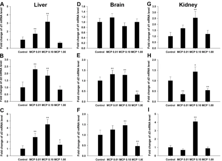
Effects of MCP pesticide on d1,d2, andd3mRNA
expression levels in the liver, brain, and kidney
Thed1mRNA levels in the liver were significantly increased by 0.72- and 2.15-fold and the hepatic d2 mRNA expression was significantly up-regulated by 1.30- and 0.84-fold after treatment with 0.01 and 0.10 mg/L MCP pesticide, respectively (P,0.01), relative to those in the control (Fig. 3A and 3B). Hepatic d3
mRNA levels were significantly higher in all the MCP-treated groups, especially in the 0.01 and 0.10 mg/L MCP pesticide-treated groups (2.66- and 4.50-fold higher, respectively), than those in the control (P,0.01; Fig. 3C).
With regard to the gene transcription level of deiodinase in the brain (Fig. 3D–3F), there was no significant difference in the d1
gene transcription in any of the MCP-exposed groups. MCP
pesticide exposure significantly stimulatedd2gene transcription in the 0.01 mg/L and 0.10 mg/L groups (P,0.01) and d3 gene transcription in the 0.10 mg/L group (P,0.01). However, the transcription of bothd2and d3genes was significantly inhibited after exposure to 1.00 mg/L MCP pesticide (P,0.01).
Transcription of thed1gene in the kidney was significantly up-regulated after exposure to 0.10 mg/L MCP (P,0.01; Fig. 3G). The transcription of d2 was significantly down-regulated after exposure to 0.01 and 1.00 mg/L MCP pesticide, but was stimulated in the 0.10 mg/L group (0.01,P,0.05; Fig. 3H). The expression levels ofd3mRNA in the kidney were significantly Figure 1. Quantification of plasma total 3,39,5-triiodo-L
-thyro-nine (T3) and total 3,39,5,59-L-thyroxine (T4) levels in female
goldfish exposed to 0, 0.01, 0.10, and 1.00 mg/L 40% mono-crotophos (MCP) pesticide.(designated C, MCP0.01, MCP0.10, and MCP1.00, respectively). The data are presented as the means 6
standard deviations (n = 9). Asterisks indicate statistically significant differences from the control group (*0.01,P,0.05, **P,0.01). doi:10.1371/journal.pone.0108972.g001
Figure 2. Quantification of plasma free 3,39,5-triiodo-L -thyro-nine (T3) and free 3,39,5,59-L-thyroxine (T4) contents and the
relative mRNA expression levels of hepatic transthyretin (ttr) in female goldfish exposed to 0, 0.01, 0.10, and 1.00 mg/L 40% monocrotophos (MCP) pesticide.(designated C, MCP0.01, MCP0.10, and MCP1.00, respectively). For panel C, fold change (Y axis) represents the expression of the target gene mRNA relative to that of the control group (equals 1 by definition). The data are presented as the means6
up-regulated only in the 0.10 mg/L MCP-exposed group (4.0-fold,P,0.01; Fig. 3I).
Effects of MCP pesticide on pituitarytshb mRNA
expression and plasma TSH levels
The mRNA expression of pituitarytshb was significantly up-regulated in both the 0.10 and 1.00 mg/L MCP pesticide-treated groups (P,0.01; Fig. 4A). In contrast, the plasma TSH levels in the 0.10 and 1.00 mg/L MCP pesticide-treated groups were markedly decreased to 0.8560.16mIU/mL (P,0.01) and 0.9160.14mIU/mL (0.01,P,0.05), respectively, compared to those in the control group (1.2460.30mIU/mL).
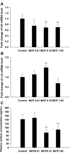
Effects of MCP pesticide on hypothalamictrh andcrh
mRNA and plasma cortisol levels
The expression of hypothalamic trh was significantly down-regulated after treatment with the MCP pesticide (Fig. 5A). The transcription of the crh gene was significantly stimulated after treatment with 0.10 mg/L MCP pesticide (P,0.01), but inhibited in the 1.00 mg/L MCP pesticide-treated group (0.01,P,0.05; Fig. 5B). The measured plasma cortisol content was 142.01624.11 ng/mL in the control female goldfish and was significantly decreased to 74.53618.26 ng/mL and
89.88622.05 ng/mL in the 0.10 and 1.00 mg/L MCP pesti-cide-treated groups, respectively (P,0.01; Fig. 5C).
The correlation coefficient for plasma THs levels and the expression of certain genes related to the HPT axis
The correlation coefficient for plasma THs levels and the expression of certain genes associated with the HPT axis are shown in Table 2. Significant correlations of the ratio of TT3to TT4 with hepatic d3 expression, TT3 with pituitary tshb expression, and FT4with hepaticttrexpression were found. Discussion
An increasing number of studies have reported that groups of pesticides, including acetochlor, amitrole, and metolachlor, have the potential to influence several steps in HPT axis homeostasis and to induce THs disturbance in adult fish, particularly with respect to sex differences occurring in response to chemical-induced thyroid system disruption [10,21,22]. For example, Li et al. [21] showed that TH-related genes such as malic enzyme and sodium iodide symporter were significantly down-regulated in the brains of the rare minnow Gobiocypris rarus, and that the expression of these genes in females was more sensitive to Figure 3. Relative mRNA expression levels of types I, II, and III deiodinases (d1,d2, andd3) in the liver, brain, and kidney of female goldfish exposed to 0, 0.01, 0.10, and 1.00 mg/L 40% monocrotophos (MCP) pesticide.(designated C, MCP0.01, MCP0.10, and MCP1.00, respectively). Fold change (Y axis) represents the expression of the target gene mRNA relative to that of the control group (equals 1 by definition). The data are presented as the means6standard deviations (n = 9). Asterisks indicate statistically significant differences from the control group (*0.01,P,0.05, **P,0.01).
acetochlor than that in males. Recently, we found that a 21-d exposure to MCP pesticide caused significant decreases in plasma TT3levels and TT3-to-TT4ratios in male goldfish [9]; however, whether similar effects occur in females is not clear.
In the present study, although the plasma levels of TT4 remained unchanged, those of TT3, FT4, and FT3 in female goldfish were significantly altered after a 21-day exposure to MCP pesticide, suggesting a failed adaptation and auto-regulation of THs homeostasis. Almost all THs circulating in the plasma are bound to transporter proteins, and the equilibrium of TH binding to the plasma proteins determines the concentration of free THs within the plasma [23]. TTR, which is primarily a secretory product of the liver, has been proposed to be the major TH-carrier protein that binds THs and transports them to target tissues in fish [24–26]. In our study, TTR gene expression was up-regulated after exposure to 0.01 and 0.10 mg/L MCP pesticide. This up-regulation might have resulted in higher TTR mRNAs and thus TTR proteins, leading to decreases in plasma FT3 and/or FT4 levels. Moreover, the expression of ttr showed a positive correlation with plasma FT4levels in females. Notably, less than 1% of plasma TT4is free with 99% reversibly bound to plasma proteins [23]. Indeed, changes of such small amount of FT4
contents may not represent a dynamic circulating TT4reservoir and vice verse. For example, in brown trout (Salmo trutta) fed diets enriched withb–Tetrabromoethylcyclohexane for 56 days, there was no significant difference among treatments in FT4, but TT4 was significantly reduced in the high dose group relative to the control [27]. In the 0.01 mg/L MCP group in particular, one possible explanation for the increased plasma FT4levels could be that feedback systems attempt to respond to the reduction in plasma TT3levels. However, the stimulatory effects of 0.01 mg/L MCP pesticide on plasma FT4levels in females were not observed in males [9], indicating that the thyroidal system in female goldfish Figure 4. Relative mRNA expression levels of pituitary
thyroid-stimulating hormone b subunit (tshb) and quantification of plasma TSH content in female goldfish exposed to 0, 0.01, 0.10, and 1.00 mg/L 40% monocrotophos (MCP) pesticide. (designated C, MCP0.01, MCP0.10, and MCP1.00, respectively). For panel A, fold change (Y axis) represents the expression of the target gene mRNA relative to that of the control group (equals 1 by definition). The data are presented as the means6standard deviations (n = 9). Asterisks indicate statistically significant differences from the control group (*0.01,P,0.05, **P,0.01).
doi:10.1371/journal.pone.0108972.g004
Figure 5. Relative mRNA expression levels of thyrotropin-releasing hormone (trh) and corticotrophin-releasing hormone (crh) in the hypothalamus glands and quantification of plasma cortisol content in female goldfish exposed to 0, 0.01, 0.10, and 1.00 mg/L 40% monocrotophos (MCP) pesticide. (designat-ed C, MCP0.01, MCP0.10, and MCP1.00, respectively). For panels A and B, fold change (Y axis) represents the expression of the target gene mRNA relative to that of the control group (equals 1 by definition). The data are presented as the means6standard deviations (n = 9). Asterisks indicate statistically significant differences from the control group (*0.01,P,0.05, **P,0.01).
is more sensitive to MCP than that of males. In the group treated with the highest dose of MCP pesticide, the total TH level was more readily responsive to the changes in free hormone levels, since TTR gene expression was not stimulated in this group.
Among the thyroid follicle secretions, T4 is the predominant circulating hormone in the blood of fish, and T3appears to be produced largely by enzymatic deiodination of T4 in the peripheral tissues [28]. Consequently, plasma T3 levels mostly decline due to a drop in thyroidal T4production and secretion and/or changes in the peripheral TH metabolism [29]. Our finding that decreases in plasma TT3 levels along with relatively stable plasma TT4levels suggested possible changes in peripheral TH deiodination or metabolism. Iodothyronine deiodinases play a crucial role in the mechanism of TH biotransformation in extra-thyroidal tissues. Conversion from T4to T3is mediated by outer-ring deiodination (ORD). Both inner-outer-ring deiodination (IRD) and ORD are involved in the inactivation of T4 to 3,39,59-triiodo-L -thyronine (rT3) and T3and rT3to 3,39-diiodo-L-thyronine (3,39 -T2) [16,30]. Three types of deiodinases have been identified in teleosts: type I (D1) has both ORD and IRD activities, whereas types II (D2) and III (D3) have ORD and IRD activity, respectively [31–34]. At the end of the 21-day exposure to 0.01 and 0.10 mg/L MCP pesticide, the transcription of all three types of deiodinases was stimulated in the liver: changes in the expression of d1 and d3 mRNA were higher than those of d2
mRNA. Further, in the 1.00 mg/L treatment, only hepatic d3
mRNA levels were significantly up-regulated. Van der Geyten et al. [35] showed that the changes in hepatic deiodinases activities tend to be consistent with those of their mRNA levels, indicating pre-translated regulation of the hepatic deiodinases. Both D1 and D3 are involved in the metabolism of THs. D3 catalyzes the degradation of T4and T3to inactive metabolites and could protect tissues from an excess of biologically active TH, namely, T3[36]. Although D1 also has the ability to degrade T3, it is less effective than D3, and its preferred substrate is rT3 [37]. Therefore, the stimulated enzymatic metabolism of T3in the liver could cause a reduction in the levels of circulating TT3. Consistently, a significant negative correlation between hepatic d3 mRNA expression and the ratio of TT3to TT4was observed. It is also worth noting that, in the highest dose group, alterations of gene expression of the three types of deiodinases in females were not consistent with those in males in which hepaticd1andd2mRNA levels were significantly down-regulated, whereasd3mRNA levels were not affected [9].
Although liver is considered to be the main peripheral source of circulating T3, TH deiodination also occurs in other extra-thyroidal tissues such as brain and kidney; however, the available T3 derived from these tissues is primarily utilized by the same tissues itself [38]. Exposure to MCP pesticide had no effect on the
d1mRNA expression in the brain, but stimulated the transcription ofd1gene in the kidney of female goldfish. The functional roles of D1 might indicate why the patterns ofd1mRNA regulation varied among tissues after exposure to MCP. Recent studies have suggested that the major role of D1 might be to clear rT3 and sulfated iodothyronines from circulation. Indeed, it functions as a scavenger enzyme to remove inactive iodothyronines and recycle iodine within the organisms [39–41]. Thus, 0.10 mg/L MCP pesticide might enhance the metabolism of THs by up-regulating the expression ofd1 mRNA in the kidney. Further, the specific patterns of deiodinase-mediated gene regulation show tissue-specific variations, probably in accordance with the distinct thyroid status and tissue-specific requirements for available THs [42]. In the brain, the transcriptions of d2 and d3 gene were stimulated after treatment with 0.01 and 0.10 mg/L MCP but inhibited by 1.00 mg/L MCP pesticide, whereas, in the kidney, the transcriptions of d2 and d3 gene were stimulated after treatment with 0.10 mg/L MCP but inhibited by treatment with 0.01 and 1.00 mg/L MCP pesticide. Such expression profiles of
d2andd3changing in a parallel way indicates that intracellular T3levels are tightly controlled [43].
THs are also regulated by TSH and TRH/CRH, but can interfere with the synthesis and release of TSH and TRH/CRH
via feedback mechanisms. This regulatory system plays an important role in maintaining the homeostatic balance along the HPT axis [44,45]. In the present study, the tshb mRNA levels were up-regulated and showed a negative correlation with plasma TT3levels. The T3feedback mechanism is known to control TSH expression in certain teleosts, and Sohn et al. [46] provided evidence that T3acted directly on the pituitary and inhibitedtshb gene expression in goldfish, probably via the negative T3 -responsive elements in the tshb gene. Hence, the decreased plasma TT3levels would feedback to stimulate the transcription of
tshb, and similar negative feedback has also been observed in males [9]. TSH is a glycoprotein secreted by the pituitary thyrotrope cells. In our previous study, Bing [47] reported direct damage of the MCP pesticide to the structure of thyrotrope cells in adenohypophysis. The partly dissolved nuclear membrane, prominent dilated rough endoplasmic reticulum, and slightly dissolved mitochondrial cristae indicated a reduction on the
Table 2.Pearson correlation coefficients for the plasma thyroid hormone levels and the expression of certain genes related to the hypothalamus-pituitary-thyroid axis.
Paired samples Correlation coefficient
TT3/TT4vs. hepaticd1 2.189
TT3/TT4vs. hepaticd2 2.232
TT3/TT4vs. hepaticd3 2.384*
TT3vs. pituitarytshb 2.469*
TT3vs. hypothalamustrh .356
TT3vs. hypothalamuscrh .025
FT3vs. hepaticttr 2.069
FT4vs. hepaticttr .396*
hormone secretion yield of thyrotrope cells. Accordingly, in the current study, although pituitarytshbmRNA levels were elevated, the reduced plasma TSH levels in the 0.10 and 1.00 mg/L MCP treatments were probably resulted from the diminished TSH protein synthesis and secretion of thyrotrope cells. In goldfish, both TRH and CRH might be involved in the regulation of the pituitary-thyroid axis, and TRH might act as a multifunctional hypophysiotropic factor. For instance, TRH is a potent stimulator of alpha melanocyte-stimulating hormone release from the pars intermedia cells in various species of teleost fish, including goldfish [48], and is also involved in the regulation of feeding and locomotor behaviors [49]. Therefore, the up-regulatedtrhmRNA expression levels caused by the MCP pesticide would not be the only response of the HPT axis. Instead, CRH might act as a TSH-releasing factor in lower vertebrates [14], and might also be mediated and/or intensified by a concomitant increase in corticosteroids [50]. It is also worth noting that CRH plays important roles in mediating the hypothalamic-pituitary-interrenal axis (HPI axis) to produce cortisol in response to stress, and that cortisol could also tightly control the release of CRHviaa negative feedback loop [51,52]. The reductions in plasma cortisol levels in the 0.10 mg/L group would be the result of MCP interference with cortisol synthesis and metabolism at some level. A recent study in our lab revealed that 0.10 mg/L MCP pesticide decreased interrenal synthesis of cortisol and further promoted the metabolism of cortisol, resulting in lower overall cortisol levels and a diminished stress capacity in female zebrafish (Danio rerio) [53]. In goldfish, whether CRH acts on thyroid activity either directly or via adrenal steroids has yet to be determined; the decreased plasma TT3and cortisol levels would also signal the up-regulation ofcrhgene expression, as observed in the 0.10 mg/L group. On the other hand, in the 1.00 mg/L MCP pesticide group, both plasma cortisol levels and hypothalamiccrhmRNA
levels were decreased, indicating that the high concentration of MCP pesticide disrupted the feedback mechanisms along the HPI axis. Some investigators have suggested that hyperactivity of the cortisol-producing cells associated with severe stress might lead to an inter-renal exhaustion with very low levels of cortisol [54,55].
Our results showed that the MCP pesticide disturbed the THs homeostasis and interfered with the transport and conversion of THs, synthesis and secretion of pituitary TSHs, and regulation of hypothalamic TRH/CRH in female goldfish. Similar to the findings in males, MCP pesticide decreased plasma TT3and FT3 levels with relatively stable plasma TT4levels, and profiles of the relative abundance of deiodinase transcripts were observed in the liver, brain, and kidneys in female goldfish; however, 0.01 mg/L MCP pesticide merely enhanced the plasma FT4levels in females, and gender differences existed in the hepatic deiodinase transcripts at the highest dose. Although the reproductive and thyroidal endocrine systems have been reported to be related to the neuroendocrine control of many complex functions, including growth, metabolism, and reproduction in teleosts [1,2,5], and many chemicals, including polychlorinated biphenyl and perfluor-ooctanoic acid, could simultaneously cause a variety of effects along both endocrine systems [56–59], further studies unraveling the interaction between the MCP-induced reproductive axis disruption and thyroidal axis disruption are necessary to explain gender differences observed in the thyroid system response to MCP exposure in goldfish.
Author Contributions
Conceived and designed the experiments: XZ HT WW SR. Performed the experiments: XZ HT. Analyzed the data: XZ HT. Wrote the paper: XZ SR.
References
1. Cyr DG, Eales JG (1996) Interrelationships between thyroidal and reproductive endocrine systems in fish. Rev Fish Biol Fish 6: 165–200.
2. Power DM, Llewellyn L, Faustino M, Nowell MA, Bjornsson BT, et al. (2001) Thyroid hormones in growth and development of fish. Comp Biochem Physiol C Toxicol Pharmacol 130: 447–459.
3. Oommen OV, Sreejith P, Beyo RS, Divya L, Vijayasree AS, et al. (2006) Thyroid hormones regulate mitochondrial respiration as well as antioxidant defense in teleosts too! J Endocrinol Reprod 102: 96–105.
4. Hurlburt ME (1977) Role of the thyroid gland in ovarian maturation of the goldfish,Carassius auratusL. Can J Zool 55: 1906–1913.
5. Habibi HR, Nelson ER, Allan ERO (2012) New insights into thyroid hormone function and modulation of reproduction in goldfish. Gen Comp Endocrinol 175: 19–26.
6. Kang Y, Zhang G (2000) Analysis of organophosphorous pesticides from source water using solid-phase extraction technique. China Environ Sci 20: 1–4. 7. Kumari B, Madan VK, Kathpal TS (2007) Pesticide residues in rain water from
Hisar, India. Environ Monit Assess 133: 467–471.
8. Anjum R, Malik A (2013) Evaluation of mutagenicity of wastewater in the vicinity of pesticide industry. Environ Toxicol Pharmacol 35: 284–291. 9. Zhang X, Tian H, Wang W, Ru S (2013) Exposure to monocrotophos pesticide
causes disruption of the hypothalamic–pituitary–thyroid axis in adult male goldfish (Carassius auratus). Gen Comp Endocrinol 193: 158–166.
10. Jin Y, Chen R, Wang L, Liu J, Yang Y, et al. (2011) Effects of metolachlor on transcription of thyroid system-related genes in juvenile and adult Japanese medaka (Oryzias latipes). Gen Comp Endocrinol 170: 487–493.
11. Tian H, Ru S, Bing X, Wang W (2010) Effects of monocrotophos on the reproductive axis in the male goldfish (Carassius auratus): potential mechanisms underlying vitellogenin induction. Aquat Toxicol 98: 67–73.
12. Tian H, Ru S, Wang W, Bing X (2010) Effects of monocrotophos on the reproductive axis in the female goldfish (Carassius auratus). Comp Biochem Physiol C Toxicol Pharmacol 152: 107–113.
13. Jahnke GD, Choksi NY, Moore JA, Shelby MD (2004) Thyroid toxicants: assessing reproductive health effects. Environ Health Perspect 112: 363–368. 14. De Groef B, Van der Geyten S, Darras VM, Kuhn ER (2006) Role of
corticotrophin releasing hormone as a thyrotropin-releasing factor in non-mammalian vertebrates. Gen Comp Endocrinol 146: 62–68.
15. Morgado I, Hamers T, Van der Ven L, Power DM (2007) Disruption of thyroid hormone binding to sea bream recombinant transthyretin by ioxinyl and polybrominated diphenyl ethers. Chemosphere 69: 155–163.
16. Orozco A, Valverde-RC (2005) Thyroid hormone deiodination in fish. Thyroid 15: 799–813.
17. Ru S, Wei X, Jiang M, Li Y (2003) In vivo and in vitro inhibitions of red drum (Sciaenops ocellatus) brain acetylcholinesterase and liver carboxylesterase by monocrotophos at sublethal concentrations. Water Air Soil Pollut 149: 17–25. 18. Wang HM, Zhang ZM (1989) Monocrotophos. In: Wang, H.M., Zhang, Z.M.
(Eds.), A New Pesticide Manual. China Agriculture Press Beijing p 19. 19. Leiner KA, Han GS, MacKenzie DS (2000) The effects of photoperiod and
feeding on the diurnal rhythm of circulating thyroid hormones in the red drum,
Sciaenops ocellatus. Gen Comp Endocrinol 120: 88–98.
20. Vandesompele J, De Preter K, Pattyn F, Poppe B, Van Roy N, et al. (2002) Accurate normalization of real-time quantitative RT-PCR data by geometric averaging of multiple internal control genes. Genome Biol 3, research0034.10– research0034.11.
21. Li W, Zhan J, Spear PA, Li Z, Yang L, et al. (2009) Changes of thyroid hormone levels and related gene expression in Chinese rare minnow (Gobiocypris rarus) during 3-amino-1,2,4-triazole exposure and recovery. Aquat Toxicol 92: 50–57. 22. Li W, Zha J, Li Z, Yang L, Wang Z (2009) Effects of exposure to acetochlor on the expression of thyroid hormone related genes in larval and adult rare minnow (Gobiocypris rarus). Aquat Toxicol 94: 87–93.
23. Brown SB, Adams BA, Cyr DG, Eales JG (2004) Contaminant effects on the teleost fish thyroid. Environ Toxicol Chem 23: 1680–1701.
24. Kawakami Y, Seoka M, Miyashita S, Kumai H, Ohta H (2006) Characteriza-tion of transthyretin in the Pacific bluefin tuna,Thunnus orientalis. Zoolog Sci 23: 443–448.
25. Power DM, Elias NP, Richardson SJ, Mendes J, Soares CM, et al. (2000) Evolution of the thyroid hormone-binding protein, transthyretin. Gen Comp Endocrinol 119: 241–255.
26. Schreiber G (2002) The evolutionary and integrative roles of transthyretin in thyroid hormone homeostasis. J Endocrinol 175: 61–73.
28. Van der Geyten S, Mol KA, Pluymers W, Ku¨hn ER, Darras VM (1998) Changes in plasma T3during fasting/refeeding in tilapia (Oreochromis niloticus)
are mainly regulated through changes in hepatic type II iodothyronine deiodinase. Fish Physiol Biochem 19: 135–143.
29. Li D, Xie P, Zhang X (2008) Changes in plasma thyroid hormones and cortisol levels in crucian carp (Carassius auratus) exposed to the extracted microcystins. Chemosphere 74: 13–18.
30. Van der Geyten S, Byamungu N, Reyns GE, Ku¨hn ER, Darras VM (2005) Iodothyronine deiodinases and the control of plasma and tissue thyroid hormone levels in hyperthyroid tilapia (Oreochromis niloticus). J Endocrinol 184: 467–479. 31. Kalsbeek A, Buijs RM, van Schaik R, Kaptein E, Visser TJ, et al. (2005) Daily variations in type II iodothyronine deiodinase activity in the rat brain as controlled by the biological clock. Endocrinology 146: 1418–1427.
32. Ko¨hrle J (1999) Local activation and inactivation of thyroid hormones: the deiodinase family. Mol Cell Endocrinol 151: 103–119.
33. Sanders JP, van der Geyten S, Kaptein E, Darras VM, Ku¨hn ER, et al. (1997) Characterization of a propilthiouracil-insensitive type I iodothyronine deiodi-nase. Endocrinology 138: 5153–5160.
34. Valverde C, Croteau W, Lafleur GJ Jr, Orozco A, Germain DL (1997) Cloning and expression of a 59-iodothyronine deiodinase from the liver ofFundulus heteroclitus. Endocrinology 138: 642–648.
35. Van der Geyten S, Toguyeni A, Baroiller JF, Fauconneau B, Fostier A, et al. (2001) Hypothyroidism induces type I iodothyronine deiodinase expression in tilapia liver. Gen Comp Endocrinol 124: 333–342.
36. Sutija M, Longhurst TJ, Joss JMP (2004) Deiodinase type III in the Australian lungfish,Neoceratodus forsteri. Gen Comp Endocrinol 136: 152–161. 37. Moreno M, Berry MJ, Horst C, Thoma R, Goglia F, et al. (1994) Activation and
inactivation of thyroid hormone by type I iodothyronine deiodinase. FEBS Lett 344: 143–146.
38. Eales JG, Morin PP, Tsang P, Hara TJ (1993) Thyroid hormone deiodination in brain, liver, gill, heart and muscle of Atlantic salmon (Salmo salar) during photoperiodically-induced parr-smolt transformation. II. Outer- and inner-ring 3,5,39-triiodo-L-thyronine and 3,39,59-triiodo-L-thyronine (reverse T3)
deiodin-ation. Gen Comp Endocrinol 90: 157–167.
39. Bianco AC, Salvatore D, Gereben B, Berry MJ, Larsen PR (2002) Biochemistry, cellular and molecular biology, and physiological role of the iodothyronine selenodeiodinases. Endocr Rev 23: 38–89.
40. Maia AL, Goemann IM, Meyer EL, Wajner SM (2011) Deiodinases: the balance of thyroid hormone: type 1 iodothyronine deiodinase in human physiology and disease. J Endocrinol 209: 283–297.
41. St Germain DL, Galton VA, Hernandez A (2009) Minireview: defining the roles of the iodothyronine deiodinases: current concepts and challenges. Endocrinol-ogy 150: 1097–1107.
42. Darras VM, Van Herck SL (2012) Iodothyronine deiodinase structure and function: from ascidians to humans. J Endocrinol 215: 189–206.
43. Becker KB, Stephens KC, Davey JC, Schneider MJ, Galton VA (1997) The type 2 and type 3 iodothyronine deiodinases play important roles in coordinating development in Rana catesbeianatadpoles. Endocr inology138: 2989–2997. 44. Ng TB, Idler DR, Eales JG (1982) Pituitary hormones that stimulate the
thyroidal system in teleost fishes. Gen Comp Endocrinol 48: 372–389.
45. Yoshiura Y, Sohn Y, Munakata A, Kobayashi M, Aida K (1999) Molecular cloning of the cDNA encoding the beta subunit of thyrotropin and regulation of its gene expression by thyroid hormones in the goldfish,Carassius auratus. Fish Physiol Biochem 21: 201–210.
46. Sohn YC, Yoshiura Y, Suetake H, Kobayashi M, Aida K (1999) Isolation and characterization of the goldfish thyrotropinbsubunit gene including the 59 -flanking region. Gen Comp Endocrinol 115: 463–473.
47. Bing X (2003) The environmental estrogen effect of monocrotophos on male goldfish, Carassius Auratus. M.Sc. Thesis, Ocean University of China. Available: http://cdmd.cnki.com.cn/Article/CDMD-10423-2003093472.htm. 48. Tran TN, Fryer JN, Bennett HP, Tonon MC, Vaudry H (1989) TRH stimulates
the release of POMC-derived peptides from goldfish melanotropes. Peptides 10: 835–841.
49. Abbott M, Volkoff H (2011) Thyrotropin Releasing Hormone (TRH) in goldfish (Carassius auratus): role in the regulation of feeding and locomotor behaviors and interactions with the orexin system and cocaine- and amphetamine regulated transcript (CART). Horm Behav 59: 236–245.
50. Decuypere E, Scanes CG, Kuhn ER (1983) Effects of glucocorticoids on circulating concentrations of thyroxine (T4) and triiodothyronine (T3) and on
peripheral monodeiodination in pre and posthatching chickens. Horm Metab Res 15: 223–236.
51. Huising MO, Metz JR, van Schooten C, Taverne-Thiele AJ, Hermsen T, et al. (2004) Structural characterisation of a cyprinid (Cyprinus carpio L.) CRH, CRH-BP and CRH-R1, and the role of these proteins in the acute stress response. J Mol Endocrinol 32: 627–648.
52. Mommsen TP, Vijayan MM, Moon TW (1999) Cortisol in teleosts: dynamics, mechanisms of action, and metabolic regulation. Rev Fish Biol Fisheries 9: 211– 268.
53. Zhong Y (2014) Effects of monocrotophos pesticide on the interrenal stress axis in zebrafish (Danio rerio). M.Sc. Thesis, Ocean University of China. In press. 54. Hontela A, Rasmussen JB, Audet C, Chevalier G (1992) Impaired cortisol stress
response in fish from environments polluted by PAHs, PCBs, and mercury. Arch Environ Contam Toxicol 22: 278–283.
55. Thangavel P, Sumathiral K, Karthikeyan S, Ramaswamy M (2005) Endocrine response of the freshwater teleost, Sarotherodon mossambicus (Peters) to dimecron exposure. Chemosphere 61: 1083–1092.
56. Desaulniers D, Leingartner K, Wade M, Fintelman E, Yagminas A, et al. (1999) Effects of acute exposure to PCBs 126 and 153 on anterior pituitary and thyroid hormones and FSH isoforms in adult Sprague Dawley male rats. Toxicol Sci 47: 158–169.
57. Ulbrich B, Stahlmann R (2004) Developmental toxicity of polychlo-rinated biphenyls (PCBs): a systematic review of experimental data. Arch Toxicol 78: 252–268.
58. Lau C, Anitole K, Hodes C, Lai D, Pfahles-Hutchens A, et al. (2007). Perfluoroalkyl acids: A review of monitoring and toxicological findings. Toxicol Sci 99: 366–394.