RESUMO: “Efeito imunoestimulante de compostos fenólicos de líquens na produção de peróxido de hidrogênio e óxido nítrico”. Os efeitos dos compostos isolados de líquens brasileiros e seus derivados na produção de NO e H2O2 foram estudados utilizando macrófagos murinos na tentativa de desvendar suas possíveis propriedades imunomodulatórias. A citotoxicidade dos compostos foi estudada utilizando o ensaio de MTT. A estimulação dos macrófagos foi avaliada através da determinação de NO (metodologia de Griess) e H2O2 (peroxidase de raíz forte/vermelho de fenol) no sobrenadante de culturas de macrófagos peritoneais de camundongos Swiss. Este estudo demonstrou atividade estimulante de alguns compostos fenólicos e seus derivados na produção de NO e H2O2. A relação estrutura atividade sugere inúmeras direções sintéticas para futuros melhoramentos da atividade imunológica.
Unitermos: líquens, peróxido de hidrogênio, óxido nítrico, macrófagos, compostos fenólicos.
ABSTRACT: The effects of isolated compounds from Brazilian lichens and their derivatives on H2O2 and NO production were studied using murine macrophages as a part of an attempt to understand their possible immunomodulatory properties. The compound cytotoxicity was studied using MTT assay. Macrophage stimulation was evaluated by the determination of NO (Griess assay) and H2O2 (horseradish peroxidase/phenol red) in supernatants of peritoneal macrophage cultures of Swiss mice. This research demonstrated stimulatory activities of some phenolic compounds isolated from lichens and their derivatives on H2O2 and NO production. Structure-activity relationships suggest several synthetic directions for further improvement of immunological activity.
Keywords: lichens, hydrogen peroxide, nitric oxide, macrophages, phenolic compounds.
the immune response macrophages are involved and they can release many compounds such as hydrogen peroxide (H2O2) and nitric oxide (NO). H2O2 is a lipid soluble molecule that crosses the cell membranes and it is scavenged by catalase and by glutathione peroxidase in biological systems (Schafer & Buettner, 2001). Studies have suggested that H2O2 plays an important role in the functions of macrophages (Ramasarma, 1990). It also has many different functions in the human body, acting as a signaling molecule and a cytotoxic agent in the defense system (Abe & Bradford, 1999). NO has cytotoxic properties and it is produced during host defense against invasive organisms and immunologic reactions. It is also involved in many physiologic processes of mammals, including neurotransmition, blood pressure control and inlammation (Costa et al., 2003; Garcia & Stein, 2006).
19(4): 847-852, Out./Dez. 2009
A
rt
igo
Received 18 August 2008; Accepted 24 November 2008
Immunostimulatory effects of the phenolic compounds from
lichens on nitric oxide and hydrogen peroxide production
Iracilda Z. Carlos,
*,1Camila B. A. Carli,
1Danielle C. G. Maia,
1Fernanda P. Benzatti,
1Flavia C. M. Lopes,
1Fernanda M. Roese,
2Márcio Watanabe,
2Ana C. Micheletti,
2Lourdes C. dos Santos,
3Wagner Vilegas,
3Neli K. Honda
21Departament of Clinical Analysis, Faculdade de Ciências Farmacêuticas de Araraquara,
São Paulo State University, Post Ofice Box 502, Araraquara-SP 14801-902, Brazil
2Departament of Chemistry, Universidade Federal do Mato Grosso do Sul, Campo Grande-MS, Brazil
3Department of Organic Chemistry, Instituto de Química de Araraquara, São Paulo State University,
Post Ofice Box 355, Araraquara-SP 14801-970, Brazil
INTRODUCTION
Lichens are a large group of organisms formed by the symbiotic association of Chlorophyta or Cyanobacteria and a fungus. Along with Cyanobacteria they play an important role as pioneer organisms in colonizing rocks. Similar to higher plants, lichens were used as natural drugs. The organisms produce secondary metabolites and many of them are known for presenting biological and/or pharmacological activities (Fahselt , 1994).
Many polysaccharides obtained from fungi are considered modiiers of the biological response and have been shown to modulate immune response. Compounds isolated from fungi had been shown to enhance immune response through the up-regulation of nitric oxide from peritoneal macrophages acting as an effective immunomodulator and an anti-tumoral agent. (Suzuki et al., 1985; Kim et al., 2004; Santos et al., 2004; Gao et al., 2005; Zheng et al., 2005). Since the symbiotic association of algae with fungus forms lichens and there are just few works about the pharmacological properties of lichens, in this research we decided to study pure compounds isolated from Brazilian lichens and their derivatives. The effects of phenolic compounds and their derivatives on H2O2 and NO production were studied using murine macrophages as a part of an attempt to understand their possible immunomodulatory properties.
MATERIAL E MÉTODOS
Lichens
The lichen Parmotrema dilatatum (Vain.) Hale Parmeliaceae was collected in Mato Grosso do Sul, state, Brazil; Ramalina sp. Ramalinaceae and Parmotrema lichexanthonicum Eliasaro & Adler Parmeliaceae were obtained from the shops of decoration products. The identiication was conduced by Profa. Dra. Mariana Fleig from UFRGS and Prof. Dr. Marcelo P. Marcelli from the Instituto de Botânica de São Paulo. A voucher specimen of each species is kept in our laboratory for future reference.
Extraction and isolation of compounds
P. dilatatum, P. lichexanthonicum and Ramalina sp. were extracted with chloroform and acetone exaustively. The extracts were concentrated in vacuo. The extract obtained from each lichen was fractionated on Si-gel CC and eluted with hexane-chloroform mixtures in crescent polarity to give lichexanthone [1] (P. lichexanthonicum). From the acetone extract of the lichens P. dilatatum and Ramalina sp. were isolated protocetraric acid [2] and norstitic acid [3], respectively. The puriication of these
compounds was conduced by treatment with small volume of acetone in an ice bath. The degree of purity for all lichen compounds was > 95% as determined by TLC, HPLC and NMR analysis. The compounds [1], [2] and [3] and the derivatives obtained from compounds [2] and [3] were studied in this research.
Derivatives
Compounds I-IX and X-XI were obtained through reactions with alcohols with the protocetraric acid [2] and norstitic acid [3], respectively. The puriication of
these compounds was conduced by sucessive treatments
with acetone in an ice bath. Methyl protocetrarate (XII): protocetraric acid [2] was treated with CH3I and K2CO3 in DMF (Garner & Park, 1992). Physodalic acid (XIII);
protocetraric acid [2] was solubilized in DMF and acetylated by glacial acetic acid (Elix & Engkaninan, 1975). Conprotocetraric acid (XIV): Protocetraric acid [2] was reduced by sodium borohydride (NaBH4) (Wolfrom & Thomson, 1963). The structures were conirmed by 1D and 2D NMR spectra and checked with literature data (Huneck & Yoshimura, 1996).
Animals
Swiss mice (six-eight weeks old, weighting 18 to 25 g) were maintained in a polycarbonate box (at 23±1 ºC, 55±5% humidity, 10-18 circulations/h and a 12 h light/ dark cycle), with water and food available ad libitum. At least ive animals were used for each experiment.
Peritoneal exsudate cells
Thioglycollate-elicited peritoneal exsudate cells (PEC) were harvested from Swiss mice using 5.0 mL of sterile phosphate-buffered saline (PBS), pH 7.4. The cells were washed twice by centrifugation at 200 xg for 5 min at 4 °C and re-suspended in appropriate medium for each test.
MTT assay
PEC (5x 10⁶ cells/mL) were resuspended in
RPMI-1640 containing 5% heat inactivated fetal bovine serum (FBS), 100 IU/mL penicillin, 100 µg/mL streptomycin and 50 mM 2-mercaptoethanol. The suspension (100 µL) and the test material (100 µL) were added to each well of a ninety six-well tissue culture plate and the plates were incubated for 24 h. MTT solution was added (100 µL) and the plate was incubated for 3 h at 37 oC with 5% CO2. Absorbance was read in a UV/Vis spectrophotometer at a wave length of 540 nm, with a reference ilter of 620 nm. The culture medium and cells alone were used as control, representing 100% viability of the macrophages (Mosmann, 1983).
H2O2 production
PEC (2x 10⁶ cells/mL) were cultivated as
of 4 N NaOH and the samples were read at 620 nm with a Multiskan Ascent ELISA reader against a blank containing phenol red solution and 4 N NaOH. The results were expressed as nanomoles of H2O2/2x 106 peritoneal cells, from a standard curve established in each test consisting of known molar concentrations of H2O2 in buffered phenol red. At least four experiments were used for the test (Pick & Keisari, 1980; Pick & Mizel, 1981).
NO production
PEC were harvested from Swiss mice using sterile PBS, pH 7.4. The cells were washed twice by centrifugation at 200 xg for 5 min and resuspended in complete RPMI-1640 culture medium containing 100 U/mL penicillin, 100 mg/mL streptomycin, 5x 10⁻² M mercaptoethanol and 5% inactivated fetal calf serum. The medium of this composition was denoted complete RPMI-1640 (RPMI-1640-C) and was used for cell counts in a Neubauer chamber (Boeco, Germany) and the cell suspension was adjusted to a concentration of 5x 10⁶ cells/mL. A 100 µL
of this suspension was added to each well of a ninety six well tissue culture plate with different tested compounds. Escherichia coli O26:B6 lipopolysaccharide (LPS) was used as a positive control. The plates were then incubated for 24 h at 37 oC under, 7.5% CO
2. The nitrite concentrations were indirectly measured by a quantitative colorimetric assay using Griess reagent system: 50.0 µL aliquots of supernatant were added to 50.0 µL of Griess reagent (1%
sulphanilamide, 0.1% naphthylethylenediamine and 3% H3PO4), incubated at room-temperature for 10 min, and the absorbance was measured at 540 nm in an Elisa microplate reader (Multiskan Ascent Labsystems). Supernatants from quadruplicate cultures were assayed in four experiments and reported as µmols NO/5x 105 cells as calibrated against solutions of known NaNO2 concentration. (Green et al., 1982).
Statistical Analysis
Data were expressed as mean±standard deviation, and the Dunnet t-test (Microcal Origin 5.0) was used to determine the signiicance of the differences between the negative control and tested compounds.
RESULTS AND DISCUSSION
Nowadays, compounds isolated from botanical sources as mushrooms, algae, lichens and higher plants have attracted a great deal of attention in the biomedical area because of their broad spectrum of therapeutic properties and relatively low toxicity (Schepetkin & Quinn, 2006).
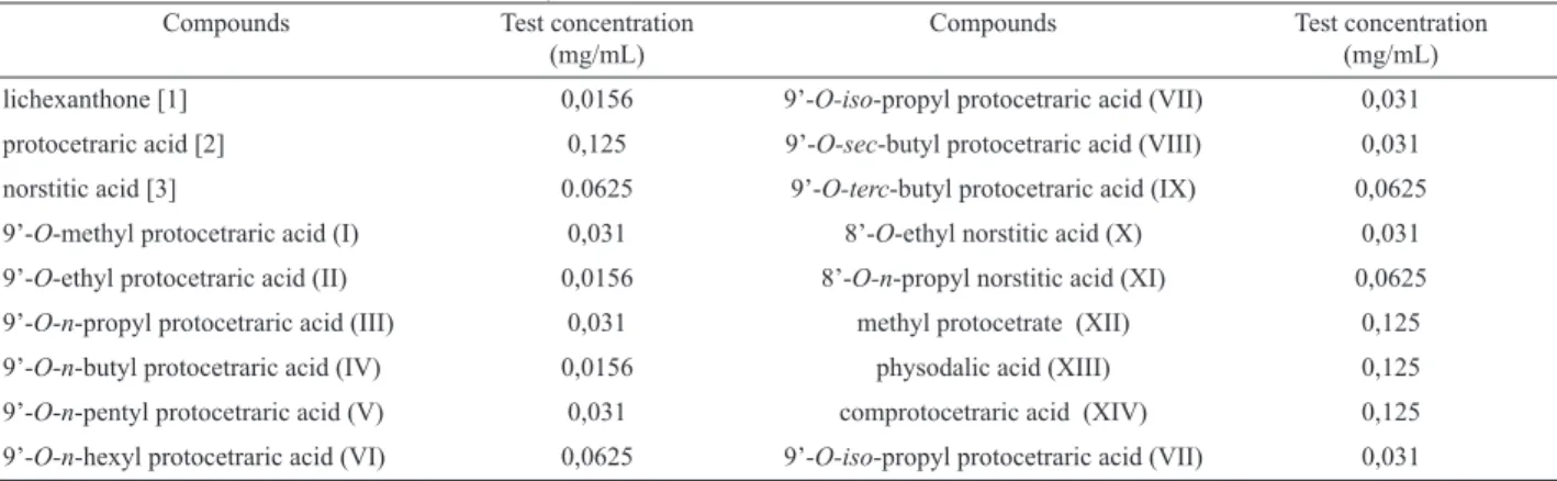
The cytotoxic effect of the compounds tested in peritoneal macrophages was assessed by the MTT assay as previously described (Table 1, Figure 1) (Mosmann, 1983). The tested concentration was show in the Table 1. It was used the concentration which the viability was higher or the same of 50%.
Table 1. Tested concentrations used in the assays.
Compounds Test concentration (mg/mL)
Compounds Test concentration (mg/mL)
lichexanthone [1] 0,0156 9’-O-iso-propyl protocetraric acid (VII) 0,031
protocetraric acid [2] 0,125 9’-O-sec-butyl protocetraric acid (VIII) 0,031
norstitic acid [3] 0.0625 9’-O-terc-butyl protocetraric acid (IX) 0,0625
9’-O-methyl protocetraric acid (I) 0,031 8’-O-ethyl norstitic acid (X) 0,031
9’-O-ethyl protocetraric acid (II) 0,0156 8’-O-n-propyl norstitic acid (XI) 0,0625
9’-O-n-propyl protocetraric acid (III) 0,031 methyl protocetrate (XII) 0,125
9’-O-n-butyl protocetraric acid (IV) 0,0156 physodalic acid (XIII) 0,125
9’-O-n-pentyl protocetraric acid (V) 0,031 comprotocetraric acid (XIV) 0,125
9’-O-n-hexyl protocetraric acid (VI) 0,0625 9’-O-iso-propyl protocetraric acid (VII) 0,031
Initial stage of the immune response macrophages is involved and they can release many mediators such as hydrogen peroxide (H2O2) and nitric oxide (NO). The killing of intracellular and extracellular pathogens by phagocytes is due in part to the production of reactive oxygen intermediate (ROIs). The H2O2 is responsible for killing internalized bacteria in phagocytosis. The NO has central functions in innate immunity and regulation of immune functions. It has important roles in inhibiting tumor cell adhesion, arresting the growth of tumor cells and contributing to
the control of microbial pathogens (Maragos et al., 1993; Kong et al., 1996; Rajan et al., 1996; Bogdan et al., 2000; Carlos et al., 2003; Chakravortty & Hensel, 2003). The antimicrobial
and cytotoxic actions of NO are enhanced by other macrophage products such as H2O2 and superoxide (MacMicking et al.,
1997).
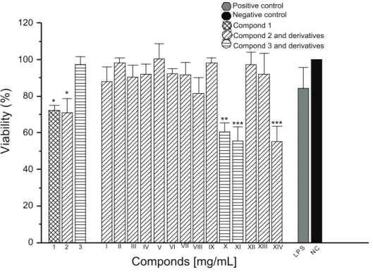
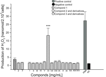
Our results showed that the compounds I, V, VIII and IX induced a small production of H2O2 (Figure 2), however the compound VI showed a high production of this mediator when it was added to the cell culture (approximately 18 nmols/2 x 105 cells). Analyzing the production of NO (Figure 3), it was observed that compounds VIII, IX and XIII induced a small production of this mediator when compared with the negative control, with IX presenting statistical difference, p < 0.05. The compounds I, II and V were more representative in the production
Figure 1. Determination of the viability in macrophages peritoneal culture of Swiss mice in the presence of phenolic substances
isolated from Brazilian lichens and their modiication structural products. * p < 0.05, **p < 0.01, ***p < 0.001
Figure 2. Determination of H2O2 in macrophages peritoneal culture of Swiss mice in the presence of presence of phenolic substances
Figure 3. Determination of NO in macrophages peritoneal culture of Swiss mice in the presence of presence of phenolic substances
isolated from Brazilian lichens and their modiication structural products. *p < 0.05, ***p < 0.001
The compound [2] (protocetraric acid) presents three
functions: alcohol, acid and aldehyde that were modiied in this
study and showed high production of NO. When the aldehyde
function was modiied for alcohol (XII) or the acid function
was modiied for ester (XIV), H2O2 and NO production was not observed. In contrast, when the alcohol function was substituted for ether, induced a small NO production, exception compound V demonstrated a higher release of this mediator. However, a small
production of H2O2 induced by compounds V, VIII and IX and a high liberation caused by compound VI can be explained due to higher liphophylic of the molecule because of the presence of the n-hexyl radical in the position 9’. The norstitic acid [3] and their derivatives (X and XI) did not induced the production of H2O2 and NO by the macrophages. The lichexanthone [1]
showed signiicative production of NO and its derivatives must
be subsequently studied.
O
O OH
OCH3 H3CO
CH3
O O CH3
HO CHO
O
OH
H3C CO2H
CH2OH
O O CH3
HO CHO
O
OH CH3
O O
H HO
O O CH3
HO
CHO O
OH CH2OR
H3C CO2H O
O CH3
HO CHO
O
OH CH3
O O
H H3CH2CO
O O CH3
HO
CHO O
OH CH3
O O
H
H3CH2CH2CO
[2] [3]
[1]
X XI
XII
XIII XIV
O O CH3
HO
CHO O
OH
H3C CO2CH3 CH2OH
O O CH3
HO
CHO O
OH
H3C CO2H CH2OCOCH3
O O CH3
HO
CH2OH O
OH
H3C CO2H CH2OH
I R = CH3 II R = CH2CH3 III R = CH2CH2CH3
IV R = CH2(CH2)2CH3
V R = CH2(CH2)3CH3
VI R = CH2(CH2)4CH3
VII R = CH(CH3)2
VIII R = CH(CH3)CH2CH3
This research demonstrated stimulatory activities of some phenolic compounds isolated from lichens on H2O2 and NO production by murine macrophages, suggesting that these compounds are activators of macrophages. The induction of H2O2 and NO may contribute in the immunoprevention of cancer and infecctious diseases.
In conclusion, structure-activity relationships suggest several synthetic directions for further improvement of immunological activity. Protocetraric acid derivatives appear to be promising as immunostimulating agents.
REFERENCES
Abe J, Bradford BC 1999. Fyn and JAK2 mediate ras activation
by reactive oxygen species. J Biol Chem 274: 21003-21010.
Bogdan C, Rollinghoff M, Diefenbach A 2000. Reactive oxygen and reactive nitrogen intermediates in innate and speciic
immunity. Curr Opin Immunol 12: 64-76.
Carlos IZ, Sgarbi DBG, Santos GC, Placeres MCP 2003.
Sporothrix schenckii lipid inhibits macrophage
phagocytosis: involvement of nitric oxide and tumor necrosis factor-α. Scand J Immunol 57: 214-220.
Chakravortty D, Hensel M 2003. Inducible nitric oxide synthase
and control of intracellular bacterial pathogens. Microbes
Infect 5: 621-627.
Costa MT, Fabeni RC, Aptekmann KP, Machado RR 2003.
Diferentes papéis do óxido nítrico com ênfase nas neoplasias. Cienc Rural 33: 967-974.
Elix JA, Engkaninan U 1975. The structure of galbinic acid. A depsidone from the lichen Usnea undulata. Aust J Chem
28: 1793-1797.
Fahselt D 1994. Secondary biochemistry of lichens. Symbiosis
16: 117-165.
Gao Y, Gao H, Chan E, Tang W, Xu A, Yang H, Huang M, Lan J, Li X, Duan W, Xu C, Zhou S 2005. Antitumor activity and underlying mechanisms of ganopoly, the reined
polysaccharides extracted from Ganoderma Lucidum, in Mice. Immunol Invest 34: 171-198.
Garcia X, Stein F 2006. Nitric oxide. Semin Pediatr Infect Dis
17: 55-57.
Garner P, Park JM 1992. 1,1-Dimethylethyl (S)- or
(R)-4-formyl-2,2-Dimethyl-3-oxazolidinecarboxylate: a useful serinal
derivative. Org Synth 70: 18-28.
Green LC, Wagner DA, Glogowski J, Skipper PL, Wishnok JS,
Tannenbaum SR 1982. Analysis of nitrate, nitrite, and
[15N] nitrate in biological luids. Anal Biochem 126: 131-138.
Huneck S, Yoshimura I 1996. Identiication of Lichen Substances.
Berlin: Springer-Verlag.
Kim GY, Choi GS, Lee SH, Park YM 2004. Acidic polysaccharide
isolated from Phellinus linteus enhances through the
up-regulation of nitric oxide and tumor necrosis factor-α
from peritoneal macrophages. J Ethnopharmacol 95:
69-76.
Kong L, Dunn GD, Keefer LK, Korthuis RJ 1996. Nitric oxide
reduces tumor cell adhesion to isolated rat postcapillary venules. Clin Exp Metastasis 14: 335-343.
MacMicking JD, Xie QW, Nathan C 1997. Nitric oxide and
macrophage function. Annu Rev Immunol 15: 323-350.
Maragos CM, Wang JM, Hrabie JA, Oppenheim JJ, Keefer LK 1993. Nitric oxide/nucleophile complexes inhibit the in vitro proliferation of A375 melanoma cells via nitric
oxide release. Cancer Res 53: 564-568.
Mosmann T 1983. Rapid colorimetric assay for cellular growth
and survival: application to proliferation and cytotoxicity assays. J Immunol Methods 65: 55-63.
Pick E, Keisari Y 1980. A simple colorimetric method for the measurement of hydrogen peroxide produced by cells in culture. J Immunol Methods 38: 161-170.
Pick E, Mizel D J 1981. Rapid microassays for the measurement of superoxide and hydrogen peroxide production by macrophages in culture using an automatic enzyme immunoassay reader. J Immunol Methods 46: 211-226.
Rajan TV, Porte P, Yates JA, Keefer L, Shultz LD 1996. Role
of nitric oxide in host defense against an extracellular, metazoan parasite. Infect Immun 64: 3351-3353. Ramasarma T 1990. H2O2 has a role in cellular regulation.
Indian J Biochem Bio 27: 269-74.
Santos LC, Honda NK, Carlos IZ, Vilegas W 2004. Intermediate
reactive oxygen and nitrogen from macrophages induced by Brazilian lichens. Fitoterapia 75: 473-479.
Schafer FQ, Buettner GR 2001. Redox state of the cell as viewed
though the glutathione disulide/glutathione couple. Free
Radical Biol Med 30: 1191-1212.
Schepetkin IA, Quinn MT 2006. Botanical polysaccharides:
Macrophage immunomodulation and therapeutic potential. Int Immunopharmacol 6: 317-333.
Schiller M, Metze D, Luger TA, Grabbe S, Gunzer M 2006.
Immune response modiiers-mode of action. Exp
Dermatol 15: 331-341.
Suzuki I, Itani T, Ohno N, Oikawa S, Sato K, Miyazaki T, Yadomae T 1985. Effect of a polysaccharide fraction from Grifola frondosa on immune response in mice. J
Pharmacobio-Dynam 8: 217-226.
Wolfrom ML, Thompson A 1963. Reduction products-reduction
with sodium borohydrate. In: Whistler RL, Wolfrom ML Methods in Carbohydrate Chemistry. New York:
Academic Press, p. 65-68.
Zheng R, Jie S, Hanchuan D, Moucheng W 2005. Characterization
and immunomodulating activities of olysaccharide from