Endogenous Murine BST-2/Tetherin Is Not a
Major Restriction Factor of Influenza A Virus
Infection
Sarah L. Londrigan1☯, Michelle D. Tate2,3☯, Emma R. Job1, Jessica M. Moffat1, Linda M. Wakim1, Christopher A. Gonelli1, Damien F. J. Purcell1, Andrew G. Brooks1, Jose A. Villadangos1,4, Patrick C. Reading1,5, Justine D. Mintern4*
1Department of Microbiology and Immunology, Peter Doherty Institute for Infection and Immunity, The University of Melbourne, Parkville, Victoria, 3010, Australia,2Centre for Innate Immunity and Infectious Diseases, MIMR-PHI Institute of Medical Research, Clayton, Victoria, 3168, Australia,3Monash University, Clayton, Victoria, 3168, Australia,4Department of Biochemistry and Molecular Biology, The University of Melbourne, Bio21 Molecular Science and Biotechnology Institute, 30 Flemington Rd, Parkville, Victoria, 3010, Australia,5WHO Collaborating Centre for Reference and Research on Influenza, Victorian Infectious Diseases Reference Laboratory, at the Peter Doherty Institute for Infection and Immunity, Melbourne, Victoria, 3000, Australia
☯These authors contributed equally to this work. *[email protected]
Abstract
BST-2 (tetherin, CD317, HM1.24) restricts virus growth by tethering enveloped viruses to the cell surface. The role of BST-2 during influenza A virus infection (IAV) is controversial. Here, we assessed the capacity of endogenous BST-2 to restrict IAV in primary murine cells. IAV infection increased BST-2 surface expression by primary macrophages, but not alveolar epithelial cells (AEC). BST-2-deficient AEC and macrophages displayed no differ-ence in susceptibility to IAV infection relative to wild type cells. Furthermore, BST-2 played little role in infectious IAV release from either AEC or macrophages. To examine BST-2 dur-ing IAV infectionin vivo, we infected BST-2-deficient mice. No difference in weight loss or in viral loads in the lungs and/or nasal tissues were detected between BST-2-deficient and wild type animals. This study rules out a major role for endogenous BST-2 in modulating IAV in the mouse model of infection.
Introduction
2 (tetherin; CD317, HM1.24) is a host cell protein of importance to viral immunity. BST-2 prevents newly generated viral particles from being released from the infected cell by forming a "tether" that retains virions at the cell surface. Viral tethering by BST-2 was first identified for human immunodeficiency virus [1,2] with subsequent studies showing activity against many enveloped viruses [3–6]. In addition to viral tethering, BST-2 can exert other immunomodula-tory functions during viral infection [7,8]. Specifically, BST-2 has been shown to modulate type 1 interferon (IFN) production [9,10] and trigger NFκB activation [11–13]. Consequently, BST-2 is a molecule of significant interest in host viral defense.
OPEN ACCESS
Citation:Londrigan SL, Tate MD, Job ER, Moffat JM, Wakim LM, Gonelli CA, et al. (2015) Endogenous Murine BST-2/Tetherin Is Not a Major Restriction Factor of Influenza A Virus Infection. PLoS ONE 10 (11): e0142925. doi:10.1371/journal.pone.0142925
Editor:Michael Schindler, Helmholtz Zentrum Muenchen—German Research Center for
Environmental Health, GERMANY
Received:May 13, 2015
Accepted:October 28, 2015
Published:November 13, 2015
Copyright:© 2015 Londrigan et al. This is an open access article distributed under the terms of the Creative Commons Attribution License, which permits unrestricted use, distribution, and reproduction in any medium, provided the original author and source are credited.
Data Availability Statement:All relevant data are within the paper and its Supporting Information files.
Influenza viruses belong to theOrthomyxoviridaefamily of enveloped viruses and are an important cause of respiratory disease worldwide. Type A influenza virus (IAV) is the major etiological agent capable of causing epidemics and pandemics in humans. Host anti-viral restriction factors have been described that can limit intracellular replication of IAV. Examples include interferon (IFN)-inducible transmembrane-3 [14,15], viperin [16] and myxovirus resistance gene A [17,18]. Currently, whether BST-2 acts to restrict the release of infectious IAV is controversial. Initial reports indicated that BST-2 limited release of IAV virus-like parti-cles (VLP) [19,20], as well as release of infectious IAV [21–23]. In contrast, several studies reported that BST-2 did not restrict IAV release from infected cells [19,24,25]. Overall, studies investigating the role of BST-2 during IAV infection have utilized cell lines engineered to over-express or suppress BST-2. Differences in BST-2 over-expression levels, the contribution of trans-formed cell lines and/or expression of BST-2 in heterologous cell lines may be important factors contributing to the conflicting data published to date. Hence, further studies are required to clarify the impact of BST-2 during IAV infection, using primary cells susceptible to IAV infection where physiologically relevant levels of endogenous BST-2 are expressed. To date, one study has examined the role of BST-2 during influenza virus infectionin vivo. Sur-prisingly, BST-2-deficient mice infected with influenza B virus (IBV) displayed a modest, but significant, reduction in lung viral titers at day 3 post-infection, although no significant differ-ences were detected at day 6 [7]. This finding is paradoxical to the proposed role of BST-2 in limiting viral release, and so far remains largely unexplained.
In the airways, primary alveolar epithelial cells (AEC) and macrophages represent two of the major cell types susceptible to IAV infection [26]. Herein, we have compared AEC and macrophages isolated from wild-type and BST-2-deficient mice [7], for their susceptibility to IAV infection and their ability to support productive IAV replicationin vitro. In addition, we have compared weight loss and viral replication following intranasal infection of wild type or BST-2-deficient mice with IAV. Together, our data indicate that endogenous BST-2 does not play a major role in host restriction of IAV in the mouse model of infection.
Materials and Methods
Mice
C57BL/6 and BST-2-deficient mice (kindly provided by M. Colonna, Washington University [7]) were bred and housed in specific pathogen-free conditions at the Bio21 Animal House Facility, The University of Melbourne. Mice (female and male) 6–10 weeks of age were used in experiments conducted in accordance with guidelines provided by National Health and Medi-cal Research Council of Australia. Experimental procedures were approved by the Animal Eth-ics Committees at the University of Melbourne (Application 1112261). Studies comply with Animal Research: Reporting In Vivo Experiment guidelines (S1 Checklist).
Cell lines, primary macrophages and primary alveolar epithelial cells
Cell lines used in this study included Madin-Darby canine kidney (MDCK) cells (American Type Culture Collection, ATCC), LA-4 mouse lung epithelial cells and the RAW264.7 macro-phage cell line (ATCC). Resident peritoneal exudate macromacro-phages were obtained from mice as previously described [27]. Macrophages were seeded into 8-well glass chamber-slides (Lab-Tek), incubated for 2 hours at 37°C and cell monolayers were washed to remove non-adherent cells. The next day, any remaining non-adherent cells were removed and the adherent macro-phages used in virus infection assays. Mouse primary lung epithelial cells were prepared as pre-viously described [28]. Briefly, lungs from mice were digested in 1.5 mg/ml Pronase (Roche, USA), 0.1 mg/ml DNase I (Sigma-Aldrich, USA) for 1 hour at 37°C in 5% CO2. Single cellCompeting Interests:The authors have declared that no competing interests exist.
suspensions were incubated with purified rat anti-mouse CD45 antibody (BD Biosciences, USA) and epithelial cells negatively enriched with BioMag goat anti-rat immunoglobulin-cou-pled magnetic beads (Qiagen, USA). Cells were cultured on collagen-coated (MP Biomedicals, USA) plates. To confirm alveolar epithelial cell purity, monolayers were detached with 3 mM EDTA and cells stained with mouse EpCAM, podoplanin (AEC type I) and anti-CD74 (AEC type II) antibodies and examined by flow cytometry.
Viruses
The representative IAV laboratory strain used in this study was HKx31, a reassortant of A/ PR8/34 (PR8, H1N1) with A/Aichi/2/68 (H3N2) bearing the H3N2 surface glycoproteins. In some experiments A/Brazil/11/78 (Brazil/78; H1N1) and A/Solomon Islands/3/2006 (Sol Is/06; H1N1) were used as representative seasonal strains. Sol Is/06 was obtained from the World Heath Organization Collaborating Centre for Reference and Research on Influenza, Mel-bourne, Australia. Virus was amplified in the allantoic cavity of 10-day-old embryonated hen’s eggs and titrated on MDCK cells by standard plaque assay [29].
Virus infection of mice
Groups of 5 mice were lightly anaesthetized (methoxyflurane) and infected with either 102or 104 PFU of HKx31 via the intranasal route. Mice were weighed daily and assessed for signs of clinical disease. Animals that had lost20% of their original body weight were euthanized by CO2
inha-lation followed by cervical dislocation. No adverse events occurred during these experiments. Lungs and nasal tissues were removed, homogenized and clarified by centrifugation. Titers of infectious virus in tissue homogenates were determined by standard plaque assays [29].
Virus infection assays
Mouse peritoneal macrophages and primary lung epithelial cells were infected with IAV and the percentage of IAV-infected cells determined as described previously for epithelial cells [30] and macrophages [31]. Briefly, cells were incubated with IAV in serum-free media for 1 hour at 37°C. Virus inoculum was removed and cells incubated at 37°C in serum-free media. IAV-infected cells were fixed with 80% vol/vol acetone at the indicated time points post-infection and stained using monoclonal antibody (mAb) MP3.10g2.1C7 (WHO Collaborating Centre for Reference and Research on Influenza, Melbourne, Australia) specific for IAV nucleopro-tein. Virus-infected cells were co-stained with 4',6-diamidino-2-phenylindole (DAPI) or propi-dium iodide (PI). A minimum of 200 cells were scored for each sample.
Virus growth assays
Cells were infected with IAV as described above. As trypsin counteracts BST-2 anti-viral activ-ity [32], this was omitted in the infection media, thereby limiting IAV replication to a single round. Cell supernatants were collected at the indicated time-points post-infection, and incu-bated with 4μg/ml L-(tosylamido-2-phenyl) ethyl chloromethyl ketone (TPCK)-treated tryp-sin (Sigma Aldrich) for 30 minutes at 37°C to facilitate cleavage of the viral hemagglutinin [33] before the titers of infectious virus were determined by standard plaque assay on MDCK cells [29].
BST-2 expression assays
non-specific staining via Fc receptors, then stained with rat anti-BST-2 mAb conjugated to FITC (generated in house) or rat IgG1k-FITC isotype control antibody (Biolegend). In some experiments, BST-2 expression was monitored post-infection with IAV (as described above) at the indicated virus doses and time-points. Cells were treated with 1000 international units (IU)/ml recombinant murine IFNα(R&D Systems) as a control to upregulate BST-2 surface expression.
Results
BST-2 expression is upregulated on murine macrophages but not
alveolar epithelial cells in response to IAV infection
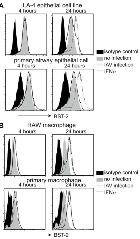
First, flow cytometry was used to examine BST-2 expression by murine AEC and macrophages that were uninfected, or had been infected 4 and 24 hours previously with IAV strain HKx31 (H3N2). BST-2 expression was analysed using the LA-4 AEC line and primary AEC, as well as using the RAW264.7 macrophage cell line and primary macrophages. Peritoneal exudate mac-rophages were used given the difficulty in obtaining sufficient numbers of alveolar macro-phages via bronchoalveolar lavage (BAL). Note that peritoneal and alveolar macromacro-phages exhibit similar susceptibility and ability to support IAV infection [34,35]. For epithelial cells, uninfected LA-4 cells and primary AEC expressed cell-surface BST-2, however levels did not increase further following IAV infection. Of interest, culture of AEC in the presence of IFNα did result in upregulation of cell-surface BST-2 (Fig 1A). In contrast to epithelial cells, macro-phages upregulated cell-surface BST-2 in response to IAV and levels were similar (RAW264.7), or higher (primary cells), than those elicited in response to IFNα(Fig 1B).
BST-2 does not modulate susceptibility to IAV infection or release of
newly synthesized virions from murine AEC and macrophages
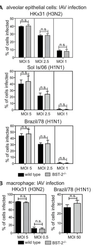
Next, we examined the impact of BST-2 on the susceptibility of primary AEC and macrophages to IAV infection using cells from wild type (WT) and BST-2-deficient animals. We confirmed expression patterns of 2 by flow cytometry for primary macrophages isolated from BST-2+/+, BST-2+/-and BST-2-/-mice (S1 Fig). Next, cells isolated from WT or BST-2-deficient mice were inoculated with IAV at different multiplicities of infection (MOI) and the percentage of IAV-infected cells was determined 6–8 hours later by detection of newly synthesized viral nucleoprotein (NP) [35]. Viral NP was not detected at 2 hours post infection, indicating its presence at later time points was due to newly synthesized viral protein and not input virus (S2 Fig). Overall, the percentage of IAV-infected AEC did not differ in the presence or absence of BST-2 at any MOI tested for the IAV laboratory strain HKx31, or the representative seasonal strains Brazil/78 and Sol Is/06 (Fig 2A). Similar to AEC, macrophages exhibited equivalent sus-ceptibility to IAV infection regardless of the presence or absence of BST-2 for both the HKx31 and Brazil/78 strains (Fig 2B). The Sol Is/06 IAV strain did not efficiently infect primary murine macrophages, and therefore was not assessed (data not shown).
Fig 1. BST-2 expression is upregulated on murine macrophages but not alveolar epithelial cells in response to influenza A virus.Monolayers of (A) the LA-4 AEC line and primary AEC, or (B) RAW264.7 macrophages and primary macrophages were incubated (i) with a MOI of 5 (HKx31) for 1 hour at 37°C and washed to remove excess virus (IAV infection, solid black line), (ii) in 1000 IU/ml recombinant mouse IFNα
(dashed line) or (iii) in media alone (no infection, grey histogram). Cells were then incubated at 37°C for a total of 4 or 24 hours and levels of cell-surface BST-2 determined by flow cytometry. For each cell type, the isotype control (solid black histograms) is shown for‘no infection’cells only but is representative of profiles obtained using IAV-infected and IFNα-treated cells. Data are representative of 3 independent experiments.
Fig 2. BST-2 expression does not modulate IAV susceptibility of murine epithelial cells and macrophages to IAV infection.(A) Primary AEC or (B) macrophages isolated from BST-2 wild type (WT) and BST-2-deficient (BST-2-/-) mice were incubated with the indicated MOI and strain of IAV for 1 hour at 37°C, washed to remove excess virus and cultured as indicated. Monolayers were fixed at 8 hours post-infection before staining by immunofluorescence to detect newly synthesized viral NP. Data show the mean (±1 SD) pooled from 3 independent experiments. n.s. = no significant difference,p=>0.05, two-way ANOVA
followed by Bonferroni analysis.
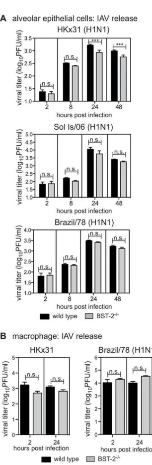
supernatants by standard plaque assays. For HKx31, at 24 and 48 hours post-infection, titres of infectious virus released from BST-2-deficient AEC were significantly reduced compared to WT AEC, although this was not the case for the Brazil/78 or Sol Is/06 IAV strains, where viral release of these strains from AEC occurred independently of BST-2 (Fig 3A). For macrophages, IAV titres did not increase between 2 and 24 hours following infection with either HKx31 or Brazil/78, and this was unaffected by the presence or absence of BST-2 (Fig 3B). In these exper-iments, MDCK cells were included as a control to confirm that the virus inoculum could give rise to productive IAV replication and release from cells (data not shown).
Lack of BST-2 does not alter the susceptibility of mice to IAV infection or
the ability of IAV to replicate in the airways
To assess the impact of BST-2in vivowe compared the susceptibility of WT BST-2 (bst-2+/+or
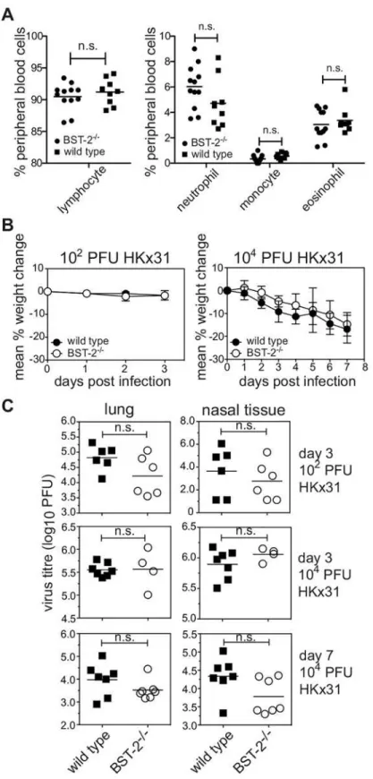
bst-2+/-) or BST-2-deficient (bst-2-/-) mice to IAV infectionin vivo. First, we assessed the status of BST-2-deficient mice under resting conditions. Proportions of lymphocytes, monocytes, neutrophils and eosinophils in the peripheral blood did not differ significantly between BST-2-deficient and wild type mice (Fig 4A) indicating that the absence of BST-2 did not elicit overt alterations in immune homeostasis. BST-2-deficient mice were inoculated with IAV strain HKx31 via the intranasal route. Infection of mice with this strain elicits a mild respiratory ill-ness and weight loss is a reliable indicator of disease severity [36,37]. Infection with HKx31 provokes a significant increase in IFNαin the respiratory tract [38] (S3 Fig) and consequently elicits conditions under which BST-2 expression is likely to be elevated. Elevated BST-2 expres-sion has also been detected on immune cells isolated from the respiratory tract on IAV-infected mice [39]. Mice infected with 102PFU of HKx31 lost little weight by day 3 post-infection whereas infection with 104PFU resulted in progressive weight loss, however no significant dif-ferences were recorded between WT or BST-2-deficient mice (Fig 4B). IAV replication in the airways was assessed in the lung and nasal tissues at day 3 and day 7 post-infection and no sig-nificant differences were recorded between WT and BST-2-deficient mice at either time point, irrespective of inoculum dose (Fig 4C). Therefore, endogenous BST-2 does not play a major role in restricting IAV infectionin vivo.
Discussion
Here we have undertaken a comprehensive analysis of the role of endogenous BST-2 in restrict-ing IAV infection by performrestrict-ing, to our knowledge, the first analysis of infection of BST-2-deficient primary murine cells with IAV and importantly, IAV infection of BST-BST-2-deficient mice. Our analyses rule out a major role for BST-2 as a host molecule that restricts IAV in the mouse model of infection.
BST-expression in AEC [21], however the ability of IAV to antagonize BST-2 has been disputed [25] and we detect no evidence for impaired BST-2 expression following IAV infection. In summary, we observe that BST-2 is expressed by cell types of importance to IAV infection and consequently has the potential to modulate IAV infection.
Analysis of IAV infection of primary cellsin vitrofailed to support a major role for BST-2 in restricting productive virus replication and release. In AEC, the role for BST-2 varied depending on the IAV strain tested. For HKx31, BST-2 promotes, rather than inhibits, the release of newly synthesized virions from IAV-infected cells. This was not the case following infection with IAV Brazil/78 or Sol Is/06, and therefore the significance of a role for BST-2 in promoting IAV release is unclear, Regardless, we detect no evidence for a major role for BST-2 in restricting IAV release in AEC under the experimental conditions tested. It is well established that macrophages do not support productive replication of seasonal IAV [27,35] and herein we demonstrate that the absence of BST-2 did not reverse this phenotype, despite increased expression of BST-2 following IAV infection of macrophages from WT mice. In summary, BST-2 is not acting as a dominant restriction factor for IAV in either murine AEC or macrophages.
Numerous studies have addressed the ability of BST-2 to restrict release of different viruses
in vitro, however less is known regarding its ability to modulate viral infectionsin vivo. The availability of BST-2-deficient mice [7,41] has allowed the impact of viral infection to be assessed in the absence of endogenous BST-2. Infection of BST-2-deficient mice with Chikun-gunya virus (CHIKV) results in increased viremia, in accordance with its capacity to tether CHIKVin vitro[42]. Similarly, infection of BST-2 deficient mice with Moloney murine leuke-mia virus elicits enhanced viral titers [41]. In contrast, BST-2 does not act according to its pre-dicted role as a viral tetherin in several infection models. Infection of BST-2-deficient mice with vesicular stomatitis virus, a target of BST-2 tetheringin vitro[6], results in reduced, rather than increased, viral titers [7]. This was also the case following infection with IBV, where BST-2-deficient mice display reduced viral titers in the lung [7]. Herein, we also report reduced lev-els of infectious IAV in cell supernatants from BST-2-deficient AEC, although this was only under specific experimental conditions andin vivoviral titers in the respiratory tract were not significantly different to those in WT animals. Increased viral titers in the presence of BST-2 is perplexing as it excludes a major role for BST-2 in restricting viral release and instead, impli-cates a pro-viral role for BST-2. One explanation for this is the potential role for BST-2 in enhancing, rather than inhibiting, viral entry. This is reported for cytomegalovirus, where BST-2 exerts a reverse-tethering mechanism to promote entry [43]. Note, however, that we find no evidence of BST-2 promoting infectious entry of IAV, given that BST-2-deficient AEC and/or macrophages are equally susceptible to infection compared to WT cells. How BST-2 acts to promote, rather than inhibit, IAV release remains to be determined. Regardless, our infection studies revealed similar kinetics of weight loss and viral replication in BST-2-deficient and WT mice. Therefore, in the mouse, BST-does not have a major impact on IAV infection outcomesin vivo.
Together, our data do not support a role for endogenous BST-2 as a major factor restricting infectious entry of primary murine IAV into target cells or in limiting the release of newly
2-deficient (BST-2-/-) mice were incubated with the indicated strain of IAV for 1 hour at 37°C, washed to remove excess virus and cultured. AEC were infected at a MOI of 1 of HKx31, Brazil/78 and Sol Is/06 and macrophages infected at a MOI 5 for HKx31 and a MOI of 50 for Brazil/78. Culture supernatants were removed at 2 hours, or at 8, 24 or 48 hours as indicated, clarified by centrifugation and titres of infectious virus were determined by plaque assay on MDCK cells. Data is displayed as viral titer (log10PFU/ml) and represent the mean (±1 SD) from triplicate samples. Data is representative of 2 independent experiments. n.s. = no
significant difference,***p<0.001, two-way ANOVA followed by Bonferroni analysis.
synthesized virions from infected cells. In conclusion, based on our studies in the moue model of infection, it is likely that murine IAV infection and replication can be inhibited by the (com-bined) action of a number of host molecules, of which BST-2 may contribute only a minor role.
Supporting Information
S1 Checklist. NC3Rs ARRIVE Guidelines Checklist.
(PDF)
S1 Fig. BST-2 expression.Primary macrophages isolated frombst-2+/+(solid black line), bst-2+/-(dashed line) andbst-2-/-(black histogram) were cultured for 24 hours with 1000 IU/ml of IFNαbefore analysis of cell-surface BST-2 expression by flow cytometry. Isotype control shown is forbst-2+/+cells and was indistinguishable from that observed forbst-2-/-andbst-2
+/-cells. (EPS)
S2 Fig. Assessment of viral nucleoprotein expression in primary murine epithelial cells and macrophages following IAV infection.Primary AEC or macrophages were incubated with HKx31 at an MOI of 5 for 1 hour at 37°C, washed to remove excess virus and cultured. Mono-layers were fixed with 80% vol/vol acetone at 2 or 8 hours post-infection before staining by immunofluorescence to detect newly synthesized viral NP (green) and with DAPI to stain the nucleus (blue). Similar results were also obtained with Brazil/78 for AEC and macrophages and Sol Is/06 for AEC (data not shown). Images were acquired with a Zeiss LSM700 or Olympus IX70 confocal microscope in conjunction with Zen2012 software.
(PDF)
S3 Fig. Influenza A virus infection elicits IFNαin the respiratory tract.Mice were infected with 104PFU HKx31 via the intranasal route. Lungs were removed 3 days following infection. IFNαin total lung homogenates was measured by enzyme-linked immunosorbent assay. Anti-bodies to IFNαused for capture (22100–1) and detection (32100–1) were from PBL Assay Sci-ence (NJ, USA). Data is pooled from two independent experiments. Each symbol represents an individual mouse and the bar indicates the mean.p<0.05, Student's unpairedt-test.
(EPS)
Acknowledgments
We acknowledge technical support from J. Corbin (Walter and Eliza Hall Institute).
Author Contributions
Conceived and designed the experiments: JDM SLL MDT PCR JAV. Performed the experi-ments: SLL MDT ERJ JMM CAG LMW. Analyzed the data: SLL MDT ERJ JMM CAG LMW. Contributed reagents/materials/analysis tools: AGB DFJP. Wrote the paper: JDM SLL MDT PCR.
intranasal route. Mice were weighed daily and the results expressed as the mean percentage weight change per group (±1 SEM) relative to original body weight. (C) Virus titres were determined in clarified homogenates
prepared from lungs and nasal tissues using a standard plaque assay on MDCK cells. Symbols show titres from individual animals and horizontal bars represent the mean virus titre. n.s. = no significant difference,
p=>0.05, Student’st-test; two-tailed.
References
1. Neil SJ, Zang T, Bieniasz PD. Tetherin inhibits retrovirus release and is antagonized by HIV-1 Vpu. Nature. 2008; 451(7177):425–30. Epub 2008/01/18. doi:10.1038/nature06553PMID:18200009. 2. Van Damme N, Goff D, Katsura C, Jorgenson RL, Mitchell R, Johnson MC, et al. The interferon-induced
protein BST-2 restricts HIV-1 release and is downregulated from the cell surface by the viral Vpu pro-tein. Cell Host Microbe. 2008; 3(4):245–52. doi:10.1016/j.chom.2008.03.001PMID:18342597; PubMed Central PMCID: PMC2474773.
3. Jouvenet N, Neil SJ, Zhadina M, Zang T, Kratovac Z, Lee Y, et al. Broad-spectrum inhibition of retroviral and filoviral particle release by tetherin. J Virol. 2009; 83(4):1837–44. Epub 2008/11/28. doi:10.1128/ JVI.02211-08PMID:19036818; PubMed Central PMCID: PMC2643743.
4. Kaletsky RL, Francica JR, Agrawal-Gamse C, Bates P. Tetherin-mediated restriction of filovirus bud-ding is antagonized by the Ebola glycoprotein. Proc Natl Acad Sci U S A. 2009; 106(8):2886–91. doi: 10.1073/pnas.0811014106PMID:19179289; PubMed Central PMCID: PMC2650360.
5. Sakuma T, Noda T, Urata S, Kawaoka Y, Yasuda J. Inhibition of Lassa and Marburg virus production by tetherin. J Virol. 2009; 83(5):2382–5. doi:10.1128/JVI.01607-08PMID:19091864; PubMed Central PMCID: PMC2643706.
6. Weidner JM, Jiang D, Pan XB, Chang J, Block TM, Guo JT. Interferon-induced cell membrane proteins, IFITM3 and tetherin, inhibit vesicular stomatitis virus infection via distinct mechanisms. J Virol. 2010; 84 (24):12646–57. doi:10.1128/JVI.01328-10PMID:20943977; PubMed Central PMCID: PMC3004348. 7. Swiecki M, Wang Y, Gilfillan S, Lenschow DJ, Colonna M. Cutting edge: paradoxical roles of BST2/
tetherin in promoting type I IFN response and viral infection. J Immunol. 2012; 188(6):2488–92. doi:10. 4049/jimmunol.1103145PMID:22327075; PubMed Central PMCID: PMC3522186.
8. Li SX, Barrett BS, Heilman KJ, Messer RJ, Liberatore RA, Bieniasz PD, et al. Tetherin promotes the innate and adaptive cell-mediated immune response against retrovirus infection in vivo. J Immunol. 2014; 193(1):306–16. doi:10.4049/jimmunol.1400490PMID:24872193; PubMed Central PMCID: PMC4163935.
9. Blasius AL, Giurisato E, Cella M, Schreiber RD, Shaw AS, Colonna M. Bone marrow stromal cell anti-gen 2 is a specific marker of type I IFN-producing cells in the naive mouse, but a promiscuous cell sur-face antigen following IFN stimulation. J Immunol. 2006; 177(5):3260–5. PMID:16920966.
10. Cao W, Bover L, Cho M, Wen X, Hanabuchi S, Bao M, et al. Regulation of TLR7/9 responses in plasma-cytoid dendritic cells by BST2 and ILT7 receptor interaction. J Exp Med. 2009; 206(7):1603–14. doi:10. 1084/jem.20090547PMID:19564354; PubMed Central PMCID: PMC2715090.
11. Galao RP, Le Tortorec A, Pickering S, Kueck T, Neil SJ. Innate sensing of HIV-1 assembly by Tetherin induces NFkappaB-dependent proinflammatory responses. Cell Host Microbe. 2012; 12(5):633–44. doi:10.1016/j.chom.2012.10.007PMID:23159053; PubMed Central PMCID: PMC3556742. 12. Tokarev A, Suarez M, Kwan W, Fitzpatrick K, Singh R, Guatelli J. Stimulation of NF-kappaB activity by
the HIV restriction factor BST2. J Virol. 2013; 87(4):2046–57. doi:10.1128/JVI.02272-12PMID: 23221546; PubMed Central PMCID: PMC3571454.
13. Cocka LJ, Bates P. Identification of alternatively translated Tetherin isoforms with differing antiviral and signaling activities. PLoS Pathog. 2012; 8(9):e1002931. doi:10.1371/journal.ppat.1002931PMID: 23028328; PubMed Central PMCID: PMC3460627.
14. Siegrist F, Ebeling M, Certa U. The small interferon-induced transmembrane genes and proteins. J Interferon Cytokine Res. 2011; 31(1):183–97. doi:10.1089/jir.2010.0112PMID:21166591. 15. Feeley EM, Sims JS, John SP, Chin CR, Pertel T, Chen LM, et al. IFITM3 inhibits influenza A virus
infection by preventing cytosolic entry. PLoS Pathog. 2011; 7(10):e1002337. doi:10.1371/journal.ppat. 1002337PMID:22046135; PubMed Central PMCID: PMC3203188.
16. Wang X, Hinson ER, Cresswell P. The interferon-inducible protein viperin inhibits influenza virus release by perturbing lipid rafts. Cell Host Microbe. 2007; 2(2):96–105. doi:10.1016/j.chom.2007.06. 009PMID:18005724.
17. Xiao H, Killip MJ, Staeheli P, Randall RE, Jackson D. The human interferon-induced MxA protein inhib-its early stages of influenza A virus infection by retaining the incoming viral genome in the cytoplasm. J Virol. 2013; 87(23):13053–8. doi:10.1128/JVI.02220-13PMID:24049170; PubMed Central PMCID: PMC3838145.
18. Pavlovic J, Zurcher T, Haller O, Staeheli P. Resistance to influenza virus and vesicular stomatitis virus conferred by expression of human MxA protein. J Virol. 1990; 64(7):3370–5. PMID:2161946; PubMed Central PMCID: PMC249583.
20. Yondola MA, Fernandes F, Belicha-Villanueva A, Uccelini M, Gao Q, Carter C, et al. Budding capability of the influenza virus neuraminidase can be modulated by tetherin. J Virol. 2011; 85(6):2480–91. doi: 10.1128/JVI.02188-10PMID:21209114; PubMed Central PMCID: PMC3067929.
21. Mangeat B, Cavagliotti L, Lehmann M, Gers-Huber G, Kaur I, Thomas Y, et al. Influenza virus partially counteracts restriction imposed by tetherin/BST-2. J Biol Chem. 2012; 287(26):22015–29. doi:10. 1074/jbc.M111.319996PMID:22493439; PubMed Central PMCID: PMC3381161.
22. Leyva-Grado VH, Hai R, Fernandes F, Belicha-Villanueva A, Carter C, Yondola MA. Modulation of an ectodomain motif in the influenza A virus neuraminidase alters tetherin sensitivity and results in virus attenuation in vivo. J Mol Biol. 2014; 426(6):1308–21. doi:10.1016/j.jmb.2013.12.023PMID: 24380762; PubMed Central PMCID: PMC3963499.
23. Dittmann M, Hoffmann HH, Scull MA, Gilmore RH, Bell KL, Ciancanelli M, et al. A serpin shapes the extracellular environment to prevent influenza A virus maturation. Cell. 2015; 160(4):631–43. doi:10. 1016/j.cell.2015.01.040PMID:25679759; PubMed Central PMCID: PMC4328142.
24. Bruce EA, Abbink TE, Wise HM, Rollason R, Galao RP, Banting G, et al. Release of filamentous and spherical influenza A virus is not restricted by tetherin. J Gen Virol. 2012; 93(Pt 5):963–9. doi:10.1099/ vir.0.038778–0PMID:22258861.
25. Winkler M, Bertram S, Gnirss K, Nehlmeier I, Gawanbacht A, Kirchhoff F, et al. Influenza A virus does not encode a tetherin antagonist with Vpu-like activity and induces IFN-dependent tetherin expression in infected cells. PLoS ONE. 2012; 7(8):e43337. doi:10.1371/journal.pone.0043337PMID:22952667; PubMed Central PMCID: PMC3428345.
26. Yu WC, Chan RW, Wang J, Travanty EA, Nicholls JM, Peiris JS, et al. Viral replication and innate host responses in primary human alveolar epithelial cells and alveolar macrophages infected with influenza H5N1 and H1N1 viruses. J Virol. 2011; 85(14):6844–55. doi:10.1128/JVI.02200-10PMID:21543489; PubMed Central PMCID: PMC3126566.
27. Reading PC, Miller JL, Anders EM. Involvement of the mannose receptor in infection of macrophages by influenza virus. J Virol. 2000; 74(11):5190–7. PMID:10799594; PubMed Central PMCID: PMC110872.
28. Thomas BJ, Porritt RA, Hertzog PJ, Bardin PG, Tate MD. Glucocorticosteroids enhance replication of respiratory viruses: effect of adjuvant interferon. Sci Rep. 2014; 4:7176. doi:10.1038/srep07176PMID: 25417801.
29. Anders EM, Hartley CA, Reading PC, Ezekowitz RA. Complement-dependent neutralization of influ-enza virus by a serum mannose-binding lectin. J Gen Virol. 1994; 75 (Pt 3):615–22. PMID:8126457. 30. Londrigan SL, Turville SG, Tate MD, Deng YM, Brooks AG, Reading PC. N-linked glycosylation
facili-tates sialic acid-independent attachment and entry of influenza A viruses into cells expressing DC-SIGN or L-DC-SIGN. J Virol. 2011; 85(6):2990–3000. doi:10.1128/JVI.01705-10PMID:21191006; PubMed Central PMCID: PMC3067946.
31. Ng WC, Liong S, Tate MD, Irimura T, Denda-Nagai K, Brooks AG, et al. The macrophage galactose-type lectin can function as an attachment and entry receptor for influenza virus. J Virol. 2014; 88 (3):1659–72. doi:10.1128/JVI.02014-13PMID:24257596; PubMed Central PMCID: PMC3911607. 32. Hammonds J, Wang JJ, Yi H, Spearman P. Immunoelectron microscopic evidence for Tetherin/BST2
as the physical bridge between HIV-1 virions and the plasma membrane. PLoS Pathog. 2010; 6(2): e1000749. doi:10.1371/journal.ppat.1000749PMID:20140192; PubMed Central PMCID: PMC2816691.
33. Skehel JJ, Wiley DC. Receptor binding and membrane fusion in virus entry: the influenza hemaggluti-nin. Annu Rev Biochem. 2000; 69:531–69. doi:10.1146/annurev.biochem.69.1.531PMID:10966468. 34. Tate MD, Brooks AG, Reading PC. Correlation between sialic acid expression and infection of murine
macrophages by different strains of influenza virus. Microbes Infect. 2011; 13(2):202–7. doi:10.1016/j. micinf.2010.10.004PMID:20974275.
35. Tate MD, Pickett DL, van Rooijen N, Brooks AG, Reading PC. Critical role of airway macrophages in modulating disease severity during influenza virus infection of mice. J Virol. 2010; 84(15):7569–80. doi: 10.1128/JVI.00291-10PMID:20504924; PubMed Central PMCID: PMC2897615.
36. Tate MD, Schilter HC, Brooks AG, Reading PC. Responses of mouse airway epithelial cells and alveo-lar macrophages to virulent and avirulent strains of influenza A virus. Viral Immunol. 2011; 24(2):77–88. doi:10.1089/vim.2010.0118PMID:21449718.
37. Tate MD, Ioannidis LJ, Croker B, Brown LE, Brooks AG, Reading PC. The role of neutrophils during mild and severe influenza virus infections of mice. PLoS ONE. 2011; 6(3):e17618. doi:10.1371/journal. pone.0017618PMID:21423798; PubMed Central PMCID: PMC3056712.
39. Moffat JM, Segura E, Khoury G, Caminschi I, Cameron PU, Lewin SR, et al. Targeting antigen to bone marrow stromal cell-2 expressed by conventional and plasmacytoid dendritic cells elicits efficient anti-gen presentation. Eur J Immunol. 2013; 43(3):595–605. Epub 2013/01/11. doi:10.1002/eji.201242799 PMID:23303646.
40. Julkunen I, Sareneva T, Pirhonen J, Ronni T, Melen K, Matikainen S. Molecular pathogenesis of influ-enza A virus infection and virus-induced regulation of cytokine gene expression. Cytokine Growth Fac-tor Rev. 2001; 12(2–3):171–80. PMID:11325600.
41. Liberatore RA, Bieniasz PD. Tetherin is a key effector of the antiretroviral activity of type I interferon in vitro and in vivo. Proc Natl Acad Sci U S A. 2011; 108(44):18097–101. doi:10.1073/pnas.1113694108 PMID:22025715; PubMed Central PMCID: PMC3207693.
42. Mahauad-Fernandez WD, Jones PH, Okeoma CM. Critical role for bone marrow stromal antigen 2 in acute Chikungunya virus infection. J Gen Virol. 2014; 95(Pt 11):2450–61. doi:10.1099/vir.0.068643–0 PMID:25053563; PubMed Central PMCID: PMC4202266.