Licence Creative Commom CC
RBCDH
1 Universidade do Estado do Rio de Janeiro. Laboratório de Ativi-dade Física e Promoção da Saúde. Rio de Janeiro, RJ. Brasil. 2 Universidade Salgado de Olivei-ra. Programa de Pós-graduação em Ciências da Atividade Física. Niterói, RJ. Brasil.
Received: 13 May 2014 Accepted: 29 October 2014
Efect of the number of sets on acute
cardiovascular responses during stretching
exercise
Efeito do número de séries nas respostas cardiovasculares
agudas durante exercício de alongamento
Tainah Lima1
Paulo Farinatti1,2
Walace Monteiro1,2
Abstract– Few studies have investigated the efects of manipulating variables of stretching exercise prescription on acute cardiovascular responses. he objective of this study was to compare the acute responses of heart rate, blood pressure and double product before, during and ater four sets of passive static stretching during unilateral hip lexion. he sample consisted of 16 adult men aged 18 to 27 years (22±2.8 years) with no experience in lexibility training. he cardiovascular variables were measured by photoplethysmog-raphy with continuous recording for 5 minutes at rest, during exercise and 10 minutes ater stretching. A diference was observed in all variables (p<0.05) when resting and post-exercise values were compared to exercise values. here was only an increase in blood pressure values between sets. Systolic blood pressure was increased in the third set compared to the irst [set 1 = 136 mmHg (±9.4); set 3 = 145 mmHg (±9.7)] and diastolic blood pressure was increased in the second set compared to the irst [set 1 = 79 mmHg (±6.7); set 2 = 84 mmHg (±9.1)]. Hypotension was not observed post-exercise. It was concluded that the stretching sets have a cumulative efect on systolic and diastolic blood pressure responses, but not on heart rate. However, the stretching protocol produced no hypotensive efect ater exercise.
Key words: Blood pressure; Muscle stretching exercise; Physical education and training.
Stretching and acute cardiovascular responses Lima et al.
INTRODUCTION
Flexibility is one of the components of health-related physical itness1,2. he
American College of Sports Medicine (ACSM)1 recommends two or three
weekly sessions of lexibility training which should consist of at least four sets involving the main muscle groups. Furthermore, it is recommended that stretching is performed to the point of discomfort in order to obtain gains in range of motion.
Although the variables of lexibility training prescription for healthy adults are well established, knowledge of the cardiovascular responses resulting from the manipulation of these variables is still insuicient. Within this context, heart rate (HR) and blood pressure (BP) can be used to control exercise-associated cardiovascular stress3. However, in contrast to strength4-6, aerobic7,8 or even concurrent exercise9,10, few studies have investigated the cardiovascular responses associated with the manipulation of variables of lexibility training prescription11-13.
Studies on the inluence of methodological aspects of lexibility ex-ercise prescription on cardiovascular responses are sparse. Many factors can afect acute cardiovascular responses to stretching exercise, including the number of sets, interval between stimuli or stimulus duration11-14. In this respect, some studies have investigated cardiovascular responses ma-nipulating these variables11-13, but important methodological limitations restrict the extrapolation of the results. Cornelius et al.11, for example, did not report the degree of joint range of motion of the subjects. here was also no mentioning of aspects of breathing control during exercise. he sum of these factors may exert diferent efects on cardiovascular responses due to the diferent levels of exercise-related stress14. Holt et al.13 used an oscillometric method and did not control the angle of movement during exercise, which may have caused a loss of tension during exercise, mini-mizing cardiovascular responses. he fact that the subjects performed and controlled their own stretching exercises may have afected the accuracy of the BP measurements since oscillometry is sensitive to movement and the arm should be held completely still during measurement15.
More recently, Farinatti et al.12 examined cardiovascular responses in a successive series of stretching exercise, controlling the level of lexibility and breathing pattern of the subjects. However, BP was measured manu-ally, a fact that could reduce accuracy of the measurement due to possible
artifacts caused by movement of the subject16. herefore, even when the
measurements were made by an experienced examiner, the BP values may have been underestimated when compared to more accurate noninvasive
methods such as photoplethysmography17.
METHODOLOGICAL PROCEDURES
Sample
Post hoc statistical power analysis was performed with the GPower 3.1 sotware considering an alpha value of 0.05, a sample of 16 subjects and an efect size of 0.25. A β value of 0.8 was obtained for F-statistics. here-fore, 16 asymptomatic men without experience in lexibility training were studied. he following exclusion criteria were adopted: use of medications interfering with cardiovascular responses; osteomyoarticular problems limiting participation in the exercise sessions; consumption of substances that could alter cardiovascular responses on the day of data collection; a high lexibility level tested by unilateral lexion of the hip with the knee extended, considering a cut-of value higher than 90o for this movement18; a positive Physical Activity Readiness Questionnaire (PAR-Q). Prior to study entry, the volunteers signed the informed consent form and the study was approved by the institutional Ethics Committee.
Experimental procedures
Data were collected during two visits. In the irst visit, the PAR-Q was applied, anthropometric measures were obtained, and lexibility was evaluated. he last assessment was necessary to verify homogeneity of the sample in terms of the range of motion observed because of the possible relationship between lexibility levels and HR and BP responses for the same
joint range of motion14. he pre-test range of motion was measured with
a Fleximeter® (Instituto Code de Pesquisa, Campinas, São Paulo, Brazil). In the second visit, HR, SBP and DBP were measured at rest and during
and ater exercise using the Finometer Pro® (Finapres Medical Systems,
Amsterdam, he Netherlands).
Stretching protocol
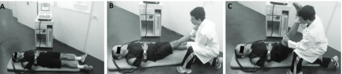
he stretching protocol was applied in a single visit. he subjects were asked not to perform any physical activity during the 24 hours prior to evaluation. During the stretching exercises, the examiner manipulated the segment of the subject until the maximum range of motion, which is characterized by the point of mechanical restriction to movement or symptoms of pain reported by the subject. he subjects were asked to breathe continuously during the stretching exercise in order to avoid the Valsalva maneuver. he acute cardiovascular variables were measured continuously from passive static stretching of the hamstring muscles during unilateral hip lexion with the knee extended. Four sets of 30 seconds each were applied, with a 30-second interval between sets.
Stretching and acute cardiovascular responses Lima et al.
position lying on the mat (A); intermediate position: the subject performed mild unilateral hip lexion in order to reduce the time between the begin-ning of the movement and achievement of the inal angle (B); position of maximum range: the movement was conducted with the knee extended until the occurrence of pain or mechanical restriction to movement (C). During the exercise, the ankle remained free to avoid the transmission of tension by the triceps surae.
Figure 1. Position adopted for rest and post-exercise measurements (A), intermediate position (B), and inal exercise position (C).
Although only one examiner manipulated the segment for application of the stretching protocol, three examiners were necessary. he irst had the task to control the time during stretching and the respective intervals, passing this information to the second examiner who applied the protocol. Finally, the third examiner held the contralateral hip in order to stabilize the movement, preventing movements in this joint. hese examiners are not shown in the igures in order to permit a better understanding of the movement phases.
Cardiovascular assessment
he cardiovascular variables were measured at three time points (rest, exercise and post-exercise). he subjects were placed in the supine position on a mat in a silent room with controlled temperature and humidity (23.5 ± 2.12 oC and 62.5 ± 10.6 g/m3, respectively). he right arm was used for the measurements in all subjects. For this purpose, the Finometer sensor was placed on the middle phalanx of the right middle inger and the forearm
was maintained lexed at 90o and supported on the trunk. Resting data
were monitored over a period of 10 minutes. he irst 5 minutes were used to calibrate the equipment, while the subsequent 5 minutes were used to calculate the mean of each cardiovascular variable. In the post-exercise situation, the mean value of each variable was calculated every 5 minutes, for a total of 10 minutes.
and the results are expressed as the mean ± standard deviation. One-way ANOVA for repeated measures followed by Fisher’s post-hoc test was used to verify diferences in the responses obtained between the diferent situa-tions. A level of signiicance of p≤0.05 was adopted. he data were analyzed using the Statistica 6.0 sotware (Tulsa, USA) and the graphs were prepared using the GraphPad Prism 5 program (San Diego, USA).
RESULTS
he age, anthropometric characteristics, cardiovascular variables at rest and lexibility level of the sample are shown in Table 1.
Table 1. Age, anthropometric measures, cardiovascular variables at rest, and lexibility level of the volunteers.
Variable Mean±standard deviation
Age (years) 22.0±2.8
Height (m) 1.75±0.10
Body weight (kg) 78.3±13.2
Body mass index (kg/m2) 25.5±4.4
Heart rate (bpm) 66.0±10.0
Systolic blood pressure (mmHg) 124.0±9.0 Diastolic blood pressure (mmHg) 70.0±4.0
Flexibility (degrees) 84.9±10.4
Flexibility: unilateral hip lexion with the dominant limb.
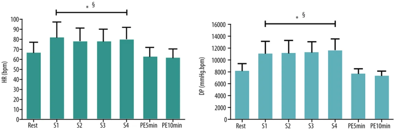
Figure 2 and 3 illustrate the SBP, DBP, HR and DP responses at rest, during exercise and post-exercise. Diferences were observed in all vari-ables at the end of each set compared to resting and post-exercise values (p<0.05). With respect to SBP and DBP at the end of the sets, signiicant diferences (p<0.05) were found from the irst to the third and fourth sets. In contrast, no diferences in HR or DP were observed between sets.
Stretching and acute cardiovascular responses Lima et al.
DISCUSSION
he present study investigated the cardiovascular responses to multiple sets of stretching exercise involving a large muscle group. he irst inding was that all cardiovascular variables studied were higher at the end of the sets when compared to resting and post-exercise values. SBP and DBP difered from the irst to the third and fourth sets of stretching exercise. Further-more, no hypotensive efect was detected ater the stretching protocol.
Few studies have investigated methodological aspects of stretching exercise prescription and their repercussions on acute cardiovascular responses. Cornelius et al.11 evaluated SBP and DBP responses to diferent proprioceptive neuromuscular facilitation (PNF) stretching techniques. Both SBP and DBP varied less than 15 mmHg compared to pre-exercise levels. his inding was probably due to the fact that the time of tension was less than 10 seconds during the isometric contraction phase of the protocol. In the present study, the application of passive static stretching resulted in a variation of 22 mmHg for SBP and of 19 mmHg for DBP compared to resting values. A possible explanation for this divergence is the diference in the duration of the stretch stimulus. Continuous stimuli lasting 30 seconds were applied in the present study, while in the study of Cornelius et al.11 the three stimuli of each set of PNF exercise were shorter.
Also using PNF techniques, Gültekin et al.20 investigated the efect of 10 sets of stretching of the upper limb in adult men. SBP, DBP, HR and DP were measured before, immediately ater the stimulus, and at the irst, third and ith minute of recovery. he protocol afected all variables studied at the end of the sets. A reduction in SBP and DP was only observed at the ith minute of recovery. It should be noted that the SBP and DBP values obtained in that study were lower than those observed here (125±10.8 versus 146±10.8 mmHg and 75±7.7 versus 88±8.3 mmHg, respectively). he same was not observed for HR and DP (108±15 versus 84±9.5 bpm and 13,500±238 versus 11,751±278 mmHg.bpm, respectively). Some factors may have inluenced these difer-ences. Although 10 sets were performed in the study of Gültekin et al.20, the stretch stimuli were reduced and intermingled with periods of relaxation as
Figure 3. Responses of heart rate (HR) (A) and double product (DP) (B) at rest, during exercise and post-exercise. S1 = irst set; S2 = second set; S3 = third set; S4 = fourth set; PE5min = 5 minutes post-exercise; PE10min = 10 minutes post-exercise. * signiicant diference in HR (p=0.005) and DP (p<0.001) obtained at the end of all sets compared to resting values; § signiicant diference in the values obtained in the
Holt et al.13, also using the PNF method, investigated SBP, DBP and HR responses of two muscle groups, erector spinae (trunk lexion performed in the sitting position) and hamstrings (unilateral hip lexion performed in the supine position). he subjects performed four sets per muscle, which was exercised at two intensities (100% and 50% of maximum voluntary isometric contraction). At the higher intensity, SBP values resulting from stretching of the erector spinae muscle difered from the second set on-wards when compared to resting and post-exercise values, with a peak of 163±37.1 mmHg. However, for hamstring stretching performed at the same intensity, a signiicant diference was only observed between the last set and resting and post-exercise values, with a peak of 159±24 mmHg. Diferences in SPB between sets were found until the fourth set. In the present study, stabilization of SBP was observed ater the third set, showing similarity between experiments. With respect to DBP, Holt et al.13 obtained similar values for the two muscle groups at the higher intensity (erector spinae: 91±25.8 mmHg; hamstrings: 88±22.4 mmHg). In the present experiment, a similar increase in DBP was observed for hamstring stretching (88±8.3 mmHg). Furthermore, the highest values were also found in the last sets, demonstrating a cumulative efect of the number of sets.
he size of the muscle group exercised, together with the characteristic of the movement, plays an important role when the efect of exercise on acute BP responses is analyzed. In this case, Holt et al.13 showed higher BP values for trunk lexion compared to unilateral hip lexion. Furthermore, the erector spinae muscles, which are basically postural muscles, are very stif21, a fact that can reduce mobility of the segment. his additional stif-ness may lead to diferent cardiovascular responses due to higher exercise-associated stress14. Moreover, abdominal compression resulting from trunk lexion can increase intra-abdominal pressure and consequently reduce venous return, decreasing illing of the cardiac chamber, with consequent loss of contractile eiciency of the myocardium (Frank-Starling Law). With the reduction in cardiac output, BP also decreases. To restore cardiac con-traction strength, the organism makes use of systemic arteriolar vasocon-striction, increasing BP levels through the discharge of catecholamines22. Still comparing the data of Holt et al.13 with the indings of the present study, HR values were lower for both types of exercise when performed at maximum range (101±33 bpm for erector spinae, 94±25 bpm for hamstrings versus 84±10 bpm in the present study). Furthermore, in the present study the highest value was observed in the second set, while in the study of Holt et al.13 peak HR was found in the fourth set. hese results suggest that HR may not be inluenced by the sum of sets such as BP, but higher values are observed when stretching involves larger muscle groups12,14.
Perhaps the study by Farinatti et al.12 is the one methodologically
Stretching and acute cardiovascular responses Lima et al.
seconds) of the hamstring and gastrocnemius muscles performed in the presence and absence of the Valsalva maneuver. he highest SBP values were observed in the fourth set (about 160 mmHg) in the presence of the Valsalva maneuver. However, SBP values for the same muscle group also increased signiicantly in the absence of the Valsalva maneuver (about 150 mmHg). his was also observed in the last set. In contrast to the present study, the SBP values across sets were suicient to trigger a signiicant increase in DP during unilateral hip lexion in the absence of the Valsalva
maneuver12. hese values were even higher (about 16,000 mmHg.bmp) when
stretching was performed in the presence of the maneuver.
here are several explanations for these diferences in the responses between experiments. One explanation is related to the diferent methods used for BP measurement. Farinatti et al.12 performed manual BP measure-ments, while in the present study BP was measured by photoplethysmog-raphy. Moreover, the intervals between sets used here were twice as long as those adopted in the experiment of Farinatti et al.12 (30 versus 15 seconds, respectively). Finally, although both studies used the same cut-of value for the deinition of reduced lexibility, it is unknown to which extent the minimum values of the two groups were similar. In the case of less lexible subjects, a greater intensity needs to be imposed to overcome muscle
stif-ness in an attempt to maintain the maximum range of motion14.
Although the volunteers were instructed to relax the quadriceps muscle during unilateral hip lexion, relex static contraction of the antagonistic musculature cannot be ruled out. However, the examiner paid careful at-tention to the occurrence of any involuntary contraction of the quadriceps muscle. Furthermore, during execution of the movement, the examiner called the subject’s attention regarding the importance of maintaining this musculature relaxed. It should be noted that there was no case of invol-untary contraction perceived by the examiner or reported by the subject.
CONCLUSIONS
In the protocol of stretching exercise prescription studied, a diference was observed in all cardiovascular variables during exercise compared to resting and post-exercise values. However, only the SBP and DBP responses exhibited a cumulative efect related to the number of sets. Furthermore, the stretching protocol produced no hypotensive efect. Further studies are needed to better elucidate the cardiovascular responses to stretching exercise. Such studies should consider diferent respiratory maneuvers, as well as the efects of stretching sessions with higher training volumes (number of sets and stimulus duration).
REFERENCES
1. American College of Sports Medicine. ACSM’s guidelines for exercise testing and prescription. 8th ed. Philadelphia: Lippincott Williams & Wilkins; 2009. 2. Yamamoto K, Kawano H, Gando Y, Iemitsu M, Murakami H, Sanada K, et al. Poor
Corresponding author
Walace Monteiro
Laboratório de Atividade Física e Promoção da Saúde.
Universidade do Estado do Rio de Janeiro (LABSAU/UERJ)
Rua São Francisco Xavier, 524/8121, 8º andar, bloco F,
20550-900 - Rio de Janeiro, RJ. Brasil. E-mail: [email protected]
4. Castinheiras-Neto AG, Costa-Filho IR, Farinatti PTV. Cardiovascular responses to resistance exercise are afected by workload and intervals between sets. Arq Bras Cardiol 2010;95(4):493-501.
5. Camara LC, Ritti-Dias RM, Forjaz CL, Greve JM, Santarem JM, Jacob-Filho W, et al. Cardiovascular responses during isokinetic muscle assessment in claudicant patients. Arq Bras Cardiol 2010;95(5):571-6.
6. Cucato GG, Ritti-Dias RM, Wolosker N, Santarem JM, Jacob Filho W, Forjaz CL. Post-resistance exercise hypotension in patients with intermittent claudication. Clinics 2011;66(2):221-6.
7. Fagard RH. Exercise characteristics and the blood pressure response to dynamic physical training. Med Sci Sports Exerc 2001;33(6 Suppl):S484-492; discussion S493-4. 8. Liu, SJ, Goodman J, Nolan R, Lacombe S, homas SG. Blood pressure responses to acute and chronic exercise are related in prehypertension. Med Sci Sports Exerc 2012;44(9):1644-52.
9. Keese F, Farinatti P, Pescatello L, Cunha FA, Monteiro WD. Aerobic exercise in-tensity inluences hypotension following concurrent exercise sessions. Int J Sports Med 2012;33(2):148-53.
10. Keese F, Farinatti P, Pescatello L, Monteiro W. A comparison of the immediate ef-fects of resistance, aerobic, and concurrent exercise on postexercise hypotension. J Strength Cond Res 2011;25(5):1429-36.
11. Cornelius WL, Jensen RL, Odell ME. Efects of PNF stretching phases on acute arterial blood pressure. Can J Appl Physiol 1995;20(2):222-9.
12. Farinatti PT, Soares PP, Monteiro WD, Duarte AF, Castro LA. Cardiovascular re-sponses to passive static lexibility exercises are inluenced by the stretched muscle mass and the Valsalva maneuver. Clinics 2011;66(3):459-64.
13. Holt LE, Pelham TW, Campagna PD. Hemodynamics during a machine-aided lexibility protocol. Can J Appl Physiol 1995;20(4):407-16.
14. Farinatti PT, Brandao C, Soares PP, Duarte AF. Acute efects of stretching exercise on the heart rate variability in subjects with low lexibility levels. J Strength Cond Res 2011;25(6):1579-85.
15. Bonnafoux P. Auscultatory and oscillometric methods of ambulatory blood pres-sure monitoring, advantages and limits: a technical point of view. Blood Press Monit 1996;1(3):181-5.
16. Cameron JD, Stevenson I, Reed E, McGrath BP, Dart AM, Kingwell BA. Accuracy of automated auscultatory blood pressure measurement during supine exercise and treadmill stress electrocardiogram-testing. Blood Press Monit 2004;9(5):269-75. 17. Polito MD, Farinatti PTV. Considerações sobre a medida da pressão arterial em
exercícios contra-resistência. Rev Bras Med Esporte 2003;9(1):25-33.
18. Kurz T. Stretching Scientiically: A Guide to Flexibility Training. 3rd ed. Island Pond: Stadion; 1994.
19. Guissard N, Duchateau J. Efect of static stretch training on neural and mechanical properties of the human plantar-lexor muscles. Muscle Nerve 2004;29(2):248-55. 20. Gültekin Z, Kin-Isler A, Sürenkök Ö. Hemodynamic and lactic acid responses to
proprioceptive neuromuscular facilitation exercise. J Sports Sci Med 2006;5:375-80. 21. Manire JT, Kipp R, Spencer J, Swank AM. Diurnal variation of hamstring and
lumbar lexibility. J Strength Cond Res 2010;24(6):1464-71.