Bloqueio Peribulbar com Ropivacaína: Influência da
Hialuronidase sobre a Qualidade do Bloqueio e a
Pressão Intra-Ocular *
Peribulbar Block with Ropivacaine: Effects of Hyaluronidase on
Blockade Quality and Intraocular Pressure
Paulo Sérgio Mateus Serzedo, TSA1, José Roberto Nociti, TSA2, Eduardo Barbin Zuccolotto, TSA1, Tatiana Lúcia Scalco3, Sérgio Borges Ferreira4
RESUMO
Serzedo PSM, Nociti JR, Zuccolotto EB, Scalco TL, Ferreira SB - Bloqueio Peribulbar com Ropivacaína: Influência da Hialuro-nidase sobre a Qualidade do Bloqueio e a Pressão Intra-Ocular
Justificativa e Objetivos - Alguns estudos têm relatado melhoria da qualidade do bloqueio peribulbar com o emprego de hialuronidase, enquanto outros concluem pela ausência de efeito. O objetivo deste estudo foi investigar a influência da hialuronidase sobre a pressão intra-ocular (PIO) e a qualidade do bloqueio peribulbar com ropivacaína a 1%.
Método- Quarenta pacientes submetidos à cirurgia de catarata foram distribuídos de forma aleatória em dois grupos e submetidos a bloqueio peribulbar com 7 ml de ropivacaína a 1% em técnica de dupla punção, com hialuronidase 50 UI.ml-1 no Grupo A (n = 20) e sem hialuronidase no Grupo B (n = 20). As medidas de PIO foram realizadas com tonômetro de aplanação de Perkins em quatro momentos: M0 = antes do bloqueio (controle); M1 = 1 min após o bloqueio; M2 = 5 min após o bloqueio; M3 = 15 min após o bloqueio. A qualidade foi avaliada pelo método de Nicoll, baseado na redução da motilidade do globo ocular.
Resultados - As médias de PIO (mmHg) antes do bloqueio foram semelhantes nos dois grupos: 16,1±2,1 (A) vs 16,4±3,3 ( B ) . A p ó s o b l o q u e i o , a s m é d i a s d e P I O f o r a m significativamente menores no Grupo A em relação ao Grupo B nos três momentos: M1 = 11,7 ± 2,4 vs 17,9 ± 3,6; M2 = 8,2 ± 1,9 vs 14,1 ± 4,0; M3 = 5,3±2,1 vs 10,2±3,1. O comportamento intragrupos também foi diferente. No Grupo A, as médias de PIO foram significativamente menores em relação ao controle nos três momentos após o bloqueio; no Grupo B a média de PIO elevou-se em M1 e foi significativamente inferior ao controle em M2 e M3. As médias para os índices de motilidade do globo ocular (Nicoll) foram significativamente menores no Grupo A em relação ao B nos três momentos: M1 = 2,55 vs 3,65; M2 = 0,25 vs 2,2; M3 = 0,00 vs 1,00.
Conclusões- Quando se emprega solução de ropivacaína a 1% adicionada de hialuronidase 50 UI.ml-1 em bloqueio
peribulbar, os valores da PIO são menores e a qualidade do bloqueio é melhor do que quando se utiliza ropivacaína a 1% sem hialuronidase.
U N I T E R M O S - A N E S T É S I C O S , L o c a l : r o p i v a c a í n a ; TÉCNICAS ANESTÉSICAS, Regional: peribulbar; TÉNICAS DE MEDIÇAO: pressão intra-ocular
SUMMARY
Serzedo PSM, Nociti JR, Zuccolotto EB, Scalco TL, Ferreira SB - Peribulbar Block with Ropivacaine: Effects of Hyaluronidase on Blockade Quality and Intraocular Pressure
Background And Objectives- Some studies have reported improved quality of peribulbar block by adding hyaluronidase to the local anesthetic solution while others claimed no beneficial effect. This study aimed at investigating the influence of hyaluronidase on intraocular pressure (IOP) and the quality of peribulbar block with 1% ropivacaine.
Methods- Participated in this study 40 patients undergoing cat-aract surgery under peribulbar block who were randomly allo-cated to one of two groups according to the nature of the local anesthetic solution: Group A (n = 20), 1% ropivacaine (7 ml) supplemented with 50 IU.ml-1hyaluronidase; Group B (n = 20), 1% plain ropivacaine (7 ml). IOP measurements were per-formed by means of a Perkins applanation tonometer in four moments: M0 = before block (control); M1 = 1 min after block; M2 = 5 min after block; M3 = 15 min after block. Quality was evaluated by Nicoll’s method based on eye motility decrease.
Results - Mean IOP values (mmHg) before block (M0) were similar for both groups: 16,1 ± 2.1 vs. 16.4 ± 3.3. After blockade, mean IOP values were significantly lower in Group A as com-pared to Group B in the three moments after block: M1 = 11.7 ± 2.4 vs. 17.9 ± 3.6; M2 = 8.2 ± 1.9 vs. 14.1 ± 4.0; M3 = 5.3 ± 2.1 vs. 10.2 ± 3.1. IOP variations were also different within each group. In group A, mean values obtained in the three moments after block were significantly lower than control; in Group B, mean values significantly increased in M1 and were lower than control in M2 and M3. Mean eye motility scores were significantly lower in Group A as compared to Group B in M1 (2.55 vs. 3.65), M2 (0.25 vs. 2.2), and M3 (0.00 vs.1.00).
Conclusions - When 1% ropivacaine supplemented with 50 IU.ml-1hyaluronidase is used in peribulbar block, IOP values are lower and blockade quality is significantly better than when 1% plain ropivacaine is used.
KEY WORDS - ANESTHETICS, Local: ropivacaine; ANES-THETIC TECHNIQUES, Regional: peribulbar; MEASURE-MENT TECHNIQUES: intraocular pressure
202 Revista Brasileira de Anestesiologia
ARTIGO CIENTÍFICO
SCIENTIFIC ARTICLE Rev Bras Anestesiol
2001; 51: 3: 202- 207
* Recebido do (Received from) CET/SBA da Santa Casa de Misericórdia de Ribeirão Preto (SCMRP), SP
1. Co-responsável pelo CET/SBA 2. Responsável pelo CET/SBA da SCMRP
3. ME2do CET/SBA da SCMRP
4. Oftalmologista Chefe do Instituto de Microcirurgia Ocular de Ribeirão Preto, SP
Apresentado (Submitted) em 28 de agosto de 2000
Aceito (Accepted) para publicação em 17 de outubro de 2000
Correspondência para (Mail to): Dr. José Roberto Nociti Rua Ayrton Roxo, 980 14025-270 Ribeirão Preto, SP E-mail: [email protected]
INTRODUÇÃO
O
uso da ropivacaína em bloqueio peribulbar para cirur-gia intra-ocular tem proporcionado bons resultados, combinando boa qualidade com baixa morbidade1-3. A hia-luronidase é associada freqüentemente ao anestésico local neste tipo de bloqueio, com a finalidade de facilitar a difusão do mesmo graças à hidrólise das ligações glicosídicas no in-terior do ácido hialurônico que forma a barreira intersticial4. Alguns estudos têm relatado efetiva melhoria da qualidade do bloqueio com o emprego de hialuronidase5,6, enquanto outros têm concluído pela ausência de efeito7.Por outro lado, já foi descrita diminuição da pressão in-tra-ocular (PIO) após bloqueio peribulbar com ropivacaína associada a hialuronidase8.
A finalidade deste estudo foi investigar a possível influência da hialuronidase sobre a qualidade do bloqueio obtido e a PIO, após bloqueio peribulbar com ropivacaína.
MÉTODO
O estudo foi aprovado pela Comissão de Ética em Pesquisa do Hospital e dele fizeram parte 40 pacientes de ambos os se-xos com idades entre 41 e 95 anos, estado físico ASA I, II ou III, encaminhados à cirurgia de catarata em regime ambula-torial. Foram excluídos portadores de glaucoma.
Não foi administrada medicação pré-anestésica e os pacien-tes foram distribuídos de forma aleatória em dois grupos, de acordo com a presença ou não de hialuronidase na solução de ropivacaína utilizada no bloqueio peribulbar:
Grupo A (n = 20) - Ropivacaína a 1% contendo hialuronida-se 50 UI.ml-1
Grupo B (n = 20) - Ropivacaína a 1% sem hialuronidase
Foram monitorizados eletrocardiograma (ECG) contínuo, oximetria de pulso (SpO2), pressão arterial sistólica (PAS), diastólica (PAD) e média (PAM), por método não invasivo. O bloqueio peribulbar foi realizado pelo método de dupla punção1, com agulha padrão 25 x 6 mm (23 G-1), volume total de solução 7 ml, injetando-se 4 ml através da pálpebra inferi-or na junção do terço lateral com os dois terços mediais da rima orbital, e 3 ml através da pálpebra superior na borda sú-pero-interna da órbita. Após as injeções, foi aplicado barof-talmo sobre o olho fechado com peso de McIntyre durante 10 minutos.
A medida da PIO foi realizada com tonômetro de aplanação de Perkins por observador independente (o próprio cirur-gião) que desconhecia a natureza da solução, em quatro mo-mentos:
M0 = Antes do bloqueio M1 = 1 min após o bloqueio M2 = 5 min após o bloqueio M3 = 15 min após o bloqueio
A avaliação da qualidade do bloqueio foi feita nos momentos M1, M2 e M3 através do método descrito por Nicoll e col1,9, baseado na redução da motilidade do globo ocular, consi-derando-se como sinal de sucesso o índice igual ou inferior a 4.
Foi pesquisada a ocorrência de possíveis eventos adversos: dor à injeção do anestésico local, hipotensão arterial (dimi-nuição da PA > 30% do controle), hipertensão arterial (au-mento da PAS > 30% do controle), bradicardia (FC£50 bpm), taquicardia (FC³120 bpm), disritmia cardíaca, náuseas e vômitos.
A comparação estatística entre as médias de idade e peso nos dois grupos foi feita pelo testetde Student; a compara-ção quanto ao sexo foi feita pelo teste do Qui-quadrado . A comparação das médias da PIO entre grupos e intragrupos foi feita pelo teste paramétrico de análise de variânciatwo way. Para comparação das médias do índice de motilidade ocular (Nicoll) entre os grupos utilizou-se o teste Mann-Whit-ney e dentro de cada grupo a prova de Friedman. Em todos os testes o nível de significância adotado foi 5% (p < 0,05).
RESULTADOS
Os grupos foram homogêneos quanto aos dados demográfi-cos, que estão apresentados na tabela I.
Tabela I - Dados Demográficos
Grupo A
(n = 20) Grupo B(n = 20) Idade (anos) Média ± DP 67,1 ± 7,8 69,1 ± 10 Amplitude 50 - 81 41 - 81 Peso (kg) Média ± DP 70,9 ± 13,4 71,1 ± 12,7
Amplitude 48 - 96 50 - 92 Sexo
Masculino 9 (45%) 10 (50%) Feminimo 11 (55%) 10 (50%)
Afreqüência dos índices de motilidade ocular (Nicoll) em am-bos os grupos nos vários momentos está expressa na tabela II e suas médias na figura 1. Foram significativas as diferen-ças tanto entre os grupos como dentro de cada grupo.
Tabela II - Índices de Motilidade Ocular (Nicoll)
Grupo A (% de pacientes) Grupo B (% de pacientes) Índice M1 M2 M3 M1 M2 M3
0 5 75 100 5 30 45
1 5 25 0 10 30 20
3 40 0 0 20 30 30
4 30 0 0 45 10 5
5 20 0 0 20 0 0
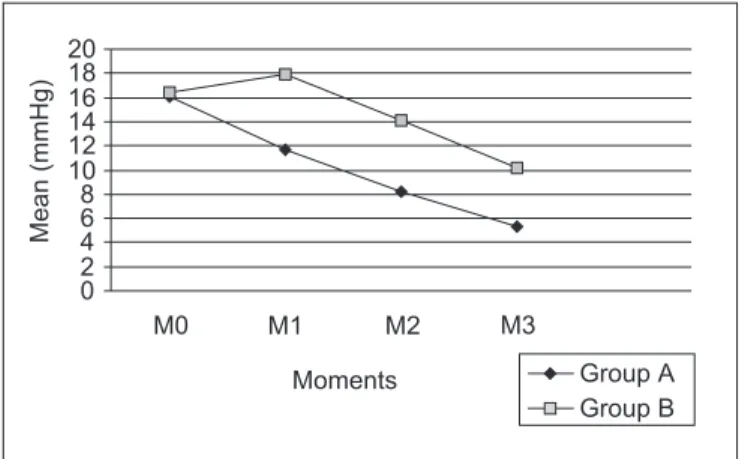
Os valores obtidos para as médias da PIO estão apresenta-dos na tabela III e na figura 2. As diferenças foram estatistica-mente significativas a partir do momento M1, tanto entre os grupos como dentro de cada grupo.
Tabela III - Medidas da Pressão Intra-Ocular (Média±DP)
Momentos Grupo A Grupo B
M0 16,1 ± 2,1 16,4 ± 3,3
M1 11,7 ± 2,4 17,9 ± 3,6*
M2 8,2 ± 1,9 14,1 ± 4,0*
M3 5,3 ± 2,1 10,2 ± 3,1*
* Análise de variância “two way”: diferença significante (p<0,001)
Registrou-se queixa de dor à injeção da ropivacaína em qua-tro pacientes do Grupo B (20%) e em um do Grupo A (5%). Ocorreram episódios de hipertensão arterial em três pacien-tes do Grupo B (15%) e em um do Grupo A (5%). Houve ne-cessidade de sedação (utilizou-se diazepam por via venosa) em um paciente de cada grupo (5%). Não ocorreram outros eventos adversos.
DISCUSSÃO
Os resultados demonstraram bloqueio motor mais efetivo e melhor qualidade do bloqueio peribulbar quando a hialuroni-dase na concentração de 50 UI.ml-1foi adicionada à solução de ropivacaína a 1%. Por outro lado, a adição de hialuronida-se influenciou também a variação da PIO. Nos pacientes sub-metidos ao bloqueio com ropivacaína adicionada de hialuro-nidase, registrou-se diminuição significativa da PIO em rela-ção ao controle logo no primeiro minuto, acentuando-se nas medidas subseqüentes. Nos pacientes submetidos ao blo-queio com ropivacaína sem hialuronidase, houve elevação significativa da PIO no primeiro minuto, para depois ocorrer diminuição nas medidas seguintes. Mas os valores obtidos para as médias de PIO nos pacientes do Grupo A (com hialu-ronidase) foram significativamente menores do que os obti-dos nos pacientes do Grupo B (sem hialuronidase), em toobti-dos os momentos após o bloqueio.
Estes resultados estão de acordo com observações de ou-tros autores, que encontraram melhoria da qualidade do blo-queio peribulbar pela adição de hialuronidase à solução do anestésico local, utilizando concentrações da enzima simila-res10ou mesmo inferiores11às empregadas neste estudo. Por outro lado, os valores mais elevados da PIO nos pacien-tes que não receberam hialuronidase foram detectados tam-bém por outros autores10, e esta ocorrência parece estar re-lacionada com a dispersão incompleta do anestésico local dentro do compartimento periocular. Sabe-se que o bloqueio peribulbar pode ocasionar elevação transitória da PIO devi-do ao aumento da pressão intraorbitária pela injeção devi-do anestésico local. Entretanto, a PIO tende a retornar ao seu valor inicial devido à dispersão da solução e ao relaxamento da musculatura extraocular (secundário ao bloqueio), dimi-nuindo a pressão externa sobre o globo ocular12.
É interessante salientar que, ao contrário do que ocorre com a bupivacaína10, a elevação inicial da PIO com a ropivacaína sem hialuronidase não se manteve nas medidas subseqüen-tes. Isto pode ser devido a um relaxamento mais completo da musculatura extraocular com a ropivacaína, já apontado em estudo anterior comparativo com a bupivacaína13. Além dis-so, é possível que a elevação inicial da PIO pela injeção do anestésico local seja contrabalanceada também por diminui-ção do volume sangüíneo intra-ocular secundário ao efeito vasoconstritor da ropivacaína14, fator inexistente com a bu-pivacaína, que é desprovida deste efeito.
Concluímos que, quando se emprega solução de ropivacaí-na a 1% adicioropivacaí-nada de hialuronidase 50 UI.ml-1em bloqueio peribulbar, os valores da PIO são menores e a qualidade do bloqueio, avaliada pelo índice de motilidade do globo ocular é melhor do que quando se usa o mesmo anestésico local sem hialuronidase. A melhor dispersão da solução de anes-tésico local no compartimento periocular proporcionada pelo efeito característico da hialuronidase parece contribuir tanto para a diminuição da PIO como para facilitar o bloqueio das estruturas nervosas perioculares pelo agente anestésico.
204 Revista Brasileira de Anestesiologia
BLOQUEIO PERIBULBAR COM ROPIVACAÍNA: INFLUÊNCIA DA HIALURONIDASE SOBRE A QUALIDADE DO BLOQUEIO E A PRESSÃO INTRA-OCULAR
Médias
(mmHg)
0 2 4 6 8 10 12 14 16 18 20
M1
M0 M2 M3
Momentos Grupo A
Grupo B
Figura 2 - Evolução das Médias da Pressão Intra-Ocular 0
1 2 3 4
M1 M2 M3
Momentos
Médias
Grupo A Grupo B
Peribulbar Block with Ropivacaine:
Effects of Hyaluronidase on Blockade
Quality and Intraocular Pressure
Paulo Sérgio Mateus Serzedo, M.D., José Roberto Nociti, M.D., Eduardo Barbin Zuccolotto, M.D., Tatiana Lúcia Scalco M.D., Sérgio Borges Ferreira, M.D.
INTRODUCTION
Ropivacaine for peribulbar block in intraocular surgeries has provided good results, combining high quality with low morbi-dity1-3. Hyaluronidase is commonly associated to local anest-hetics in this type of blockade aiming at facilitating its spread thanks to the hydrolysis of glucoside bindings within the hya-luronic acid forming the interstitial barrier4. Some studies have reported a marked improvement in blockade quality with the use of hyaluronidase5,6while others concluded for the absence of such effect7.
On the other hand, intraocular pressure (IOP) decrease after peribulbar block with ropivacaine associated to hyaluronida-se has also been described8.
This study aimed at investigating the possible influence of hyaluronidase on blockade and IOP after peribulbar block with ropivacaine.
METHODS
After the Hospital Research Ethics Committee approval par-ticipated in this study 40 patients of both genders, aged 41 to 95 years, physical status ASA I, II or III, scheduled for outpati-ent cataract surgery. Patioutpati-ents with glaucoma were excluded. No premedication was administered and patients were ran-domly distributed in two groups, according to the presence or not of hyaluronidase in the ropivacaine solution used for peri-bulbar block.
Group A (n=20) - 1% ropivacaine with 50 IU.ml-1 hyaluroni-dase
Group B (n=20) - 1% ropivacaine without hyaluronidase
Monitoring consisted of continuous ECG, pulse oximetry (SpO2), non invasive systolic (SBP), diastolic (DBP) and mean blood pressure (MBP).
Peribulbar block was performed by the double puncture tech-nique1with a 25 x 6 mm needle (23 G-1) in a total volume of 7 ml injecting 4 ml through the lower eyelid in the junction of the lateral third with the two medial thirds of the orbital rime, and 3 ml through the upper eyelid in the upper-internal orbital bor-der. After injection, an oculopression was applied on the clo-sed eye with a Mcintyre weight for 10 minutes.
IOP was measured in four moments with a Perkins applanati-on tapplanati-onometer by an independent observer (the surgeapplanati-on him-self) who did not know the solution’s composition:
M0 - before blockade M1 - 1 minute after blockade M2 - 5 minutes after blockade M3 - 15 minutes after blockade
Blockade quality was evaluated in moments M1, M2 and M3 through the method described by Nicoll et al1,9based on de-creased eye motility, considering 4 or below a successful rate.
Possible adverse events were looked for: pain at local anesthe-tics injection, hypotension (below 30% of baseline), hyper-tension (above 30% of control), bradycardia (HR£50 bpm), tachycardia (HR³120 bpm), dysrhythmia, nausea and vomi-ting.
Student’sttest was used for statistical comparison between mean age and weight for both groups; Chi-square test com-pared gender. The two way analysis of variance parametric test was used to compare mean IOP between groups and wit-hin groups. To compare mean eye motility score (Nicoll) Mann-Whitney test was used between groups and Friedman test within each group. The level of significance of 5% (p < 0.05) was adopted for all tests.
RESULTS
Demographics data, which were homogeneous between groups, are shown in table I.
Table I - Demographics Data
Group A
(n = 20) Group B(n = 20) Age (years) Mean ± SD 67.1 ± 7.8 69.1 ± 10
Amplitude 50 - 81 41 - 81 Weight (kg) Mean ± SD 70.9 ± 13.4 71.1 ± 12.7
Amplitude 48 - 96 50 - 92 Gender
Male 9 (45%) 10 (50%) Female 11 (55%) 10 (50%)
The frequency of eye motility scores (Nicoll) for both groups in all moments is shown in table II and their means in figure 1. Differences were significant both between and within groups.
Table II - Eye Motility Scores (Nicoll)
Group A (% of patients) Group B (% of patients)
Score M1 M2 M3 M1 M2 M3
0 5 75 100 5 30 45
1 5 25 0 10 30 20
3 40 0 0 20 30 30
4 30 0 0 45 10 5
5 20 0 0 20 0 0
Statistical Comparison: Within groups (Friedman) - significant as of M2; Between groups (Mann-Whitney) - significant in all moment s
IOP mean values are shown in table III and figure 2. Differen-ces were statistically significant as of M1 both between and within groups.
Table III - Intraocular Pressure Measurements (Mean±SD)
Moments Group A Group B
M0 16.1 ± 2.1 16.4 ± 3.3 M1 11.7 ± 2.4 17.9 ± 3.6*
M2 8.2 ± 1.9 14.1 ± 4.0* M3 5.3 ± 2.1 10.2 ± 3.1* * “Two way” analysis of variance: significant difference (p<0.001)
Four Group B (20%) and one Group A (5%) patients complai-ned of pain at injection. Three Group B (15%) and one Group A (5%) patients had hypertension episodes. One patient of each group (5%) needed sedation (intravenous diazepam). There were no other adverse effects.
DISCUSSION
Results have shown a more effective and better peribulbar block when 50 IU.ml-1hyaluronidase were associated to 1% ropivacaine. The addition of hyaluronidase has also influen-ced IOP variations. In patients submitted to the
ropivacai-ne-hyaluronidase blockade, there has been a significant de-crease in IOP as compared to the control group already in the first minute, which was more marked in subsequent measure-ments. In patients submitted to blockade with hyaluronida-se-free ropivacaine, there has been a significant increase in IOP in the first minute which then decreased in subsequent measurements. But IOP means obtained for Group Apatients (with hyaluronidase) were significantly lower as compared to Group B (without hyaluronidase) in all moments after blocka-de.
Such results are in line with other authors who observed a bet-ter peribulbar block with the addition of hyaluronidase to local anesthetics, using enzyme concentrations similar10or even lower11than those used in this study.
Higher IOP levels in patients not receiving hyaluronidase were also detected by other authors10, and this fact seems to be more related to the incomplete spread of local anesthetics within the periocular compartment. It is known that peribulbar block may cause a transient IOP increase due to the increase in intraorbital pressure caused by the anesthetic injection. However, IOP tends to return to its baseline value due to solu-tion spread and extraocular muscles relaxasolu-tion (secondary to blockade), thus decreasing external pressure on the eye
12.
It is interesting to highlight that, as opposed to bupivacaine10, the initial IOP increase with hyaluronidase-free ropivacaine was not maintained in subsequent measurements. This may be due to a more complete extraocular muscles relaxation with ropivacaine already observed in a previous comparative study with bupivacaine13. Moreover, it is possible that the ini-tial IOP increase caused by local anesthetics injection is furt-her counterbalanced by a decrease in intraocular blood volu-me, secondary to the vasoconstrictor effect of ropivacaine14, which is not true for bupivacaine which lacks such effect. We concluded that when 1% ropivacaine is associated to 50 lU.ml-1hyaluronidase for peribulbar block, IOP values are lo-wer and blockade quality evaluated by eye motility score, is better than when the same local anesthetics is used without hyaluronidase. The better local anesthetic spread within the periocular compartment provided by the characteristic effect of hyaluronidase, seems to contribute both for IOP decrease and to facilitate periocular nervous structures blockade by the anesthetic agent.
REFERÊNCIAS-REFERENCES
01. Serzedo PSM, Nociti JR, Zuccolotto EB et al - Ropivacaína em bloqueio peribulbar: estudo comparativo com bupivacaína. Rev Bras Anestesiol, 1998;48:258-263.
02. Nicholson G, Sutton B, Hall GM - Ropivacaine for peribulbar anesthesia. Reg Anesth Pain Med, 1999;24:337-340. 03. Luchetti M, Magni G, Marraro G - A prospective randomized
dou-ble-blind controlled study of ropivacaine 0.75% versus bupiva-caine 0.5%-mepivabupiva-caine 2% for peribulbar anesthesia. Reg Anesth Pain Med, 2000;25:195-200.
206 Revista Brasileira de Anestesiologia
PERIBULBAR BLOCK WITH ROPIVACAINE: EFFECTS OF HYALURONIDASE ON BLOCKADE QUALITY AND INTRAOCULAR PRESSURE
Mean
(mmHg)
0 2 4 6 8 10 12 14 16 18 20
M1
M0 M2 M3
Moments Group A
Group B
Figure 2 - Intraocular Pressure Mean Values Evolution
0 1 2 3 4
M1 M2 M3
Moments
Mean
Group A Group B
04. Watson D - Hyaluronidase. Br J Anaesth, 1993;71:422-425. 05. Sarvela J, Nikki P - Hyaluronidase improves regional ophthalmic
anaesthesia with etidocaine. Can J Anaesth, 1992;39:920-924. 06. Morsman CD, Holden R - The effects of adrenaline,
hyaluronida-se and age on peribulbar anaesthesia. Eye, 1992;6: 290-292. 07. Brydon CW, Basler M, Kerr WJ - An evaluation of two
concentra-tions of hyaluronidase for supplementation of peribulbar anaes-thesia. Anaesthesia, 1995;50:998-1000.
08. Serzedo PSM, Nociti JR, Zuccolotto EB et al - Pressão intraocu-lar durante bloqueio peribulbar com ropivacaína a 1%. Rev Bras Anestesiol, 2000;50:251-253.
09. Nicoll JM, Treuren B, Acharya PA et al - Retrobulbar anesthesia: the role of hyaluronidase. Anesth Analg, 1986;65:1324-1328. 10. Dempsey GA, Barrett PJ, Kirby IJ - Hyaluronidase and
peribul-bar block. Br J Anaesth, 1997; 78:671-674.
11. Kallio H, Paloheimo M, Maunuksela EL - Is hyaluronidase effecti-ve as an adjuvant in retrobulbar/peribulbar block? Internat Moni-tor, 1998;10:50.
12. Johnson RW, Forrest FC - Anaesthesia for Ophthalmic Surgery, em: Prys-Roberts C, Brown Jr BR - International Practice of Anaesthesia, Oxford, Butterworth Heinemann, 1996;110:1-29. 13. Serzedo PSM, Nociti JR, Zuccolotto EB et al - Pressão intraocular durante bloqueio peribulbar com ropivacaína ou bupivacaína: estudo comparativo. Rev Bras Anestesiol, 2000;50:341-344. 14. Ishiyama T, Dohi S, Iida H et al - The effects of topical and
intra-venous ropivacaine on canine pial microcirculation. Anesth Analg, 1997;85:75-81.
RESUMEN
Serzedo PSM, Nociti JR, Zuccolotto EB, Scalco TL, Ferreira SB - Bloqueo Peribulbar con Ropivacaína: Influencia de la Hialuro-nidasa sobre la Calidad del Bloqueo y la Presión Intra-Ocular
Justificativa y Objetivos - Algunos estudios han relatado mejoria de la calidad del bloqueo peribulbar con el uso de hialuronidasa, en cuanto otros han concluido por la ausencia del efecto. El objetivo de este estudio fue investigar la influencia de la hialuronidasa sobre la presión intra-ocular (PIO) y la calidad del bloqueo peribulbar con ropivacaína a 1%.
Método- Cuarenta pacientes sometidos a cirugía de catarata fueron distribuidos de forma aleatoria en dos grupos y sometidos a bloqueo peribulbar con 7 ml de ropivacaína a 1% en técnica de dupla punción, con hialuronidasa 50 UI.ml-1en el Grupo A (n = 20) y sin hialuronidasa en el Grupo B (n = 20). Las medidas de PIO fueron realizadas con tonómetro de aplanación de Perkins en cuatro momentos: M0 = antes del bloqueo (control); M1 = 1 min después del bloqueo; M2 = 5 min después del bloqueo; M3 = 15 min después del bloqueo. La calidad fue evaluada por el método de Nicoll, fundamentado en la reducción de la motilidad del globo ocular.
Resultados- Las medias de PIO (mmHg) antes del bloqueo fueron semejantes en los dos grupos: 16,1±2,1 (A) vs 16,4±
3,3 (B). Después del bloqueo, las medias de PIO fueron significativamente menores en el Grupo A en relación al Grupo B en los tres momentos: M1 = 11,7 ± 2,4 vs 17,9 ± 3,6; M2 = 8,2 ± 1,9 vs 14,1 ± 4,0; M3 = 5,3 ± 2,1 vs 10,2 ± 3,1. El compor-tamiento intragrupos también fue diferente. En el Grupo A, las medias de PIO fueron significativamente menores en relación al control en los tres momentos después del bloqueo; en el Grupo B la media de PIO se elevó en M1 y fue significativamente inferior al control en M2 e M3. Las medias para los índices de motilidad del globo ocular (Nicoll) fueron significativamente menores en el Grupo A en relación al B en los tres momentos: M1 = 2,55 vs 3,65; M2 = 0,25 vs 2,2; M3 = 0,00 vs 1,00.
Conclusiones- Cuando se usa solución de ropivacaína a 1% adicionada de hialuronidasa 50 UI.ml-1en bloqueo peribulbar, los valores de la PIO son menores y la calidad del bloqueo es mejor de que cuando se utiliza ropivacaína a 1% sin hialuronidasa.