MULTIPLE SCLEROSIS IN BRAZIL
ANALYSIS OF CEREBROSPINAL FLUID BY STANDARD METHODS
MARZIA PUCCIONI-SOHLER*, FABIOLA PASSERI*, CRISTIANE OLIVEIRA**, CARLOS OTÁVIO BRANDÃO*, REGINA PAPAIZ-ALVARENGA***
ABSTRACT - The demonstration of intrathecal IgG synthesis has been used as an important laboratory parameter to support the diagnosis of multiple sclerosis(MS). The Committee for European Concerted Action for Multiple Sclerosis has recommended a protocol for the assessment of intrathecal IgG synthesis. We applied this methodology to determine the cerebrospinal (CSF) profile of 128 Brazilian patients with MS. We detected hypercytosis lower than 35 cells/mm3 in 97%, protein lower than 80mg/dl in 99%, normal blood-CSF barrier function in 76%, increased IgG local production around 53% and oligoclonal IgG bands by isoelectric focusing in 85% of the definite MS patients. The diagnostic accuracy of the quantitative analysis was lower than the qualitative. The detection of oligoclonal bands was especially important in the cases of normal quantitative assays of IgG. In addition, we found a lower frequency of inflammatory reaction in CSF in our MS cases, in comparison to some European studies.
KEY WORDS: multiple sclerosis, cerebrospinal fluid, oligoclonal bands, isoelectric focusing.
Esclerose múltipla no Brasil: análise do líquido cefalorraquidiano por métodos padronizados
RESUMO – A demonstração da síntese intratecal de IgG tem sido usada como importante parâmetro de apoio laboratorial ao diagnóstico de esclerose múltipla (EM). O Comitê Europeu de Ação para Esclerose Múltipla recomenda um protocolo para avaliação de síntese intratecal de IgG. Esta metodologia foi utilizada para determinar o perfil do líquido cefalorraquidiano (LCR) de 128 pacientes com o diagnóstico de esclerose múltipla. Verificou-se pleocitoVerificou-se menor do que 35 células/mm3 em 97%, produção local de IgG por métodos quantitativos em 53% e presença de bandas oligoclonais em 85% dos casos de EM clinicamente definida. A análise qualitativa apresentou maior acurácia diagnóstica do que a avaliação quantitativa de IgG. A detecção de bandas oligoclonais foi especialmente importante nos casos de analise quantitativa de IgG. Observamos menor frequência de reação inflamatória no LCR, em relação aos estudos europeus.
PALAVRAS-CHAVE: esclerose múltipla, líquido cefalorraquidiano, bandas oligoclonais, focalização isoelétrica.
The majority of multiple sclerosis (MS) studies have reported differences in the geographic distribution1. It seems to be related with different aspects as: genetic and environmental factors2.The
frequency and distribution of multiple sclerosis in the Brazilian population is unknown, but the disease is not rare in Brazil3.
The role of cerebrospinal fluid (CSF) analysis has been well documented as an aid to the diagnosis of MS4-6.The most important aspect of CSF study is to demonstrate the inflammatory
Laboratório Especializado de LCR (Neurolife) Rio de Janeiro, Projeto Atlântico Sul, Rio de Janeiro, Brazil: *Neurologista; **Estagiária; ***Coordenadora do Projeto Atlântico-Sul. Aceite: 21-agosto-1999.
origin of the disease in the central nervous system (CNS) through the detection of a local immune response4. The “Committee for European Concerted Action for Multiple Sclerosis”, (CECAMS)
suggested that the routine CSF analysis should include the evaluation of: cytology, blood-CSF barrier function and intrathecal synthesis of IgG4. These tests may be useful not only in supporting the
diagnosis, but also in some cases to exclude MS7-9.The combination of quantitative and qualitative
methods of analysis should be performed for the intrathecal synthesis of total IgG4: The use of the
CSF/serum quotient was considered to be of great value to determine the IgG synthezised in CNS, differentiating it from the fraction derived from blood10. The analysis of oligoclonal IgG bands by
isoelectric focusing is the most sensitive test to detect intrathecal synthesis of IgG11-13.It is considered
essential to the routine examination of the CSF4.The detection of the intrathecal synthesis of specific
antibodies (against measles, rubella, varicella zoster) may also contribute to the diagnosis of MS 5,14.
The objective of this study was to standardize the CSF analysis in patients with MS in Brazil. This resulted in the delineating of CSF profile of Brazilian MS patients, using different tests for the evaluation of intrathecal IgG synthesis, based on pre-established reference values. The quantitative IgG evaluation included IgG index and IgG loc (locally synthezised)(mg/l)8,9. In addition, we also
studied the IgG% and gamma globulin% of total protein in CSF 6,15. These tests are also usually
performed as a laboratory routine. Oligoclonal IgG bands were sought in both CSF and serum.
METHOD
Paired CSF and serum samples of 128 patients from different parts of Brazil, seen in Rio de Janeiro and registered in the South Atlantic Project, were collected during the period of March 1995 to June 1998. All the cases were examined by neurologists of the MS Brazilian Group3. The diagnosis was based on the Poser et al. criteria1: 103 were clinically definite and 25 clinically probable MS. The presence of rheumatologic and infectious diseases, and B12 deficiency were excluded by routine serological evaluation. All had negative serology for HIV-1 and HTLV-I.
CSF analysis
The CSF examination included white blood cells (WBC) count, glucose and protein analysis. Albumin and IgG concentrations were determined in serum and CSF by nephelometry, using the QM300 Protein Analysis System (Sanofi Diagnostics Pasteur Instruments, USA). The CSF-blood barrier function was based on the albumin quotient:
Qalb = albuminCSF/albumin serum (Reference Value: Qalb < 8 x 10 -3).
The quantitative evaluation of intrathecal IgG synthesis was based on:
1. IgG index (Olsson and Pettersson11). An IgG index below 0.7 was considered normal. · IgG index =QIgG/ /Qalb
2. IgG (loc) (mg/l) (Reiber & Felgenhauer9). Reiber and Felgenhauer elaborated a diagram based on a hyperbolic function to determine the relation between the CSF/serum quotient and the individual CSF-blood barrier function.
· IgG (loc) (mg/l)= [QIgG - QLim (IgG)]. IgG serum
3. IgG% = CSF-IgG/total protein ratio (Fishman15). The normal ratio of the IgG and protein in CSF should be not higher than 15%.
4. Gamma globulin % fraction of the total protein in CSF by eletrophoresis on cellulose acetate with previous concentration. The upper normal limit was considered 14% of the total protein15.
The qualitative method for the evaluation of local synthesis consisted in the detection of oligoclonal IgG bands by isoelectric focusing (IEF) of serum and CSF on polyacrilamide gel (Pharmacia) with silver staining according to a previously published method12.The presence of oligoclonal bands restricted or with additional bands in CSF and serum was considered as indicative of local synthesis of IgG, as recommended by the CECAMS4.
Statistical analysis
RESULTS
Patient Characteristics
The group of patients included 89 women and 39 men, ranging in age from 14 to 62 years. The duration of symptoms was greater in the clinically definite cases [median age (min-max) 60 (4-348) months], than in the probable MS cases, 6.5 (0.4-204) months (p=0.007).There was a signifi-cant difference in the number of attacks between both groups of individuals: [median (min-max) 3(2-14)], for the definite and 1 (1-3), for the probable MS (p=0.0001).
CSF analysis
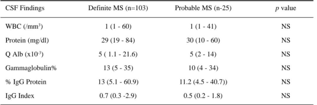
The CSF data of definite and probable MS patients are shown in Tables 1 and 2. There were no differences in the cytology, protein and albumin quotient between the groups (Figs 1, 2, 3). WBC counts higher than 35/mm3 were observed in 3% (3/
98) of the definite MS cases and 4% (1/25) of the probable. Levels of protein higher than 80 mg/dl were detected in only one case of definite MS (84 mg/dl).
The IgG Index and the IgG loc displayed higher sensitivity to detect the intrathecal synthesis of IgG, in comparison to IgG% and gamma globulin% of protein. The detection of oligoclonal IgG bands by isoelectric focusing was the most sensitive test to demonstrate intrathecal synthesis of IgG. Oligoclonal bands were detected in all cases with IgG index > or = 0.7 or IgG loc > or = 0.
A significantly lower frequency of the local IgG synthesis was observed in the group of probable MS (Tables 1 and 2).
DISCUSSION
We have determined the profile of the intrathecal humoral immune response of Brazilian MS patients, using a combination of quantitative and qualitative measures. The IgG index and IgG loc showed no significant differences to detect the intrathecal synthesis of IgG, considering that the majority of the patients had no blood-CSF barrier dysfunction. The IgG loc formula correlates the IgG and the albumin quotient, using a hiperbolic function5,9,10. It avoids the lost of sensitivity in cases
of more severe CSF-blood barrier dysfunction, compared to the IgG index9. The % IgG protein is
the simplest test. The % IgG protein and the Fig 1. Cytology examination in CSF of 128 MS
patients. WBC, White blood cells.
Fig 3. Albumin Quotient evaluation (Q Alb) in 128 MS patients. The black line indicates the median. No difference was observed in both groups. The Albumin Quotient was used as a parameter of blood-CSF barrier function. Q Alb higher than 8.0 x 10⁻³ was a parameter of Blood-CSF barrier
dysfunction. The majority of the cases displayed a normal Albumin quotient in both groups.
eletrophoresis on cellulose acetate had a lower sensitivity in the IgG profile and cannot differentiate the IgG blood fraction from IgG synthezised in CNS in cases of blood-CSF barrier dysfunction or those with elevated concentration of IgG in serum.
According to other reports4, the identification of oligoclonal bands by IEF was the most
sensitive technique able to detect the intrathecal synthesis of IgG. The interpretation of the bands is occasionally difficult and depends on the observer’s experience. In this sense, the IEF with immunodetection seems to be more objective, avoiding false-positive results13. The greater importance
of this procedure was to demonstrate local IgG production not identified by quantitative methods. It was especially important in the probable MS cases. The high frequency of negative results for quantitative analysis of IgG in such group, may be related to the use of steroids. It decreases the IgG values but not so frequently the presence of oligoclonal bands16. An other reason for this observation
may be related to the significant shorter duration of disease in the probable MS cases. Perhaps some of these cases are really not MS. It will be necessary follow up studies to the determine the evolution of these patients.
The significance of the presence of oligoclonal bands in MS patients is not clear. The pattern of absence or presence of these bands may persist during the lifetimes and is not related to the Table 2. Correlation between the MS Brazilian Groups and the frequency of the CSF findings.
CSF Findings Definite MS Probable MS p
Oligoclonal IgG Band in CSF 62/73 (85%) 6/10 (60%) NS
IgG (loc) >0 49/98 (50%) 4/21 (19%) <0.05
IgG Index >=0.7 50/98 (51%) 4/21 (19%) <0.05
% IgG protein >=15% 42/102 (41%) 7/25 (28%) NS
Gammaglobulin% > 14 40/96 (42%) 3/24 (12.5%) < 0.05
Q Alb >= 8 x10⁻³ 14/98 (14%) 4/21 (19%) NS
Protein >40 mg/dl 23/103 (22%) 5/25 (20%) NS
WBC > 4/ mm3 17/103 (16.5%) 3/25 (12%) NS
IgG (loc)>0 according to Reiber & Felgenhauer, 1987. IgG Index according to Tibbling et al.8. The frequencies were compared
using the chi-square/ Fisher exact test.
Table 1. CSF findings of 128 Brazilian MS patients.
CSF Findings Definite MS (n=103) Probable MS (n-25) p value
WBC (/mm3) 1 (1 - 60) 1 (1 - 41) NS
Protein (mg/dl) 29 (19 - 84) 30 (10 - 60) NS
Q Alb (x10-3) 5 ( 1.1 - 21.6) 5 (2 - 14) NS
Gammaglobulin% 13 (5 - 35) 10 (4 - 34) NS
% IgG Protein 13 (5.1 - 60.9) 11.2 (4.5 - 40.7)) NS
IgG Index 0.7 (0.3 -2.9) 0.5 (0.2 - 1.8) NS
Values are shown as median (min-max); Q Alb (Albumin Quotient); a normal blood-CSF barrier function was indicated as Q Alb <= 8 x10⁻³ ; normal IgG index <=0.7; the Mann-Whitney test was used to compare the median between both groups. NS`,
disease activity17. Autopsy studies have demonstrated a relationship between the lack of oligoclonal
bands in CSF and the absence of plasma cells in the meninges and plaques17.
The present study demonstrated that 85% of the definite MS patients presented oligoclonal bands restricted to the CSF. Although CECAMS reported a sensitivity higher than 95% for oligoclonal bands in definite MS cases 4 but lower frequencies (40-60%) have been usually found in routine
hospital laboratories6. The observation of lower IgG production in our MS cases may be related to
the used method or may represent a characteristic of our population. Of special interest is also the finding of lower frequency of hypercytosis in our MS cases (16.5% in comparison to 50% of the clinically definite MS cases of the European study4). These data suggest that the inflammatory
reaction in CSF of Brazilian MS cases seems to be less frequent than the other studies.
In conclusion, the evaluation of intrathecal IgG synthesis, using the CECAMS protocol was technically reproducible in Brazil. The combination of quantitative and qualitative procedures yields important results that aided in the diagnosis of MS18.
Acknowledgement: We would like to thank Dr. Charles Poser for his kindly review of this article. We
also thank the group of neurologists of Hospital da Lagoa, Hospital Gafree Guinle (Chief: Prof. Regina Alvarenga) and Hospital Universitário Clementino Fraga Filho –Universidade Federal do Rio de Janeiro (HUCFF-UFRJ) (Chief: Prof. Sérgio Novis).
REFERENCES
1. Poser C, Paty DW, Scheinberg L, et al. New diagnostic criteria for multiple sclerosis: guidelines for research protocols. Ann Neurol 1983;13:227-231.
2. Poser CM. The dissemination of multiple sclerosis: a viking saga? A historical essay. Ann Neurol 1994;36:S231-S243. 3. Papais-Alvarenga R, Leon AS, Miranda C, et al. Characteristics of multiple sclerosis in Brazil: a multicentric study in a
prevalence COHORT -South Atlantic Project Phase I. Abstr J Neurol Sci 1997;150(Suppl):S229.
4. Anderson M, Alvarez-Cermeno J, Bernardini G, et al. Cerebrospinal fluid in the diagnosis of multiple sclerosis: a consensus report. J Neurol Neurosurg Psychiatry 1994;57:897-902.
5. Reiber H, Ungefehr S, Jacobi C. The intrathecal, polyspecific and oligoclonal immune response in multiple sclerosis. Multiple Sclerosis 1998;4:11-117.
6. Poser CM. The unfortunate triumph of mechanodiagnosis in multiple sclerosis: a clinician’s lament. Cl Neurol Neurosurg 1992;94 (Suppl): S139-S142.
7. Puccioni-Sohler M, Kitze B, Felgenhauer K, et al. The value of CSF analysis for the differential diagnosis of HTLV-I associated myelopathy and multiple sclerosis. Arq Neuropsiquiatr 1995;53:760-765.
8. Tibbling G, Link H, Öhman S. Principles of albumin and IgG analysis in neurological disorders: I. Establishment of reference values. Scand J Clin Lab Invest 1977;37:385-390.
9. Reiber H, Felgenhauer K. Protein transfer at the blood cerebrospinal fluid barrier and the quantification of the humoral immune response within the central nervous system. Clin Chim Acta 1987;163:319-328.
10. Reiber H. External quality assessment in clinical neurochemistry: survey of analysis for cerebrospinal fluid (CSF) proteins based on CSF/serum quotients. Clin Chem 1995;41/2:256-263.
11. Olsson JE, Petttersson B. A comparisson between agar gel electrophoresis and CSF serum quotients of IgG and albumin in neurological diseases. Acta Neurol Scand 1976;53:308-322.
12. Pirtilla T, Mathila K, Frey H. Cerebrospinal fluid proteins in neurological disorderes analyzed by immobilized pH gradient isoeletric focusing using narrow pH gradients. Acta Neurol Scand 1991;83: 43-40.
13. Sellbjerg F, Christiansen M. Qualitative assessment of intrathecal IgG synthesis by isoeletric focusing and immunodetection: interlaboratory reproducibility and interobserver agreement. Scand J Clin Lab Invest 1996;56: 135-143.
14. Felgenhauer K, Reiber H. The diagnostic significance of antibody specificity indices in multiple sclerosis and herpes virus induced diseases of the nervous system. Clin Investig 1992;70:28-37.
15. Fishmann RA. Cerebrospinal fluid in diseases of the nervous system. 2Ed. Philadelphia: Saunders, 1992:197-212. 16. Lamers KJB, Frequin STJM, Hommes OR. Effects of immunotherapeutic strategies on cerebrospinal fluid parameters in
multiple sclerosis. In: Thmpson EJ, Trojano M, Livrea P. Cerebrospinal fluid analysis in multiple sclerosis. Milano: Springer-Verlag, 1996:113-122.
17. Farrel MA, Kaufman JCE, Gilber JJ, Noseworthy JH, et al. Oligoclonal bands in multiple sclerosis: clinical-pathological correlation. Neurology 1985;35: 212-218.