RBCCV 44205-1587 DOI 10.5935/1678-9741.20140118
Graft pathology at the time of harvest: impact on
long-term survival
Patologia do enxerto no momento d
a coleta:
impacto na sobrevida a longo prazo
Shi-Min Yuan
1, MMed, PhD; Yun Li
2, MD, PhD; Yan Hong Ben
3,RN; Xiao Feng Cheng
3, MMed,MD;
Da Zhu Li
4, MD; De Min Li
3, MMed, PhD; Hua Jing
3, MMed, MD
1The First Hospital of Putian, Teaching Hospital, Fujian Medical University, Putian, People's Republic of China.
2Department of Thoracic Surgery, Provincial Hospital Afiliated to Shan -dong University, Ji'nan, Shan-dong Province, People's Republic of China. 3Department of Cardiothoracic Surgery, Jinling Hospital, School of Clinical Medicine, Nanjing University, Jiangsu Province, People's Republic of China. 4First Department of Surgery, Fourth People’s Hospital of Lu’an, Lu'an, An-hui Province, People's Republic of China.
This study was carried out at Jinling Hospital, School of Clinical Medicine, Nanjing University, Jiangsu Province; and First Hospital of Putian, Teaching Hospital, Fujian Medical University, Putian, Fujian Province, People’s Re-public of China.
No inancial support.
Correspondence address: Shi-Min Yuan
Longdejing Street, 389 - Chengxian District, Putian, Fujian Province, Peo-ple’s Republic of China
E-mail: [email protected]
Article received on July 20th, 2014 Article accepted on October 12th, 2014 Abstract
Objective: This study aims to present the graft pathology at the time of harvest and its impact on long-term survival.
Methods: The remnants of the bypass grafts from 66 consec-utive patients with coronary artery disease receiving a coronary artery bypass grafting were investigated pathologically, and pertinent predictive risk factors and survival were analyzed.
Results: Medial degenerative changes with or without intimal proliferation were present in 36.8%, 37.8% and 35.6% of left internal mammary artery (IMA), radial artery and saphenous vein grafts. There were 2 (3.0%) hospital deaths and 9 (14.1%) late deaths. Multinomial logistic regression revealed left IMA pathological changes, dyslipidemia, history of percutaneous transluminal coronary angioplasty/stent deployment and Y-graft
were signiicant predictive risk factors negatively inluencing the
patients’ long-term survival. Kaplan-Meier survival analysis revealed that the long-term survival of patients with left IMA
pathological changes were signiicantly reduced compared with
those without (74.1% vs. 91.4%, P=0.002); whereas no differences were noted in long-term survivals between patients with and without pathological changes of the radial arterial or saphenous vein grafts.
Conclusion: Pathological changes may be seen in the bypass
graft at the time of harvest. The subtle ultrastructural modii
-cations and the expressions of vascular tone regulators might be responsible for late graft patency. The pathological changes of the left IMA at the time of harvest rather than those of the
radial artery or saphenous vein graft affect signiicantly
long-term survival. Non-traumatic maneuver of left IMA harvest, well-controlled dyslipidemia and avoidance of using composite grafts can be helpful in maintaining the architecture of the grafts.
Descriptors: Coronary Artery Bypass. Pathology. Survival Analysis. Mammary Arteries.
Resumo
Objetivo: Este estudo tem como objetivo apresentar a patolo-gia do enxerto no momento da coleta e do impacto na sobrevida a longo prazo.
Métodos: Os remanescentes de pontes de safena de 66 pacien-tes consecutivos com doença arterial coronária que receberam uma cirurgia de revascularização coronariana foram investigados patologicamente, e os fatores de risco preditivos e a sobrevivência foram analisados.
The patient demographics and the extents of coronary lesions by coronary angiography were listed in Tables 1 and 2.
Harvesting
Conventional harvesting of the IMA (pedicled), RA and SVG were applied in all patients.
Specimens
The remnants of the grafts were collected at the comple-tion of CABG from these 66 consecutive patients. The samples were cut into 1 mm3 in size and immersed in 10% methanal solution in appropriate bottles for pathological inspections. Hematoxylin and Eosin staining was performed on 4 μm paraf -in-embedded sections. The pathological changes of the grafts were observed and evaluated by an experienced pathologist.
Deinitions
Pathologies of the grafts are classiied as: normal, prolif -eration and eration (of the smooth muscle cells), degen-eration (of the intima), atherosclerosis, calciication, vascular wall hemorrhage and inlammatory cell iniltration, uneven vascular wall thickness and varicose (of the SVG). Prolifer-ation is deined as remarkable growth of vascular cells either in the intima or smooth muscle cells. Destructive changes present in the vascular cells are termed as degeneration. A form of arteriosclerosis is characterized by the deposition of atheromatous plaques containing cholesterol and lipids on the innermost layer of the vessel walls. Vessels permeated with calcium are deined as calciication.
Ethics
This study complies with the Declaration of Helsinki. Informed consent was obtained from each patient and the Institutional Ethics Committee has approved the research protocol.
Abbreviations, acronyms & symbols
CABG Coronary artery bypass grafting eNOS Endothelial nitric oxide synthase IMA Left internal mammary artery
LAD Left anterior descending coronary artery
RA Radial artery
SVG Saphenous vein graft
INTRODUCTION
The left internal mammary artery (IMA) has gained pop-ularity as an arterial graft for coronary artery bypass grafting (CABG) as it provides the gold standard left IMA-left de-scending coronary artery (LAD) bypass[1]. The radial artery (RA) has become a graft of choice for CABG since its irst clinical use by Carpentier in 1971[2]. The graft low patterns have been evaluated in several studies intraoperatively[3] or postoperatively[4]. Even though the RA graft showed better low than the IMA and saphenous vein graft (SVG)[3], the patency of the RA graft was inferior to that of the IMA and SVGs[5]. The RA has been recommended to be anastomosed to the circumlex artery or right coronary artery[6]. Use of the RA graft may improve patients’ survival and decrease the in-cidence of cardiac-related events within the irst postopera -tive years[7]. The low morbidity rate associated with the RA has urged the use of this conduit as a supplement of the right IMA in the IMA-to-LAD bypass[7]. However, controversies still exist on the graft choices in CABG. The pathological changes of bypass grafts at the time of harvest have not been frequently investigated and the relations between the pathol-ogy and graft patency and long-term survival are scanty. The present study was designed to evaluate the graft pathology at the time of harvest and its impact on long-term survival.
METHODS
Patients’ information
From 2004 to 2011, 66 patients with coronary artery dis-ease receiving CABG had their remnants of bypass grafts for pathological inspections. There were 55 males and 11 females with a mean age of 68.3±8.1 (range: 50-84; median, 73) years. No difference was found in patient age between males and females (68.8±7.9 years, vs. 65.8±9.0 years, P=0.2608). (ATIE), artéria radial e veia safena. Houve dois (3,0%) óbitos hospitalares e nove (14,1%) óbitos tardios. A regressão logística multinomial revelou que alterações patológicas na ATIE, disli-pidemia, história de angioplastia/stent implantação coronariana
transluminal percutânea e Y-enxerto foram signiicativos fatores de risco preditivos que inluenciam negativamente a sobrevivência
a longo prazo dos pacientes. Análise de sobrevida de Kaplan-Meier revelou que a sobrevivência a longo prazo de pacientes com
alterações patológicas da ATIE foi signiicativamente reduzida em
comparação com aqueles sem (74,1% vs. 91,4%, P=0,002), consi-derando que não foram observadas diferenças na sobrevivência de longo prazo entre pacientes com e sem alterações patológicas dos enxertos da artéria radial ou de veia safena.
Conclusão: As alterações patológicas podem se desenvolver
na revascularização no momento da coleta. As modiicações
ultraestruturais sutis e as expressões de reguladores do tônus vascular podem ser responsáveis pela patência tardia do enxerto. As alterações patológicas da ATIE no momento da coleta, em vez do enxerto da artéria radial ou da veia safena, podem afetar
signiicativamente a sobrevida de longo prazo. Manobra não trau -mática da ATIE na coleta, bom controle da dislipidemia e para evitar uso de enxertos compostos pode ser útil na manutenção da arquitetura dos enxertos.
Table 1. Patient demographics. Variables
Sex, male/female Age, year Course of disease Major symptom, n (%)
Chest pain/chest distress Shortness of breath Palpitation
Upper abdominal discomfort Associated disorder, n (%)
Pacemaker Cerebral infarction Pyelonephritis Others Comorbiditiy, n (%) Hypertension
Diabetes mellitus Dyslipidemia Alcohol Smoker
New York Heart Association class Cardiovascular agent, n (%)
Angiotensin converting enzyme inhibitor ß-blocker
Calcium channel antagonist Diuretics
Vasodilator Statin
Myocardial infarction, n (%)
Acute myocardial infraction
Non-ST segment elevation myocardial infarction Non-Q wave myocardial infarction
Percutaneous transluminal coronary angioplasty/stent, n (%) Coronary artery disease, n (%)
2-vessel disease 3-vessel disease
Left main+1-vessel disease Left main+2-vessel disease Left main+3-vessel disease Coronary stenosis on angiography, % Left main coronary artery Left anterior descending artery
Circumlex artery
Right coronary artery Hospitalization, day
Postoperative complications, n (%)
Results (n=66) 55/11 68.3±8.1 (range, 50-84) 5.9±7.6 year (range, 1 day-30 year)
48 (72.7) 6 (9.1) 10 (15.2)
2 (3.0)
3 (4.6) 6 (9.1) 1 (1.5) 1 (1.5)
58 (87.9) 16 (24.2) 7 (10.6) 9 (13.6) 21 (31.8) 2.2±0.4 (range, 2-3)
11 (16.7) 16 (24.2) 27 (40.9) 13 (19.7) 17 (25.8) 2 (3.0)
24 (36.4) 20 (30.3)
3 (4.6) 1 (1.5) 5 (7.6)
9 (13.6) 27 (40.9)
2 (3.0) 2 (3.0) 26 (39.4)
61.8±21.8 (range, 20-95) (n=30) 91.0±11.4 (range, 70-100) (n=58) 87.5±14.9 (range, 30-100) (n=52) 84.0±19.2 (range, 20-100) (n=51) 22.0±15.5 (range, 3-70) (n=66)
4 (6.1)
Table 2. Number or extent of the coronary lesions.
Coronary artery
Left main coronary artery
Left anterior descending coronary artery
Circumlex artery
Right coronary artery
No lesion n (%) 35 (53.0)
1 (1.5) 8 (12.1) 7 (10.6)
Solitary n (%) 26 (39.4) 20 (30.3) 19 (28.8) 12 (18.2)
2 n (%) 3 (4.6) 11 (16.7) 12 (18.2) 5 (7.6)
≥3
n (%) 0 (0) 6 (9.1) 7 (10.6) 11 (16.7)
Long lesion n (%)
0 (0) 8 (12.1)
4 (6.1) 9 (13.6)
Diffuse lesion n (%) 1 (1.5) 16 (24.2) 12 (18.2) 17 (25.8)
Statistics
Data were expressed as mean ± standard deviation. Un-paired t-test was made for intergroup comparisons of the quantitative data. Fisher exact test and logistic regression analysis were performed for the postoperative outcomes and the pathology of the grafts. Patients’ survival was evaluated with Kaplan-Meier survival analysis. A two-tailed P value of <0.05 was considered of signiicance.
RESULTS
The CABG procedures were performed under CPB in 55 (83.3%), off-pump in 10 (15.2%) and a stepped off-pump and beating heart revascularization under CPB in 1 (1.5%) patient, respectively. Ten (15.2%) patients had a secondary procedure during the operations including mitral valve replacement or re-pair in 5 (50%), aortic valve replacement, left ventricular pseu-doaneurysmectomy, left ventricular pseudoaneurysmectomy with mitral valve repair, left ventricular pseudoaneurysmec-tomy with the aid of intraaortic balloon pump and intraaortic balloon pump alone in 1 (10%) patient each.
There were totally 236 grafts bypassed in 66 patients with a mean of 3.6±0.9 (range: 2-5; median, 3) grafts per patient including 103 (43.6%) SVGs, 57 (24.2%) RAs, 74 (31.4%) left IMAs (inclusive of all distal anastomoses of the sequen-tial grafts) and 2 (0.8%) right IMAs. A sequensequen-tial graft was performed in 34 (51.5%) patients, a Y-graft in 2 (3.0%) pa-tients and only independent grafts in the remaining 30 (45.5%) patients. The donor vessels for the LAD were left IMA in 63 (96.9%), RA in 1 (1.5%) and SVG in 1 (1.5%), respective-ly (χ2=177.4, P=0.0000). The coronary arteries receiving an SVG were left main coronary artery in 1 (1.0%), LAD in 1 (1.0%), the irst diagonal branch in 21 (20.4%), intermediate artery in 3 (2.9%), circumlex artery in 1 (1.0%), obtuse mar -ginal branch in 41 (39.8%), right coronary artery in 5 (4.9%), posterior descending artery in 26 (25.2%) and posterior left ventricular branch in 4 (3.9%), totaling 103 grafts. Apart from one (1.0%) bypassed to the left main coronary artery, there were 25 (24.3%), 42 (40.8%) and 35 (34.0%) SVGs grafted to the LAD, circumlex and RCA systems, respectively (χ2=6.4, P=0.0399). The receipt vessels of the left IMA were LAD in
56 (75.7%), the irst diagonal branch in 1 (1.4%), sequentially LAD and irst diagonal branch in 8 (10.8%) and the great car -diac vein for the purpose of venous arterialization in 1 (1.4%) patient, respectively. An RA graft was used in 51 (77.3%) pa-tients, bypassed to the irst diagonal branch in 6 (11.8%), LAD in 1 (2.0%), intermediate artery in 1 (2.0%), circumlex artery in 1 (2.0%), obtuse marginal branch in 23 (45.1%) and pos-terior descending artery in 19 (37.3%) patients, respectively. They were in a sequential fashion in 4 (7.8%) patients (one RA graft for two distal anastomoses in each patient), a Y-graft in 2 (3.9%) patients and an independent graft in 41 (80.4%) patients. The 2 right IMA grafts were bypassed to the interme-diate artery and right coronary artery, respectively.
Of the 236 grafts, 161 (68.2%) remnants were collect-ed and inspectcollect-ed pathologically. There were 57 (35.4%) left IMAs, 45 (28.0%) RAs and 59 (36.7%) SVGs. Types of grafts used did not differ between gender.
The dimensions of the lumens of the three grafts were all within normal ranges. The lengths and lumen diameters of the SVG were larger than those of the IMAs and RAs, how-ever, no statistical differences were noted (Table 3).
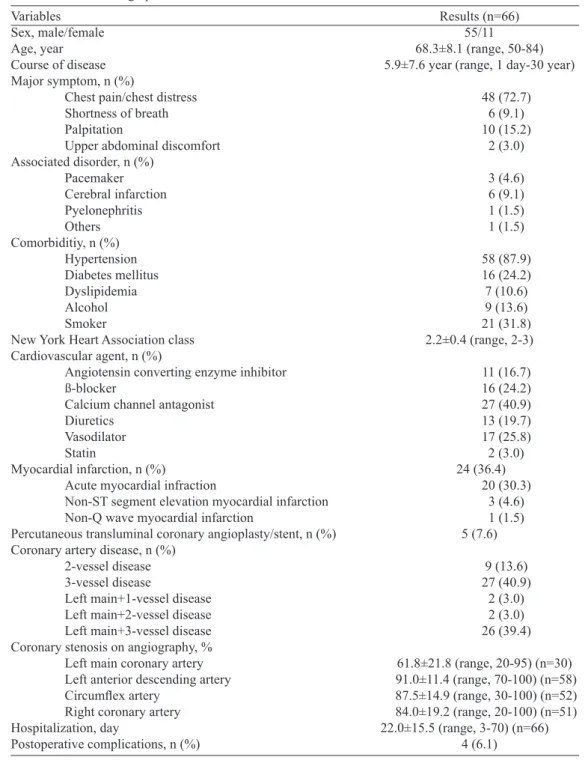
Of this patient setting, half of the patients had normal graft vessels as evidenced by histological observations (Ta-ble 4). The prevalence of pathological changes did not reach a statistical signiicance between the IMA, RA and venous grafts (47.4% vs. 46.7% vs. 45.8%, χ2=0.0, P=0.985). The prevalence of the medial degeneration did not differ be-tween the three grafts, either (36.8% vs. 37.8% vs. 35.6%, χ2=0.1, P=0.973). The microscopic views of the grafts were shown in Figure 1.
There were 2 hospital deaths with an early mortality of 3.0%. Two patients were lost for follow-up. The remaining 62 patients were followed up for an average of 30.2±8.8 (range, 6.9-55; median, 15.8) months.
Twenty-four patients had coronary angiography examined during follow-up, 1 (4.2%) patient had left IMA and 1 (4.2%) pa-tient had RA graft occlusion requiring reoperations and 2 (8.3%) patients had SVG occlusion warranting a reintervention by per-cutaneous angioplasty. There were 9 (14.1%) late deaths due to heart dysfunction (n=3), acute myocardial infarction (n=2), renal failure (n=2), malignant cancer (n=1) and unknown reason (n=1).
Table 3. Measurements of the inspected remnants of the grafts. Pathology
Lenght (cm)
Lumen diameter (cm)
Saphenous vein graft (n=59) 4.9±3.4 (range, 0.4-9) (n=42)
0.4±0.1 (0.2-0.9) (n=54)
Left internal mammary artery (n=57)
2.1±1.1 (range, 0.5-5) (n=38) 0.3±0.1 (range, 0.1-0.5)
(n=40)
Radial artery (n=45) 2.7±1.7 (range, 0.5-5) (n=33) 0.4±0.1 (range, 0.2-0.8)
Multinomial logistic regression revealed that patholo-gy of the left IMA (P=0.017), the presence of dyslipidemia (P=0.033), history of percutaneous transluminal coronary an-gioplasty/stent deployment (P=0.001) and Y-graft (P=0.006) were signiicant predictive risk factors of long-term survival.
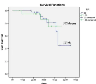
Kaplan-Meier survival analysis revealed that the long-term survival of the patients with left IMA pathological changes was signiicantly reduced in comparison with those without (74.1% vs. 91.4%, P=0.002) and that the pathological changes of the radial arterial or saphenous vein graft were unlikely to signiicantly affect patients’ long-term survival (Figures 2-4).
DISCUSSION
The left IMA-LAD graft is appraised for its promis-ing patency rate and patients' long-term survival[8]. The left IMA is usually used to bypass the anterior circula-tion and the RA graft, to bypass the coronary arteries of the inferior or the lateral territory[9]. Recent randomized
Table 4. Pathology of the inspected remnants of the grafts. Pathology
Normal
Proliferation and degeneration of smooth muscle cells Intimal degeneration
Atherosclerosis
Calciication
Vascular wall hemorrhage & inlammatory cell iniltration
Uneven vascular wall thickness Varicose (saphenous vein)
Left internal mammary artery (n=57)
n (%) 30 (52.6) 21 (36.8) 2 (3.5) 2 (3.5) 1 (1.8) 1 (1.8)
--Radial artery (n=45)
n (%) 24 (53.3) 17 (37.8) 3 (6.7)
0 (0) 1 (2.2)
0 (0)
--Saphenous vein graft (n=59)
n (%) 32 (54.2) 21 (35.6) 1 (1.7)
0 (0) 0 (0) 0 (0) 3 (5.1) 2 (3.4)
Fig. 1 - Microspcopic views of the grafts: (A) saphenous vein grafts with normal structures; (B) saphenous vein grafts showing medial degenerative changes with intimal proliferation; (C) radial arterial grafts with normal structures; (D) radial arterial grafts with medial degenerative changes with intimal proliferation; (E) internal mammary arteries with roughly normal structures; and (F) internal mammary arteries with mild degeneration of the media with intimal proliferation. Hematoxylin and Eosin staining × 200
Fig. 2 - Kaplan-Meier analysis revealed that the pathological changes of the left internal mammary graft artery were a risk factor
that negatively inluenced the survival rate. The long-term survival
ROOBY trial proved that off-pump CABG was associated with lower patency for arterial and venous graft conduits than on-pump CABG[10]. Superior long-term survival rate was observed in the on-pump CABG than in the off-pump CABG patients[11-13].
Clinical observations revealed that the RA grafts had a lower patency rate (51.3%) than the left IMAs (90.3%) or the SVGs (64.0%)[4]. At a mean of 7.7±1.5 year follow-up after CABG, RAs had much lower functional (12.0% vs. 19.7%, P=0.03) and complete graft occlusion rates (8.9% vs. 18.6%, P=0.002) compared with the SVGs[14]. Hata et al.[15] reported that the cumulative graft patency rates at 8 years were 74.3% for the RA and 64.7% for the SVG, re-spectively. RA patency rate varied with territory and bypass modes, with 79.4% in the left circumlex coronary artery, 72.7% in the LAD and 50% in the right coronary artery; the occlusion rate was 20.0% among free grafts, 18.2% among sequential grafts and 20.0% among composite grafts[16]. The RA patency rate showed a gender predilection, which was higher in men than in women (38.9% vs. 56.1%, P=0.025) at a mean follow-up of 565±511 days[17].
Non-severe stenosis of the target coronary arteries can be a potential risk factor leading to a higher diffuse narrowing rate of the RA grafts in comparison to the SVGs[9]. Several studies agreed that the history of open heart surgery or percu-taneous coronary intervention can be a signiicant risk factor of increased mortality[18,19]. Severe congestive heart failure, advanced age, postinfarction myocardial rupture, cardio-genic shock, CPB, pulmonary hypertension and increased creatinine can be the risk factors of in-hospital mortality[20]. Long-term mortality was also associated with female gender, non-Hispanic black race, small body surface area, extreme body mass index values, left main coronary disease, mul-tivessel disease, reduced ejection fraction, history of myo-cardial infarction, unstable hemodynamic state/shock and the presence of comorbidities[21].
The research of graft pathology started from the 1970’s. By light microscopy, minimal to moderate mediointimal i -brosis and graft occlusion due to recent thrombosis were noted in 31.7% (13/41) grafts early (<20 days) and old thrombus, intimal leiomyocellular proliferation, or intimal phlebosclerosis were noted in 19.5% (8/41) patients in the late group (3-39 months); whereas a vein graft showed ath-erosclerotic changes[22].
A histological study of the SVGs showed grade 1 to 2 intimal proliferation in the early group (14 days) and grade 2 to 3 or even grade 4 intimal proliferation in the late group (34 months) in all grafts[23]. In a prospective study involving 365 consecutive patients undergoing isolated CABG, signiicant lesions in the SGVs were observed in 71 (19.5%) patients[24]. Structures of the SVG, particularly the tunica media and smooth muscle cells had a signiicant impact on the late out -come after CABG[25]. In patients with postoperative coronary
Fig. 3 - Kaplan-Meier analysis revealed that pathological changes
of the radial artery graft were not a signiicant risk factor affecting the survival rate. The long-term survival rate was 83.6%, 82.9% and
85.0% for overall, patients with and without pathological changes of the radial artery.
RA= Radial artery; With= With pathological changes; Without= Without pathological changes
Fig. 4 - Kaplan-Meier analysis revealed that the pathological
changes of the saphenous vein graft were not a signiicant risk factor affecting the survival rate. The long-term survival rate was 83.6%, 78.9% and 91.3% for overall, patients with and
without pathological changes of the saphenous vein.
artery disease progression and severe venous graft disease, tunica media thickening was noted, and it was taken as a risk factor for late venous graft failure[25].
In recent years, graft pathology has been carried out in deep-going ways concerning ultrastructural and signaling pathway studies. Saphenous tunica media thickening and chunky smooth muscle cell nuclei were identiied as inde -pendent risk factors for graft disease[24]. High lipid expo-sure may be prone to early graft failure especially of the vein graft[26]. Morphometric studies of the RA disclosed mild or moderate intimal hyperplasia but no medial cal-ciication in both young and elderly patients[27]. Matrix vesicles and calciications were frequently present in the media of both the RA and IMA[28]. Fewer endothelial de-nudations, similar medial lipid deposition and submicro-scopic calciication were observed in the RA comparing to other arteries[28].
The higher proliferative potential of the smooth muscle cells and more actively secretory status of endothelial cells of the RA enhance the remodeling process and predispose a reduced long-term patency[28]. In diabetic patients, the foam cells and tendency of migration from the smooth muscle cells to the intima were more frequently observed in the RA than in non-diabetic patients as observed by electron microscopy[29].
Chronic hypoxia increases the activity of vascular en-dothelial growth factor, a potent mitogenic molecule for the smooth muscle cells[30]. Li et al.[31] discovered that platelet-de-rived growth factor, another potent mitogenic molecule, is active exclusively in the epithelioid-like smooth muscle cells isolated from the human IMA but not in the spindle-like cells. Endothelial nitric oxide synthase (eNOS) type III was expressed in the intima of the IMA, RA and SVG and in the media of the IMA and RA. However, the IMA showed a higher intensity of eNOS type III expression, particularly within the media. This may provide an histologic explanation for the better results of the IMA graft[32]. Increased expres-sions of eNOS in the intima and media were also observed in the RA grafts irrespective of patients’ age[27].
A recent study revealed that Gas6/Axl pathway cytokines are more expressed in the left IMA than in other arteries, in-dicating that the IMAs were more resistant to atherosclerotic changes[33]. Independent predictors of late RA graft failure were native coronary stenosis <75% and peripheral vascular disease. Independent predictors of late SV graft failure were use of only one anti-platelet agent and low-density/high-den-sity lipoprotein cholesterol ratio >2.5[15].
The vasa vasorum of the veins is more pronounced than in the arteries and hence the vasa vasorum plays an important role in enhancing SVG patency when harvested along with surrounding tissue for restoring medial blood low from the nutrient microvessels[34]. Upregulated in-lammation biomarkers including scavenger receptors A
and B, toll-like receptors 2 and 4, platelet endothelial cell adhesion molecule, vascular cell adhesion molecule and intercellular cell adhesion molecule have been noted in the SVGs and were thus taken as a possible mechanism of graft failure[35]. An increased expression of cytokeratin 8 and weak expressions of calponin in the tunica media of the SVGs might be useful markers of unfavorable long-term prognosis in CABG patients[36].
The skeletonization of the IMA with an ultrasonic scal-pel had no deleterious effect on the endothelium. All IMA specimens showed a completely conluent endothelium, and no endothelial injury was observed by a scanning electron microscopic study[37]. A metal clamp can cause serious in-timal injury which disrupts the internal elastic lamina, and thus should be avoided for the temporary clamping of the skeletonized IMA. A ibrous jay clamp, however, hardly ever causes intimal injury, and its clinical use for the temporary clamping of the ultrasonically skeletonized IMA is therefore recommended[37].
Retention of perivascular tissue on the SVG prepared for CABG by “no-touch” technique protects against dis-tension-induced damage, preserves vessel morphology and maintains eNOS activity[38]. In no-touch vessels, eNOS is highly expressed as compared with conventionally harvest-ed ones[39]. The RAs harvested by “no-touch” technique are associated with better preservation of the endothelial cells warranting a long-term patency[28].
In the present study, the dimensions of the lumens of the three grafts were within normal ranges, with reference to their normal limits of 3.1 to 8.5 mm[40], 2.1 mm[41] and 2.6 mm[42] for the SVG, IMA and RA, respectively, as reported in the literature. Pathologically, over half of the patients had normal graft vessels; while others showed medial degener-ative changes with or without intimal proliferation account-ing for 35.6%, 36.8% and 37.8% in three grafts, respec-tively. In line with previous reports[11,19], the present study revealed left IMA pathological changes, dyslipidemia, his-tory of PTCA/stent and Y-graft were signiicant predictive risk factors negatively inluencing the patients’ long-term survival.
The nonrandomized nature and the limitations in patient selection were likely to be the major drawbacks of this study. A multicenter study in the future would be helpful in obtain-ing more accurate results.
CONCLUSION
-tients' long-term survival. Non-traumatic maneuver of IMA harvest, well-controlled dyslipidemia and avoidance of us-ing composite grafts can be helpful in maintainus-ing the ar-chitecture of the IMA graft and may therefore improve the long-term outcomes of the patients.
REFERENCES
1. Karthik S, Fabri BM. Left internal mammary artery usage in coronary artery bypass grafting: a measure of quality control. Ann R Coll Surg Engl. 2006;88(4):367-9.
2. Carpentier A, Guermonprez JL, Deloche A, Frechette C, DuBost C. The aorta-to-coronary radial artery bypass graft. A technique avoiding pathological changes in grafts. Ann Thorac Surg. 1973;16(2):111-21.
3. Goel P, Dubey S, Makker A, Kohli VM. Evaluation of coronary artery bypass grafts by intraoperative transit time flow measurement. Ind J Thorac Cardiovasc Surg. 2003;19(2):108-12.
4. Pizzuto F, Voci P, Mariano E, Puddu PE, Aprile A, Romeo F.
Evaluation of low in the left anterior descending coronary
artery but not in the left internal mammary artery graft predicts
signiicant stenosis of the arterial conduit. J Am Coll Cardiol.
2005;45(3):424-32.
5. Khot UN, Friedman DT, Ellis SG. Radial-artery coronary bypass grafts. N Engl J Med. 2005;352(9):941-2; author reply 941-2.
6. Ennker J, Wanner M, Gehle P, Ennker IC, Rosendahl U. Postoperative evaluation of radial artery grafts for coronary artery
bypass grafting by transit-time Doppler low measurements.
Thorac Cardiovasc Surg. 2001;49(6):365-8.
7. Acar C, Cook RC. Letter regarding article by Khot et al, "Radial artery bypass grafts have an increased occurrence of angiographically severe stenosis and occlusion compared with left internal mammary arteries and saphenous vein grafts". Circulation. 2005;111(1):e6-9.
8. Leavitt BJ, O’Connor GT, Olmstead EM, Morton JR, Maloney
Authors’ roles & responsibilities
SMY Main Author
YL Carrying out operations and/or experiments YHB Carrying out operations and/or experiments XFC Carrying out operations and/or experiments DZL Carrying out operations and/or experiments DML Carrying out operations and/or experiments HJ Carrying out operations and/or experiments
CT, Dacey LJ, et al. Use of the internal mammary artery graft and in-hospital mortality and other adverse outcomes associated with coronary artery bypass surgery. Circulation. 2001;103(4):507-12.
9. Desai ND, Cohen EA, Naylor CD, Fremes SE; Radial Artery Patency Study Investigators. A randomized comparison of radial-artery and saphenous-vein coronary bypass grafts. N Engl J Med. 2004;351(22):2302-9.
10. Hattler B, Messenger JC, Shroyer AL, Collins JF, Haugen SJ, Garcia JA, et al; Veterans Affairs Randomized On/Off Bypass (ROOBY) Study Group. Off-Pump coronary artery bypass surgery is associated with worse arterial and saphenous vein graft patency and less effective revascularization: Results from the Veterans Affairs Randomized On/Off Bypass (ROOBY) trial. Circulation. 2012;125(23):2827-35.
11. Shroyer AL, Grover FL, Hattler B, Collins JF, McDonald GO, Kozora E, et al; Veterans Affairs Randomized On/Off Bypass (ROOBY) Study Group. On-pump versus off-pump coronary-artery bypass surgery. N Engl J Med. 2009;361(19):1827-37.
12. Kim JB, Yun SC, Lim JW, Hwang SK, Jung SH, Song H, et al. Long-term survival following coronary artery bypass grafting: off-pump versus on-pump strategies. J Am Coll Cardiol. 2014;63(21):2280-8.
13. Filardo G, Grayburn PA, Hamilton C, Hebeler RF Jr, Cooksey WB, Hamman B. Comparing long-term survival between patients undergoing off-pump and on-pump coronary artery bypass graft operations. Ann Thorac Surg. 2011;92(2):571-7.
14. Deb S, Cohen EA, Singh SK, Une D, Laupacis A, Fremes SE; RAPS Investigators. Radial artery and saphenous vein patency more than 5 years after coronary artery bypass surgery: results from RAPS (Radial Artery Patency Study). J Am Coll Cardiol. 2012;60(1):28-35.
15. Hata M, Yoshitake I, Wakui S, Unosawa S, Kimura H, Hata H, et al. Long-term patency rate for radial artery vs. saphenous vein grafts using same-patient materials. Circ J. 2011;75(6):1373-7.
16. Crusco F, Antoniella A, Papa V, Menzano R, Di Lazzaro D, Di Manici G, et al. Midterm follow-up of patients receiving radial artery as coronary artery bypass grafts using 16-detector-row CT coronary angiography. Radiol Med. 2007;112(4):538-49.
17. Khot UN, Friedman DT, Pettersson G, Smedira NG, Li J, Ellis SG. Radial artery bypass grafts have an increased occurrence of angiographically severe stenosis and occlusion compared with left internal mammary arteries and saphenous vein grafts. Circulation. 2004;109(17):2086-91.
18. Wu C, Camacho FT, Wechsler AS, Lahey S, Culliford AT, Jordan D, et al. Risk score for predicting long-term mortality after coronary artery bypass graft surgery. Circulation. 2012;125(20):2423-30.
Surgeons Adult Cardiac Surgery Database (the ASCERT study). Circulation. 2012;125(12):1491-500.
20. Mejía OA, Lisboa LA, Tiveron MG, Santiago JA, Tineli RA, Dallan LA, et al. Coronary artery bypass grafting in acute myocardial infarction: analysis of predictors of in-hospital mortality. Rev Bras Cir Cardiovasc. 2012;27(1):66-74.
21. Hannan EL, Racz MJ, Walford G, Jones RH, Ryan TJ, Bennett E, et al. Long-term outcomes of coronary-artery bypass grafting versus stent implantation. N Engl J Med. 2005;352(21):2174-83.
22. Thiene G, Valente ML, Miazzi P, Casarotto D, Bortolotti U, Gallucci V. [Pathology of the aorto-coronary bypass by autologous saphenous vein (author's transl)]. G Ital Cardiol. 1979;9(6):549-56.
23. Lawrie GM, Lie JT, Morris GC Jr, Beazley HL. Vein graft patency and intimal proliferation after aortocoronary bypass: early and long-term angiopathologic correlations. Am J Cardiol. 1976;38(7):856-62.
24. Perek B, Malinska A, Stefaniak S, Ostalska-Nowicka D, Misterski M, Zabel M, et al. Predictive factors of late venous aortocoronary graft failure: ultrastructural studies. PLoS One. 2013;8(8):e70628.
25. Davies AH. Vein factors that affect the outcome of femorodistal bypass. Ann R Coll Surg Engl. 1995;77(1):63-6.
26. Zhu YY, Hayward PA, Hare DL, Reid C, Stewart AG, Buxton BF. Effect of lipid exposure on graft patency and clinical outcomes: arteries and veins are different. Eur J Cardiothorac Surg. 2014;45(2):323-8.
27. Shen LZ, Chen XJ, Chen X, Xu M, Wang LM, Jiang YS. [The morphometry and eNOS expression of radial artery in elderly patients with coronary atherosclerotic heart disease]. Zhonghua Wai Ke Za Zhi. 2010;48(11):825-9.
28. Wang HY, Meng Y, Lou XJ, Chu Y, Xu XL, Sun HS, et al. [Comparative study on the ultrastructures of radial and internal mammary arteries used for coronary artery bypass grafting]. Zhonghua Bing Li Xue Za Zhi. 2005;34(8):528-32.
29. Zou L, Chen XJ, Xu M, Chen W, Wang LM, Huang FH, et al. [Comparative study on the ultrastructure of radial artery in elderly patients underwent coronary artery bypass grafting with diabetes mellitus]. Zhonghua Wai Ke Za Zhi. 2011;49(12):1109-13.
30. Hubbell MC, Semotiuk AJ, Thorpe RB, Adeoye OO, Butler SM, Williams JM, et al. Chronic hypoxia and VEGF differentially modulate abundance and organization of myosin heavy chain isoforms in fetal and adult ovine arteries. Am J Physiol Cell Physiol. 2012;303(10):C1090-103.
31. Li S, Fan YS, Chow LH, Van Den Diepstraten C, van Der Veer E, Sims SM, et al. Innate diversity of adult human arterial smooth muscle cells: cloning of distinct subtypes from the internal thoracic artery. Circ Res. 2001;89(6):517-25.
32. Gaudino M, Toesca A, Maggiano N, Pragliola C, Possati G. Localization of nitric oxide synthase type III in the internal thoracic and radial arteries and the great saphenous vein: a comparative immunohistochemical study. J Thorac Cardiovasc Surg. 2003;125(6):1510-5.
33. Lee CH, Shieh YS, Tsai CS, Hung YJ, Tsai YT, Lin CY. Expression
of growth arrest-speciic protein 6 and Axl molecules in the left
internal mammary artery of patients undergoing coronary artery bypass grafting. J Clin Pathol. 2014;67(6):506-11.
34. Dreifaldt M, Souza DS, Dashwood MR. Comparable patencies of the radial artery and right internal thoracic artery or saphenous vein beyond 5 years: results from the Radial Artery Patency and Clinical Outcomes trial. J Thorac Cardiovasc Surg. 2010;140(3):727-8.
35. Khaleel MS, Dorheim TA, Duryee MJ, Durbin HE Jr, Bussey WD, Garvin RP, et al. High-pressure distention of the saphenous vein
during preparation results in increased markers of inlammation:
a potential mechanism for graft failure. Ann Thorac Surg. 2012;93(2):552-8.
36. Perek B, Malińska A, Ostalska-Nowicka D, Puślecki M, Ligowski
M, Misterski M, et al. Cytokeratin 8 in venous grafts: a factor of unfavorable long-term prognosis in coronary artery bypass grafting patients. Cardiol J. 2013;20(6):583-91.
37. Yoshikai M, Ito T, Kamohara K, Yunoki J. Endothelial integrity of ultrasonically skeletonized internal thoracic artery: morphological analysis with scanning electron microscopy. Eur J Cardiothorac Surg. 2004;25(2):208-11.
38. Dashwood MR, Savage K, Tsui JC, Dooley A, Shaw SG, Fernández Alfonso MS, et al. Retaining perivascular tissue of human saphenous vein grafts protects against surgical and distension-induced damage and preserves endothelial nitric oxide synthase and nitric oxide synthase activity. J Thorac Cardiovasc Surg. 2009;138(2):334-40.
39. Tsui JC, Souza DS, Filbey D, Karlsson MG, Dashwood MR. Localization of nitric oxide synthase in saphenous vein grafts harvested with a novel "no-touch" technique: potential role of nitric oxide contribution to improved early graft patency rates. J Vasc Surg. 2002;35(2):356-62.
40. Fazan VP, Borges CT, Da Silva JH, Caetano AG, Filho OA.
Supericial palmar arch: an arterial diameter study. J Anat.
2004;204(4):307-11.
41. Canham PB, Finlay HM, Boughner DR. Contrasting structure of the saphenous vein and internal mammary artery used as coronary bypass vessels. Cardiovasc Res. 1997;34(3):557-67.