Prognostic Criteria of Success and Recurrence in Circumferential
Ablation for the Treatment of Atrial Fibrillation
Washington Maciel, Eduardo Andréa, Nilson Araújo, Hecio Carvalho, Luiz Gustavo Belo, Leonardo Siqueira, Claudio
Munhoz, Rodrigo Cosenza, Fabiana Mitidieri, Jacob Atié
Clínica São Vicente, Instituto Estadual de Cardiologia Aloysio de Castro e Universidade Federal do Rio de Janeiro - Rio de Janeiro, RJ, Brazil
Summary
Objectives: To analyze the success of circumferential ablation of atrial fibrillation and to investigate possible clinical and electroanatomic predictors of recurrence of cardiac arrhythmia.
Methods: 104 consecutive patients free of structural heart disease and refractory to at least two antiarrhythmic drugs, and undergoing circumferential ablation for the treatment of paroxysmal/persistent atrial fibrillation were analyzed. Seventy two patients were males and the mean age of the group was 58.6 + 10.9 years. The procedure consisted of a single transseptal puncture and three-dimension mapping using the CARTO™ system to acquire points in the left atrium and pulmonary veins. Radiofrequency applications were performed encircling the pulmonary vein ostea, up to a > 80% reduction of the atrial potential amplitude. One additional line was created in the mitral isthmus and another in the cavotricuspid isthmus. Total left atrium volume, area ablated around the pulmonary veins, and completeness of the ablation line (complete or incomplete line) were analyzed. A line was considered complete when the distance between two contiguous radiofrequency application points was lower than 10mm.
Results: In a mean 18-month follow-up, 87 patients were in sinus rhythm (84%), and 17 patients presented recurrence (16%). In the multivariate analysis, only the left atrial volume (p<0.0001) and complete ablation (p<0.05) were independent predictors of recurrence.
Conclusion: The results suggest that the left atrial volume and the presence of complete ablation are predictive of recurrence of atrial fibrillation.
Key words: Atrial fibrillation, electroanatomic mapping, catheter ablation.
Mailing Address: Washington Andrade Maciel •
R. Barão de Lucena, 76/501 – 22260-020 – Rio de Janeiro, RJ, Brazil E-mail: [email protected]
Manuscript received January 3, 2006; revised manuscript received May 13, 2006; accepted June 1, 2006.
Introduction
Atrial fibrillation is the most frequent sustained arrhythmia in the clinical practice. It is diagnosed by both general practitioners and cardiologists, it is seen in every outpatient clinic, and its prevalence increases with age1-6.
Maintenance of the sinus rhythm with antiarrhythymic drugs and successive cardioversions, as shown in t==he AFFIRM trial and in the subsequent subtrials7-9, proved to
be a strategy that did not provide benefit for the patients as regards mortality. Actually, the AFFIRM trial demonstrated an increase in mortality in the rhythm control group when compared to the simple maintenance of ventricular rate, with p=0.08.
The non-pharmacological treatment in the different approaches proposed produces satisfactory results, that is, maintenance of the sinus rhythm with a success rate ranging
from 60% to 90%10. This wide-ranging variation depends on
factors such as the population studied, the method used, success criteria, and the team experience.
The identification of criteria possibly predictive of success and failure prior to and during the intervention may be useful to define the extension of the ablation, the need for additional radiofrequency lines or approach to abnormal atrial potentials.
Another practical aspect based on the knowledge of predictors of success and recurrence is the assessment of the possible outcome of radiofrequency ablation of atrial fibrillation. Thus, an expectation can be created as regards realistic long-term outcomes, without unnecessary pessimism or ungrounded optimism.
immediately performed in the adjacent region. Radiofrequency energy release time for obtaining a reduction of the potential amplitude ranges, in average, between 5 and 15 seconds for each point.
The objective of the procedure was to encircle the left
DQGULJKWYHLQVLQDVLQJOHZD\VRDVQRWWROHDYHDPP
interval between radiofrequency points.
Therefore, the assessment of the radiofrequency line completeness was determined by measuring the distance between adjacent radiofrequency points. The line was
FRQVLGHUHG LQFRPSOHWH ZKHQ D PP GLVWDQFH ZDV
observed between adjacent points. Otherwise, the ablation line was considered complete.
The 10-mm interval was assumed as the value from which a radiofrequency application line would be considered incomplete, that is, with a gap. This value was chosen considering that when a 4-mm-tip ablation catheter releases radiofrequency energy with temperature set at 60° and maximum power at up to 50W in contact with the cardiac muscle, it produces a lesion that may range from 2 to 11 mm11,12. Therefore, a greater-than-10-mm distance between
adjacent points allows the occurrence of non-ablated muscle; thus, there would be possible passageways for electrical impulses coming from circumferential ablation areas.
The measurement of the distance between the points is provided by the CARTO™ system itself. Some patients also presented typical atrial flutter, electrocardiographically documented. Flutter was not induced for assessment of its circuit; however, a bidirectional isthmus conduction block was created in all patients.
Sheath removal (two venous and one arterial) was performed only after the procedure, when the activated clotting time reached values lower than 180s.
Left atrial volume, volume of the area ablated around the right and left veins, circum ferential radiofrequency application
Methods
One hundred and four consecutive patients with recurrent paroxysmal atrial fibrillation or persistent atrial fibrillation, and undergoing circumferential ablation of the pulmonary veins guided by electroanatomic mapping using the CARTO™ System were analyzed from March, 2002 to July, 2003.
Mean age of the group studied was 58.6 + 10.9 years, ranging from 20 to 80 years. Seventy two patients were males and 32 were females.
After informed consent, we included patients with persistent or paroxysmal atrial fibrillation for more than one year with the following characteristics: symptomatic, recurrent, documented with electrocardiographic records, and refractory to two or more antiarrhythmic drugs, including amiodarone. Patients that could not use Warfarin chronically or those with severe valvular heart disease, heart failure, or associated
FRPRUELGLWLHVDVZHOODVWKRVHZLWKDHMHFWLRQIUDFWLRQ
and/or intracardiac thrombus were excluded.
Prior to the procedure, the patients underwent a CT angiography or magnetic resonance angiography of the left atrium and pulmonary veins to define the anatomy of these structures. They were also treated with Warfarin for at least three weeks to maintain their INR at two to three; this treatment was discontinued 7 days prior to the intervention and replaced by Enoxaparin (1mg/kg) every 12 hours, until 24 hours prior to the procedure. A transesophageal echocardiogram was performed within 24 and 48 hours prior to the procedure.
The intervention was performed under conscious sedation with low doses of midazolam combined or not with propofol.
Following transseptal puncture, the patients received unfractioned heparin in escalating doses, so as to maintain an activated clotting time (ACT) > 250sec.
Mapping and ablation - After a single transseptal puncture, a mapping catheter (4-mm-tip NAVI-STAR™ Cordis-Webster) was inserted for spatial reconstruction of the left atrium and pulmonary veins. The limit between the vein and the atrium was defined by the sudden decrease in impedance with appearance of a significant atrial potential and of the fluoroscopic image of a catheter “falling” inside the atrial chamber.
For the procedure used in this study, only the anatomic map - which defines the spatial structure of the chamber analyzed, the left atrium, the mitral valve, and the pulmonary veins - was considered. This map records the atrium monochromatically and distinguishes the pulmonary veins and the radiofrequency application points only by using a different color (Fig. 1).
After anatomic reconstruction of the left atrium, the same mapping catheter was used for radiofrequency emission. Energy was set for temperature-controlled ablation (maximum temperature set at 60°C) and power limited at 50W. The radiofrequency instrument used was the Stokert™ (Cordis-Webster). Energy application was considered efficient when
WKHORFDOSRWHQWLDOKDGDGHFUHDVHLQUHODWLRQWRWKH
level recorded prior to the ablation, so this site was marked as a radiofrequency point, and then the application was
Fig. 1 -Reconstruction of the Left Atrial Chamber
completeness, number of energy application points and the relationship between the circumferential volume of radiofrequency application of the left and right veins and the left atrial volume were analyzed in the electroanatomic maps.
Age, gender, presence of hypertension, and coronary artery disease were the clinical variables analyzed.
The presence of coronary artery disease was defined by demonstrating, with coronary angiography, significant lesions requiring clinical or non-pharmacological treatment.
Hypertension was defined by clinical history and pressure
OHYHOVPP+JLQDYLVLWSULRUWRWKHLQWHUYHQWLRQ
Two echocardiographic variables were analyzed: the presence of left ventricular hypertrophy, defined as a septum and posterior wall thicker than the maximum normal limit of 1.1cm, and the ejection fraction measured using the Teichholz formula.
The left atrial volume measurement was automatically obtained by the CARTO™ System itself when the atrial chamber reconstruction was finished.
The pulmonary vein circumferential ablation is quasi circular-shaped, and naturally irregular given the complexity of the anatomy of the atrium.
The only measure that is necessary for the calculation of the circumferential ablation area is its radius, since it is almost a circle. In this study, a mean radius was calculated using the mean of the segments cutting the atrium and passing through its geometric center.
The mean of the four segments intercepting the figure was used as the mean diameter, and half this diameter was used as
WKHPHDQUDGLXV7KHDUHDRIWKHILJXUHLVWKHQ$UHD ¼
(a+b+c+d)2, where a, b, c and d are the lengths of the line
segments that intercept the atrium in its geometric center. This mapping system enables the visualization of the ablation area with and without the veins, making it easier to measure the mean radius. Since the image can be analyzed
from various angles, we sought the best image of the ablation area in relation to the atrial surface, as shown in Figure 2.
The patients studied were discharged 24 hours after the electrophysiologic intervention with Warfarin, Enoxaparin, and the same antiarrhythmic drug they had been using prior to the ablation, although this was considered inefficient due to pre-ablation recurrence.
The follow-up of the patients consisted of visits after 15 days, 30 days, three and six months, and successive visits with
DPRQWKLQWHUYDO
Recurrence of paroxysmal or persistent atrial fibrillation 45 days after ablation, and symptoms suggestive of arrhythmia, regardless of electrocardiographic confirmation of atrial fibrillation irresponsive to the initiation of antiarrhythmic drugs were considered failures.
Ablation was considered successful after 45 days, when the absence of atrial fibrillation electrocardiographically documented or absence of palpitations suggestive of tachyarrhythmia was verified. Cases of documented atrial fibrillation, or tachycardic palpitations occurring after discontinuation of the antiarrhythmic drugs that resolved with reinitiation of the antiarrhythmic drug were also considered successful.
The occurrence of atrial fibrillation within the first 45 days was not considered a failure, because it seems to be related to the inflammatory process produced by radiofrequency application, and it does not have a prognostic value. In these cases reversion is performed and the antiarrhythmic drug is maintained.
Investigation of postablation arrhythmia, in each follow-up assessment, was performed by analyzing the postablation clinical history, electrocardiogram and 24-hour Holter monitoring (at least two during the follow-up period).
Symptomatic patients were encouraged to attend an unbooked appointment to have an electrocardiogram and 24-hour Holter monitoring performed.
In the evaluation at 3 months, asymptomatic patients with stable sinus rhythm, and ECG and 24-hour Holter monitoring with no episodes of atrial fibrillation had their antiarrhythmic drug withdrawn. Warfarin was discontinued 6 months after ablation in patients with a successful procedure, but only in those without any risk factors for thromboembolism.
In patients who maintained sinus rhythm, subsequent assessments consisted of history taking, physical examination, ECG and Holter monitoring. No antiarrhythmic medication was initiated.
Magnetic resonance angiography or CT angiography was repeated between three and six months in all patients.
No patients were lost to follow-up. However, five patients were communicated by telephone that they had a recurrence. They were seen in other services and referred for an additional ablation. These patients were included in group II (failure).
The SPSS (11.0) statistical package and the following tests were used: Student’s t, Mann-Whitney, chi-square, Fisher’s exact tests, Cox Multivariate analysis, Kaplan-Meyer curves – plotted and stratified according to the variables and compared by the Log-rank test, and ROC curve. The significance level considered was 5% = p<0.05.
Fig. 2 -Measurements for the ablation area analysis
7KHDUHDRIWKHILJXUHLVFDOFXODWHGXVLQJWKHHTXDWLRQ$ ôDEFG2,
Results
All 104 patients underwent a single ablation procedure which had a mean duration of 120±40 minutes and a mean exposure time to radioscopy of 20+9 minutes. Two groups of patients were identified during and at the end of the follow-up: one group considered successful (Group I) comprising 87 patients, and a second group considered unsuccessful (Group II), with 17 patients.
The mean age of the sample population was 58.6+0.9 years; 72 patients (69.2%) were males, and 32 patients (30.8%) were females.
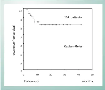
Recurrences occurred predominantly in the first 8 months. The overall success rate was 84% (Fig. 3).
Systemic hypertension was present in 56 patients (53.8%), left ventricular hypertrophy in 31 patients (29.8%), and coronary artery disease in 34 patients (33.6%). No statistical difference was observed between the groups in relation to these variables (Tab. 2).
The mean follow-up period was 18 months, ranging from two to 42 months.
Follow-up months
50 40 30 20 10 0
re
cu
rre
n
ce
-f
re
e
su
rvi
va
l
1.0
.9
.8
.7
.6
.5
.4
.3
Fig. 3 -Event-free survival curve of the population studied
We can observe that most of the recurrences occurred within the first eight months.
Kaplan-Meier 104 patients
Considering sustained sinus rhythm as the primary endpoint, we observed two groups of patients. Group I (success) was comprised of 87 patients, of which 76 (87.4%) had no recurrence of symptoms or even asymptomatic arrhythmias in the Holter monitoring, and 11 patients (12.6%) had recurrence of arrhythmia or symptoms suggestive of arrhythmia, which was fully suppressed with reinitiation of the antiarrhythmic drug.
At the end of the follow-up, all these patients were asymptomatic and with no documented arrhythmia.
The presence of palpitation - although without documentation of atrial fibrillation irresponsive to antyarrhythmic treatment - was considered a failure criterion.
Group II (failure) comprised 17 patients who remained with atrial fibrillation despite the pharmacological attempt to maintain a sinus rhythm.
Some patients underwent a second ablation procedure; however, the analysis was performed only with data regarding the initial ablation.
No statistical difference was observed between the groups as regards age and gender. There was not a normal distribution as regards age, which was analyzed using the medians (Table 1).
Group AGE
median
GENDER M/F
Success 59 60/27
Failure 61 12/5
Overall 59.5 72/32
p 0.484 0.42
Table 1 - Sample population distribution stratified by groups, according to age and gender
Groups SH
n
CAD n
LVH
n EF %
Success
Group I 45 28 27 58.68
Failure
Group Ii 11 6 4 56.65
p 0.33 0.8 0.54 0.13
n = number of patients
Table 2 - Sample population distribution stratified by groups, according to the presence of systemic hypertension, coronary artery disease, ejection fraction, and left ventricular hypertrophy.
Table 3 shows the results of left atrial volume, right and left circumferential ablation area and radiofrequency line completeness.
We can observe that the left atrial volume was significantly lower in group I, with success (p < 0.001).
Ablated areas were larger in the group with failure; however, this group had large left atria. Therefore, this information alone does not represent the true area in relation to the total area.
It is more important to analyze the ablated area in relation to the atrial volume, because although the circumferential area may be identical to a second circumferential area if one atrium is normal and the other is enlarged, the area relative to the normal-sized atrium will be larger. In other words, the percentage of the ablated area in the smaller atrium will be higher.
Circumferential line completeness in Group I (success) was obtained in 80/87 patients and in 320/348 pulmonary veins. In Group II (failure) completeness was verified in 12/17 meaning 51/58 veins. This difference was statistically significant with p = 0.025 for the patients and 0.0038 for the veins. The number of radiofrequency application points was not significantly different (Tab. 4).
When only the completeness of ablation is observed, we can verify that the group with incomplete ablation reached only 43% of success, and the group with complete lines obtained 94% success at the end of the follow-up period (Fig. 4).
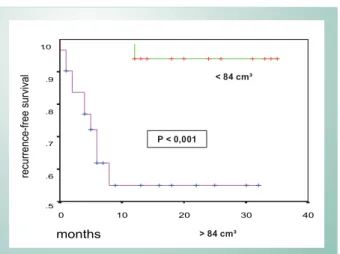
Left atrial volume was confirmed as an important parameter to stratify the groups. When analyzed separately, it has a clear correlation with prognosis. Thus, from the analysis of the ROC curve the value of 84cm³ was determined as the cut-off point, identifying the groups of lower volume as those that benefited the most (Fig. 5).
The analysis of the Kaplan-Meier event-free survival curve in patients with left atrial volume higher than or lower than 84cm³ confirmed that the group with left atria < 84cm³ had a higher probability of success (p < 0.001) (Fig. 6).
When data regarding atrial volume and circumferential ablation completeness are associated, we can observe that the group with left atrial volume lower than 84cm³ and complete circumferential ablation achieved therapeutic success, that is, sustained sinus rhythm, in 99% of the patients. On the other hand, when both variables were negative (volume higher than
Variables Analyzed Success
N = 87
Failure
N = 17 p
LA volume
(cm³) 69.50±21.81 105.10±20.20 <0.001
Right PVA (mm²)
Mean Rank 49.75
Mean Rank
66.59 0.035
Left PVA (mm²)
Mean Rank 46.21
Mean Rank
84.68 <0.001
R/LA Area 0.83±0.32 0.53±0.13 <0.001
L/LA Area 0.84±0.33 0.52±0.18 <0.001
Table 3 - Measurements of left atrial volume, ablation area, and ablated area in relation to the left atrial volume in the groups studied
Variables analyzed G I (Success)
(n=87)
G II (Failure) (n=17)
p 95.1% CI
% of complete lines/veins 320/348
(92%)
51/68
(75%) 0.0038
Patients with complete lines 80/87 12/17 0.025
No. of points in right vein Rank Sum Mean Rank
5520 70
Rank Sum Mean Rank
429 55 0.7
No. of points in left vein 8140 108 1128 142 0.45
Table 4 - Relationship of group I (success) and group II (failure) with line completeness and number of ablation points
Fig. 4 -Kaplan-Meier curve comparing recurrence between the groups with complete and incomplete ablation.
Follow-up
40 30
20 10
0
R
ecu
rre
nce
-fre
e
su
rvi
va
l
1.0 .9 .8 .7 .6 .5 .4
Fig. 5 -ROC curve.
ROC curve
- Specificity
1.0 .8
.5 .3
0.0
S
e
n
s
it
iv
it
y
1.0
.8
.5
.3
84cm³ and incomplete line), the success rate was only 34%. The combination of a positive variable with a negative variable, whatever it was, led to a success rate of 75% (Fig. 7).
Complications observed - Two patients had acute cardiac
tamponade during manipulation inside the left atrium. In both cases a pericardial puncture with drainage and interruption of bleeding was performed with the patients still in the electrophysiology laboratory. The patients were discharged within 72 hours, with no clinical sequelae.
One of the patients presented tamponade during the atrial mapping, therefore prior to ablation, and the procedure was interrupted. He progressed with recurrence of the atrial fibrillation and was included in the group of failure. This patient underwent ablation in another moment, and the intervention was successful. Considering the objectives of this study, one single procedure was taken into account.
The second patient presented tamponade at the end of the intervention, after ablation was performed, and progressed with no recurrence (success).
Three patients had manifestations suggestive of a mild cerebral embolism. One of them presented mild speech impairment; another presented decreased right arm strength, and the third had decreased right hand strength. All manifestations resolved completely within the first 24 hours, with maintenance of intravenous heparin, and neither neurological sequelae nor alterations in the brain magnetic resonance remained.
All patients underwent CT angiography or magnetic resonance angiography of the pulmonary veins between three and six months post-ablation.
In two patients a lower than 30% reduction in the diameter of one of the pulmonary veins with no clinical manifestations was observed. These patients remained asymptomatic, and CT angiography was not repeated.
Discussion
In the present study, all 104 patients were taking antiarrhythmic drugs prior to the procedure, however without
control of the arrhythmia. After ablation, 87/104 patients (84%) were in sinus rhythm by the end of 18 months, and only 11/87 (12.6%) were taking antiarrhythmic medication, previously ineffective. No deaths were observed and no patients had significant pulmonary vein stenosis.
Despite the use of CARTO™ instrumentality, not all groups showed the same outcomes. The Johns Hopkins University School of Medicine (Baltimore, USA) team recently published their experience with the same procedure with a 76% success rate, of which 56% without antiarrhythmic drugs and 20% with antiarrythmic drugs13.
In another study by the same team, they reported success in 65% of the patients (52% without antiarrhythmic drugs and 13% with antiarrhytmic drugs) using an irrigated-tip ablation catheter with radiofrequency and pulmonary vein isolation associated with the cavotricuspid line. To achieve this outcome, 14.6% of the patients needed a second ablation procedure. With this approach, only permanent, persistent atrial fibrillation, and age > 50 years were criteria predictive of recurrence14.
We should point out that the success rate shown (84%) in this study refers to a single procedure, which is profoundly different from the venous disconnection proposed by Haissaguerre because in his best result, in which he adds left atrial isthmus ablation with an occasional radiofrequency application inside the coronary sinus, 32% of the patients needed a second intervention. When venous disconnection is performed without isthmal block, 49% of the patients need a second intervention. The outcomes with these high figures of reintervention are: 87% of success in the group with isthmal block and 69% in the group without isthmal block15.
Although located in the pulmonary veins in most of the cases, the so-called “primary triggers” can also lie in the superior vena cava, coronary sinus, ligament of Marshall, or even at some point in the right or left atrium. For this reason, a procedure that only isolates the pulmonary veins will logically be inferior to another that encompasses part of the perivenous atrial muscles, including the origin of the ligament of Marshall.
months
R
e
cu
rre
n
ce
-f
re
e
su
rvi
va
l
1.0 .9 .8 .7 .6 .5 .4 .3
40 30
20 10
0
Fig. 7 -Kaplan-Meier recurrence-free survival curve associating volume with completeness of ablation.
complete + vol.< 84
one variable
incomplete + vol> 84
months
re
cu
rre
n
ce
-f
re
e
su
rvi
va
l
1.0 .9 .8 .7 .6 .5
40 30
20 10
0
Fig. 6 -Kaplan-Meier recurrence-free survival curves stratified by left atrial volume.
P < 0,001
< 84 cm³
The so-called “rotors” or “secondary triggers” are reentry areas, and are primarily located in the posterior wall, close to the pulmonary veins, playing a critical role in the perpetuation of atrial fibrillation16. For this reason, circumferential ablation
is not restricted to the approach to the pulmonary veins. The best technique for ablation of fibrillation should consider the primary and secondary triggers, and the atrial substrate. In a randomized study, the Michigan team demonstrated that left atrial ablation with lines encircling the pulmonary veins (circumferential ablation) is approximately 20% more efficient than pulmonary vein isolation in the treatment of paroxysmal atrial fibrillation17.
Recently, the pioneer in circumferential ablation, Carlo Pappone18, demonstrated that the patients had a lower
recurrence rate when the ablation area was larger, in agreement with the findings of the present study. Pappone’s study also showed that vagal denervation, which occurred in part of the patients, had an additional protective effect as regards the recurrence of atrial fibrillation.
Twelve per cent of the atrial fibrillations are known to have their inducing trigger in the vagotonia and show poor response to conventional ablation with electrical venous disconnection, even if the four main veins are isolated19.
Although electrical venous disconnection had not been sought, we observed a higher recurrence rate in the group of patients where the circumferential ablation left a > 10mm interval between adjacent points. This is consistent with the multifactor theory of atrial fibrillation.
The purely anatomic procedure does not aim to disconnect the pulmonary veins electrically. Pappone et al20 demonstrated
that there was no difference in the frequency of recurrence using circumferential ablation among patients, whether isolation was present or not.
More recently, Stabile et al (2003) also using electroanatomic mapping reported the absence of recurrence in 80.4% of the patients, although only 40% of them had criteria for electrical isolation in the electrophysiologic study.
The presence of a > 10mm discontinuity in the ablation line correlated to a higher recurrence rate is a datum that does not identify the origin of the recurrence (whether inside the veins or in the perivenous region). Knowing that both the veins and the posterior wall are involved in the genesis and perpetuation of atrial fibrillation, and that in the electroanatomic procedure the pulmonary vein isolation does not have a predictive value in relation to recurrence, the whole region should be regarded as an arrhythmogenic “complex” to be modified.
The complete radiofrequency application line does not only decrease recurrence of atrial fibrillation, but also reduces the post-ablation occurrence of iatrogenic atrial tachycardia, created by the electrical remodeling resulting from the radiofrequency lines21,22.
In this study, the circumferential ablations more frequently incomplete were those related to the right veins. This is due to an occasional difficulty in encircling the right veins without losing the transseptal puncture. The left veins are right in front of the catheter after transseptal puncture; however, the thin wall that separates the auricle from the left superior vein makes
the applications in this area very difficult.
On the atrial volume - We observed a relation between the atrial volume and the final outcome. This is only an additional confirmation of what had already been established: larger atria are more prone to arrhythmias and atrial fibrillation.
Remodeling is the designation given to the phenomenon that modifies the electric, geometric, and, finally, genetic characteristics of the atrial myocardium.
The remodeling extends from modulation of membrane channels to genetic modulation23 that results from the fact
that the loss of muscle contractile activity produces (within minutes or hours) a genetic adjustment, leading to an inversion of cell differentiation demonstrable by the identification of suppression of the adult differentiated state with “derepression” of the genes expressed during the development – known as fetospecific expression.
All these modifications are seen more intensely in larger atria, and certainly this explains a lower success rate in atria with higher volumes, as observed in this study. Pappone24,
using echocardiographic measurements, observed a significant difference in the left atrial diameter of patients without recurrence (40.3mm + 4.5 mm) in relation to the group that had recurrence (46.8mm + 6.7 mm) with p< 0.001.
Hakan Oral et al17, comparing pulmonary vein disconnection
and circumferential ablation, observed that the size of the left atrium was an independent predictor of recurrence of atrial fibrillation, just as circumferential ablation was predictive of a better outcome.
Thus, it is clear that it is important to eliminate atrial fibrillation while the remodeling does not change significantly the left atrium, i.e., during the phase of normal or slightly enlarged left atrium.
On the circumferential ablation area - When correlated
with atrial volume, the circumferential ablation area showed a significant difference between the groups in the univariate analysis. Obviously an absolute value of circumferential area lacks significance, because atrial volumes may show a wide variation. In other words, any given area value in a small atrium is much more important than the same value in a large atrial volume.
The importance of an ablation area encompassing part of the posterior wall is obvious, considering that the larger the area covered, the greater the number of primary and secondary “triggers” and reentrant circuits isolated from the rest of the atrium, and the lower the chance of recurrence of atrial fibrillation.
On the uni and multivariate analyses - In the univariate
analysis, the left atrial volume, the circumferential ablation area, the area related to the left atrial volume, and the presence of complete lines were predictive of recurrence.
In the multivariate analysis, in turn, only the left atrial volume and complete ablation were predictive of recurrence. The ROC curve identified the 84cm³ cut-off point. Thus, patients with an atrial volume lower than 84cm³ and complete ablation showed a high success rate.
existence of gaps in the energy application line.
This latter aspect has currently become more relevant because of the atrio-esophageal fistulae observed25. With
the detailed observance of the position of the esophagus and radiofrequency application outside the area of atrio-esophageal contact, some lines show to be incomplete. Future outcomes are expected to be poorer than the current ones.
On the complications - Another important aspect is the
absence of significant venous stenoses observed in the sample population, even with magnetic resonance angiography or CT angiography controls. This has been a constant in the studies with circumferential ablation26.
Considering that only 56% of the patients show the classical anatomy of four independent pulmonary veins27, the CT
angiography or magnetic resonance angiography used are considered valuable tools to prevent undue radiofrequency application in the ostium of any vein not identified by radioscopy, thus reducing the risk of venous stenosis.
Although no irreversible complication had occurred, some potentially severe events were recorded: two cardiac tamponades required pericardial drainage and three embolic accidents were medically treated and did not leave any sequelae.
The incidence of embolic complications in the case series of the Cleveland Clinic Foundation was 1.5%, and not all were completely reversible. Cardiac tamponades were also observed, and reached 2% in the group of patients > 60 years28.
Embolic complications occurred in three patients in this study (2.8%) and were transient, therefore with no sequelae. In the first few patients of the sample, the activated clotting times were maintained around 250; however, when the embolic complications occurred, we chose to maintain them closer to 300, and currently even above this value.
In Pappone’s series, cardiac tamponades were observed in up to 4% of the patients20.
On the limitations - Although these 104 patients were
highly symptomatic and had several ECG and 24-hour Holter monitoring performed, it is not possible to know whether any asymptomatic episode was missed during the periods of non-monitoring. All invasive procedures are known to have an unpredictable placebo effect.
The SOPAT trial which compared placebo with quinidine combined with verapamil and sotalol and used a 1-minute transtelephonic transmission daily is what we consider the closest to the perfect follow-up29. Even in this case, the
1-minute-per-day monitoring means the non-monitoring of 1439 minutes the same day.
Implantation of an event monitor for 12 months for the real assessment of the presence of arrhythmia would incur other problems because the ventricular response in atrial fibrillation with medication or atrioventricular node disease may be normal and would not activate the recording of events. Obviously, ethical limits do not allow such a procedure in asymptomatic patients. Therefore, symptoms are still the universally accepted criterion to control the efficacy of the procedure.
The study was conducted in a group of selected patients who were relatively young, with paroxysmal and persistent atrial fibrillation, and normal ventricular function. These outcomes could be different in other groups with older patients with permanent fibrillation or ventricular dysfunction.
The mean age of the sample group was different from the age at which the highest prevalence of atrial fibrillation occurs in the general population (the sample population of this study was younger); however, it is precisely at this age that atrial fibrillation used to be more symptomatic and to cause greater losses in the quality of life. Additionally, younger individuals will be exposed to antiarrhythmic drugs for rhythm or rate control for a longer period of time, which leads to a higher incidence of collateral effects. And since the antiarrhythmic protection is incomplete, the risk of embolic events is higher and the anticoagulation time is longer. Therefore, this is a population that can greatly benefit from the treatment with radiofrequency ablation.
The results of this study were obtained from the follow-up after a single ablation procedure. Most of the studies are known to report their results with some patients undergoing two or three procedures. Experience shows that repeated ablation improves outcomes; however, the objective of this study was to analyze the outcomes of one single procedure.
Based on the results observed, we concluded that circumferential ablation of atrial fibrillation using the CARTO™ System resulted in sustained sinus rhythm in the mid-term with a single intervention in 84% of the cases. The enlarged volume of the left atrium, the size of the ablated area, and complete ablation were predictive of a higher recurrence in the univariate and multivariate analyses. The volume of the left atrium and complete ablation were independent predictors of a procedural success. The rate of relevant clinical complications was 4.8%.
Potential Conflict of Interest
No potential conflict of interest relevant to this article was reported.
References
1. Benjamin EJ, Wolf PA, D’Agostinho RB, Silbershatz H, Kannel WB, Levy D. Impact of atrial fibrillation on the risk of death: The Framingham Heart Study. Circulation. 1998; 98: 946-952.
2. Krahn AD, Manfreda J, Tate RB, Mathewson FAL, Cuddy TE. The natural history of atrial fibrillation: incidence, risk factors and prognosis in the Manitoba follow-up study. Am J Med 1995; 98: 476-484.
3. Psaty BM, Manolio TA, Kuller LH, et al. Incidence of and risk factors for atrial fibrillation in older adults. Circulation. 1997; 96:2455-2461.
5. Stewart S, Hart CL, Hole DJ, McMurray JJV. A population-based study of the long term risks associated with atrial fibrillation: 20-year follow-up of the Renfrew/Paisley study. Am J Med. 2002; 113: 359 - 364.
6. Go AS, Hylek EM, Phillips KA, et al. Prevalence of diagnosed atrial fibrillation in adults. National implications for rhythm management and stroke prevention: The Anticoagulation and Risk Factors in Atrial Fibrillation (ATRIA) Study. JAMA. 2001; 285: 2370-2375.
7. AFFIRM Investigators. A comparison of rate control and rhythm control in patients with atrial fibrillation. N Engl J Med. 2002; 347: 1825-1833.
8. AFFIRM Investigators. Relationships between sinus rhythm, treatment, and survival in the atrial fibrillation follow-up investigation of rhythm management (AFFIRM) study. Circulation. 2004; 109: 1509 -1513.
9. Steinberg JS, Sadaniantz A, Kron J, et al. and the AFFIRM Investigators. Analysis of cause-specific mortality in the atrial fibrillation follow-up investigation of rhythm management (AFFIRM) study. Circulation. 2004; 109: 1973-1980.
10. Cappato R, Calkins H, Chen SA, et al. Worldwide survey on the methods, efficacy, and safety of catheter ablation for human atrial fibrillation. Circulation. 2005; 111: 1100-1105.
11. Nath S, DiMarco J, Haines D. Basic aspects of radiofrequency catheter ablation. J cardiovasc Electrophysiol. 1994; 5 (10): 863-876.
12. Dorwarth U, Fiek M, Remp T, et al. Radiofrequency catheter ablation; different cooled and noncooled electrode systems induce specific lesion geometries and adverse effect profiles. PACE. 2003; 26 (I): 1438-1445
13. Vasamreddy CR, Dalal D, Eldadah Z, et al. Safety and efficacy of circumferential pulmonary vein catheter ablation of atrial fibrillation. Heart Rhythm. 2005; 2: 42-48.
14. Vasamreddy CR, Lickfett L, Jayam VK, et al. Predictors of recurrence following catheter ablation of atrial fibrillation using an irrigated-tip ablation catheter. J Cardiovasc Electrophysiol. 2004; 15 (6): 692-697.
15. Jaïs P, Hocini M, Hsu LF, et al. Technique and results of linear ablation at the mitral isthmus. Circulation. 2004; 110:2996-3002.
16. Jalife J. Rotors and spiral waves in atrial fibrillation. J Cardiovasc Electrophysiol. 2003; 14: 776 – 780.
17. Oral H, Scharf C, Chugh A, et al. Catheter ablation for paroxysmal atrial fibrillation: Segmental pulmonary vein ostial ablation vs. left atrial ablation. Circulation. 2003; 108: 2355 – 2360.
18. Pappone C, Santinelli V, Manguso F, et al. Pulmonary vein denervation enhances long-term benefit after circumferential ablation for paroxysmal atrial fibrillation. Circulation. 2004c; 109: 327 – 334.
19. Oral H, Morady F. Ablation of atrial fibrillation. J Cardiovasc Electrophysiol. 2004; 15: 112 – 113.
20. Pappone C, Rosanio S, Oreto G, et al. Circumferential Radiofrequency ablation of pulmonary vein ostia. A new anatomic approach for curing atrial fibrillation. Circulation. 2000; 102: 2619-2628.
21. Pappone C, Rosanio S. Pulmonary vein isolation for atrial fibrillation. In: Douglas Zipes, José Jalife. Cardiac electrophysiology: from cell to bedside. 4ed. Philadelphia (Pennsylvania): Elsevier; 2004b: 1039-1052
22. Cha TJ, Ehrlich JR, Zhang L, Chartier d, Leung TK, Nattel S. Atrial tachycardia remodeling of pulmonary vein cardiomyocytes. Comparison with left atrium and potential relation to arrhythmogenesis. Circulation. 2005; 111:728-735.
23. Pacifico A, Henry PD. Class I or class III agents for atrial fibrillation: Are we asking the right question? PACE. 2003; 26(II): 1613-1619.
24. Pappone C, Oreto G, Rosanio S, et al. Atrial electroanatomic remodeling after circumferential radiofrequency pulmonary vein ablation. Efficacy of an anatomic approach in a large cohort of patients with atrial fibrillation. Circulation. 2001; 104: 2539-2544.
25. Pappone C, Oral H, Santinelli V, et al. Atrio-esophageal fistula as a complication of percutaneous transcatheter ablation of atrial fibrillation. Circulation. 2004a; 109:2724-2726.
26. Ellenbogen KA, Wood MA. Ablation of atrial fibrillation: Awaiting the new paradigm. J Am Coll Cardiol. 2003; 42 (2): 198 – 200.
27. Mansour M, Holmvang G, Sosnovik D, et al. Assessment of pulmonary vein anatomic variability by magnetic resonance imaging: Implications for catheter ablation techniques for atrial fibrillation. J Cardiovasc Electrophysio. 2004; 15: 387-393.
28. Bhargava M, Marrouche N, Martin DO, et al. Impact of age on the outcome of pulmonary vein isolation for atrial fibrillation using circular mapping technique and cooled-tip ablation catheter: A retrospective analysis. J Cardiovasc Electrophysiol. 2004;15:8 –13.