INTRODUCTION
Methacrylate-based composite resins release uncured monomers to the tissues (1), which have been associated with hypersensitivity and cytotoxicity (2), genotoxicity (3), estrogenicity (4), and immune system alterations (5).
Cell culture studies have demonstrated that methacrylate and dimethacrylate monomers, commonly used in the restorative polymeric technology, may affect
Filtek
TM
Silorane and Filtek
TM
Supreme XT Resins:
Tissue Reaction After Subcutaneous Implantation
in Isogenic Mice
Esther RUIZ DE CASTAÑEDA1
Léa A. B. SILVA2
Patrícia GATON-HERNÁNDEZ3
Alberto CONSOLARO4
Encarnación Gonzalez RODRIGUEZ5
Raquel A. B. SILVA2
Alexandra M. QUEIROZ2
Paulo NELSON-FILHO2
1La Societad Catalana d’odontoestomatologia de l’Acadèmia de Ciències Mèdiques I de la Salut
de Catalunya I de Balears, Barcelona, Spain
2Department of Pediatric Clinics, Preventive and Community Dentistry, Ribeirão Preto Dental School,
USP - University of São Paulo, Ribeirão Preto, SP, Brazil
3University of Barcelona, Barcelona, Spain
4Pediatric DentistryPostgraduate Program, Department of Pediatric Clinics, Preventive and Community Dentistry,
Ribeirão Preto Dental School, USP - University of São Paulo, Ribeirão Preto, SP, Brazil
5Facultad Odontología, University of Granada, Granada, Spain
The aim of this study was to evaluate the tissue compatibility of a silorane-based resin system (FiltekTM Silorane) and a
methacrylate-based nanoparticle resin (FiltekTM Supreme XT) after implantation in the subcutaneous connective tissue of isogenic mice. One hundred
and thirty five male isogenic BALB/c mice were randomly assigned to 12 experimental and 3 control groups, according to the implanted material and the experimental period of 7, 21 and 63 days. At the end of each period, the animals were killed and the tubes with the surrounding tissues were removed and processed for microscopic analysis. Samples were subjected to a descriptive and a semi-quantitative analyses using a 4-point scoring system (0-3) to evaluate the collagen fiber formation and inflammatory infiltrate. Data were statistically analyzed using the Kruskal Wallis test (a=0.05). The results showed that there was no significant difference between the experimental and control groups considering the three evaluation periods (p>0.05). The silorane-based and the methacrylate-based nanoparticle resins presented similar tissue response to that of the empty tube (control group) after subcutaneous implantation in isogenic mice.
Key Words: subcutaneous tissue, tissue reaction, nanocomposite, nanometric particles, mathacrylate-based resin, silorane-based resin.
the recruitment of leukocytes in inflammation sites by decreasing the expression of intercellular adhesion molecules and inducing enzymatic activity and expression of growth factors and cellular cytokines (6). In addition, resin monomers suppress the mitochondrial activity of macrophages and alter their inflammatory responses (4,7). In this way, reactive components released from conventional composite resins, such as unreacted monomers (1) and oxidation products, may induce toxicity or inflammatory tissue reactions (8). Adhesive
system components may also unchain an unfavorable tissue reaction, avoiding the occurrence of healing (9). In the last few years, methacrylate-based resins with modifications in their composition and structure, such as FiltekTM Supreme XT (3M ESPE, St. Paul, MN, USA) have been developed. This resin is a nanocomposite that contains only nanomeric particles and nanoclusters as inorganic fillers. The nanomeric particles are monodispersed, nonagglomerated silica particles, while the nanoclusters are spheroidal agglomerates consisting of nano-sized silica and zirconia particles. The micron-sized porous cluster is infiltrated with silane coupling agents to allow chemical bonding with the organic matrix (10). However, though adequate physical properties have been attributed to these resins (e.g.: improved surface polishing) (10), they have shown toxic effects in cell cultures (1).
On the other hand, the occurrence of polymerization shrinkage and the release of chemical products from methacrylate-based systems have led to the development of silorane-based composite resins, such as the resin FiltekTM Silorane (3M ESPE). The silorane matrix is formed by the cationic ring-opening polymerization of the silorane monomers. The silorane molecule represents a hybrid that is made of both siloxane and oxirane structural moieties (11). The silorane technology has afforded a highly hydrophobic restorative material with reduced polymerization shrinkage, more balanced volumetric stress, high ambient light stability (11), and insolubility in biological fluids (12).
Although recent studies have evaluated physicochemical properties of silorane-based resins (13,14), research regarding their biological effects, mostly based on in vitro cell culture studies (1,3), is quite limited. To date, there is no study evaluating the in vivo
compatibility of these new restorative systems with the connective tissue. The present study evaluated the tissue compatibility of a silorane-based resin system (FiltekTM Silorane) and a methacrylate-based nanoparticle resin (FiltekTM Supreme XT) after implantation in the subcutaneous connective tissue of isogenic mice.
MATERIAL AND METHODS
All experiments were conducted according to the guidelines of the Animal Care and Use Committee of the University of São Paulo, Ribeirão Preto Campus, Brazil. The study design and histological parameters for tissue reaction evaluation were in accordance with the
ISO 10993-6 standard (15).
One hundred and thirty five male isogenic BALB/c mice aged 6-8 weeks and weighing 15 to 20 g were obtained from the Laboratory Animal Care Facility of the Medical School of Ribeirão Preto, University of São Paulo, Brazil, and were randomly assigned to 12 experimental (n=10 animals/group) and 3 control groups (n=5 animals/group). Each animal received one polyethylene tube implant (10 mm long x 1 mm internal diameter) on the dorsal region. All tubes had their ends closed by using sterile heated clinical tweezers, and were then sterilized with ethylene oxide, and filled with the materials under aseptic conditions in a laminar flow chamber immediately prior to use and implantation as follows: Group I, II and III: FiltekTM Silorane at 7, 21 and 63 days, respectively; Groups IV, V and VI: FiltekTM Silorane adhesive bond at 7, 21 and 63 days; Groups VII, VIII and IX: FiltekTM Silorane primer at 7, 21 and 63 days; Groups X, XI and XII: FiltekTM Supreme XT at 7, 21 and 63 days; and Groups XIII, XIV and XV (control groups): empty tube at 7, 21 and 63 days, respectively. Before tube implantation, the animals were anaesthetized with an intraperitoneal injection of 10% chloral hydrate (1mL/100 g body weight), and had the dorsal region shaved and cleaned with a 1% chlorhexidine solution. A 1-cm-wide incision was made on the dorsal region followed by tissue divulsion with Kelly forceps, and the tube was inserted into the connective tissue and the skin borders were closed with 4-0 silk sutures (Vicryl; Johnson & Johnson: Ethicon Inc., New Brunswick, NJ, USA). The surgery was performed under aseptic conditions to minimize the trauma to the implant area. The animals were kept with free access to standard chowand water during the experimental periods and were periodically observed for local, systemic and behavioral abnormalities.
semi-quantitative microscopic analyses of tissue reaction (16) to the tested materials and the empty tubes at the different experimental periods were done. In the semi-quantitative analysis, a four-point (0-3) scoring system was used to evaluate the number and density of collagen fibers within the tissue surrounding to the tested material and the concentration of polymorphonuclear neutrophils.
- Collagen fiber formation: score 0 = absent; score 1 = mild: individual collagen fiber arrangement resembling a normal connective tissue, permeated by negative spaces indicative of non-fibrous components of the extracellular matrix; score 2 = moderate: areas with individual collagen fibers alternated with areas of eosinophilic extracellular matrix without typical linear and undulate formations; and score 3 = intense: collagen fibers merged within an eosinophilic extracellular matrix without the typical linear and undulate formations, not allowing to distinguish the individual fibers.
- Inflammatory infiltrate: score 0 = absent; score 1 = mild (1 to 10 PMN); score 2 = moderate (11 to 20 PMN); and score 3 = intense (>21 PMN).
Higher scores related to collagen fibers formation, and inflammatory infiltrate, represented the worst tissue compatibility of the materials. The results from the semi-quantitative analysis were analyzed statistically by the nonparametric Kruskal Wallis test at 5% significance level using the GraphPad 5.0 statistical software (GraphPad Software Inc., San Diego, CA, USA).
RESULTS
7 days
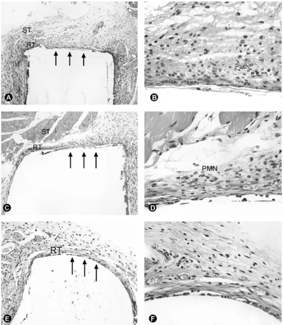
Tissue reaction was predominantly formed by young, ovoid, fusiform fibroblasts with slightly organized thin collagen fiber bundles. Macrophages were found between the fibroblasts. PMN and occasional eosinophils were diffusively distributed in the tissue. There were also a large number of newly formed blood vessels. Multinucleated foreign body giant cells (FBGC), usually with 4 to 5 nuclei each, were found at the interface between the reactionary tissue and the material (Figs. 1A,B and 2A). The thickness of the reactionary tissue ranged from 10 to 30 cells.
21 days
It was observed slightly more collagen fiber formation than in the 7-day period with more well defined
collagen fiber bundles with capsular organization. Young fibroblasts and macrophages were present. The vascular component was moderately present (Figs. 1C,D, and 2B). There was a discrete and diffuse PMN infiltrate and a few eosinophils . The thickness of the tissue reaction ranged from 10 to 20 cells. Multinucleated FBGC at the tissue/material interface had 4 to 5 nuclei each. In one specimen of group V (FiltekTM Silorane adhesive bond) there was a well organized foreign body granuloma surrounding a material fragment that extruded to the circumjacent tissue.
63 days
It was observed a thin and well organized with a fibrous capsule with well defined margins of collagen fiber bundles permeated by predominantly mature fusiform and thin fibroblasts with well condensed nuclei. The vascular component decreased and only some vessels persisted as part of the tissue structure. Several specimens did not show PMN or eosinophil infiltrate. When present, the infiltrate was mild and diffuse. At the at the reactionary tissue/material interface there were a few multinucleated FBGC with 3 to 4 nuclei each (Figs. 1E,F, and 2C). The thickness of the tissue reaction ranged from 5 to 15 cells.
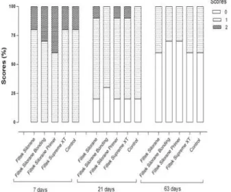
In the semi-quantitative microscopic analysis there was no statistically significant difference (p>0.05) among the groups at any of the periods according to the mean scores values obtained after analysis of collagen fiber formation and inflammatory infiltrate (Figs. 3 and 4).
DISCUSSION
irritation than conventional methacrylate-based resins. The biological response to dental restorative polymer composites is mediated by the release of uncured residual monomers. Methacrylate-free resin composite formulations have claimed to reduce unpolymerized residual mass. FiltekTM Silorane is one of these materials, based on an innovative monomer system that combines important characteristics, such as low polymerization shrinkage and high hydrophobicity
(11). Siloranes are stable materials that are insoluble in different biological fluids (12) and have little water sorption and low diffusion coefficient compared to conventional methacrylate-based resins. According to Hahel et al. (13), the macro-, micro- and nano-mechanical properties of silorane-based resins are comparable to those of methacrylate-based resins.
However, only few cell culture studies have investigated biological properties of silorane-based
Figure 1. Microscopic aspects of the tissue reaction (RT) at 7 days (A, B), 21 days (C, D) and 63 days (E, F) at the interface (arrows) between the implanted materials and the surrounding tissue (ST). The intensity of collagen fiber formation increased and the presence of polymorphonuclear neutrophils (PMN) decreased gradually with time. (A, B = group IV - bonding agent of FiltekTM Silorane at 7
days, HE ×25 and ×100 magnification; C, D = group II - FiltekTM Silorane at 21 days, HE ×25 and ×100 magnification; E, F = group
resins, showing no cytotoxic effects and low mutagenic potentials (17). Another study (1) evaluating the in vitro
cytotoxicity of different resins in Balb/c 3T3 mouse fibroblast cell cultures during 8 weeks found that the methacrylate-free resin Hermes, precursor of FiltekTM
Figure 2. Microscopic aspects of the reactionary tissue (RT) at 7 days (A), 21 days (B) and 63 days (C) at the empty tube (control group) end (arrows) in contact with the surrounding tissue. It can be observed that a serofibrinous exudate (SE) was formed at the tube lumen interface (TL) in the 7-day period. It can also be noticed that the presence of polymorphonuclear neutrophils decreased gradually with time (A, B and C = HE, ×24 magnification).
Figure 3. Score distribution of the experimental and control groups regarding collagen fiber formation.
Figure 4. Score distribution of the experimental and control groups regarding inflammatory infiltrate.
low capacity of this material inducing unfavorable tissue reactions, and it was in agreement with Brackett et al. (1) that showed that materials with new polymerization modes are promising for reducing the release of uncured components and, consequently, their cytotoxicity.
Under the tested conditions, the silorane-based (FiltekTM Silorane) and the methacrylate-based nanoparticle (FiltekTM Supreme XT) resins presented tissue response similar to that of the control group (empty tube) after implantation in the subcutaneous connective tissue of isogenic mice. The results of the present study may have great significance because, in addition to its claimed low polymerization shrinkage, FiltekTM Silorane showed tissue compatibility in vivo. Further research is needed to support the clinical use of these new materials.
RESUMO
O objetivo do presente estudo foi avaliar a compatibilidade tecidual de um sistema resinoso à base de silorane (FiltekTM Silorane) e
de uma resina nanoparticulada à base de metacrilato (FiltekTM
Supreme XT), após implantação no tecido conjuntivo subcutâneo de camundongos isogênicos. Um total de 135 camundongos isogênicos BALB/c machos foram randomicamente divididos em 12 grupos experimentais e em 3 grupos controles, de acordo com o material implantado e com o período experimental (7, 21 e 63 dias). Ao final de cada período, os animais foram mortos, sendo os tubos removidos com o tecido circundante e processados para análise microscópica. As lâminas foram submetidas a análise descritiva e análise semi-quantitativa empregando um sistema de escores de 4 pontos (0-3), a fim de avaliar a formação de fibras colágenas e o infiltrado inflamatório. Os dados obtidos foram submetidos à análise estatística por meio do teste de Kruskal Wallis (µ=0,05). Os resultados mostraram que não houve diferença estatisticamente significante entre os grupos experimentais e controles, considerando os três períodos de avaliação (p>0,05). As resinas à base de silorane e à base de metacrilato apresentaram resposta tecidual semelhante à do tubo vazio (controle), após implantação no tecido conjuntivo de camundongos isogênicos.
REFERENCES
1. Brackett MG, Bouillaguet S, Lockwood PE, Rotenberg S, Lewis JB, Messer RLW, Wataha JC. In vitro cytotoxicity of dental composites based on new and traditional polymerization chemistries. J Biomed Mater Res Part B: Appl Biomater 2007;81B:397-402.
2. Chang MC, Chen LI, Chan CP, Lee JJ, Wang TM, Yang TT, et
al.. The role of reactive oxygen species and hemeoxygenase-1 expression in the cytotoxicity, cell cycle alteration and apoptosis of dental pulp cells induced by BisGMA. Biomaterials 2010;31:8164-8171.
3. Urcan E, Scherthan H, Styllou M, Haertel U, Hickel R, Reichl FX. Induction of DNA double-strand breaks in primary gingival fibroblasts by exposure to dental resin composites. Biomaterials 2010;31:2010-2014.
4. Wada H, Tarumi H, Imazato S, Narimatsu M, Ebisu S. In vitro estrogenicity of resin composites. J Dent Res 2004;83:222-6. 5. Andersson J, Dahlgren U. Effects on mouse immunity of long-term
exposure in vivo to minute amounts of HEMA. Eur J Oral Sci 2011;119:109-114.
6. Krifka S, Petzel C, Hiller KA, Frank EM, Bosl C, Spagnuolo G, et al.. Resin monomer-induced differential activation of MAP kinases and apoptosis in mouse macrophages and human pulp cells. Biomaterials 2010;31:2964-2975.
7. Rakich DR, Wataha JC, Lefebvre CA, Weller RN. Effects of dentin bonding agents on macrophage mitochondrial activity. J Endod 1998;24:528-533.
8. Jontell M, Hanks CT, Bratel J, Bergenholtz G. Effects of unpolymerized resin components on the function of accessory cells derived from the rat incisor pulp. J Dent Res 1995;74:1162-1167. 9. Silva LAB, Freitas AC, Carvalho FK, Queiroz AM, Nelson-Filho P, Porto-Neto ST. Direct pulp capping with a self-etching adhesive system: histopathologic evaluation in dogs’ teeth. Oral Surg Oral Med Oral Pathol Oral Radiol Endod 2009;108:e34-e40. 10. Mitra SB, Wu D, Holmes BN. An application of nanotechnology
in advanced dental materials. J Amer Dent Assoc 2003;134:1382-1390.
11. Weinmann W, Thalacker C, Guggenberger R. Siloranes in dental composites. Dent Mater 2005;21:68-74.
12. Grégoire G, Dabsie F, Delannée M, Akon B, Sharrock P. Water permeability, hybrid layer long-term integrity and reaction mechanism of a two-step adhesive system. J Dent 2010;38:526-533.
13. Hahnel S, Henrich A, Bürgers R, Handel G, Rosentritt M. Investigation of mechanical properties of modern dental composites after artificial aging for one year. Oper Dent 2010;35:412-419.
14. Ilie N, Hickel R. Investigations on a methacrylate-based flowable composite based on the SDR™ technology. Dent Mater 2011;27:348-355.
15. International Standard ISO 10993-6. Biological evaluation of medical devices- Part 6: Tests for local effects after implantation. 1994:1-11.
16. Silva LA, Leonardo MR, Oliveira DS, Silva RA, Queiroz AM, Hernández PG, et al.. Histopathological evaluation of root canal filling materials for primary teeth. Braz Dent J 2010;21:38-45. 17. Krifka S, Seidenader C, Hiller KA, Schmalz G, Schweikl H.
Oxidative stress and cytotoxicity generated by dental composites in human pulp cells. Clin Oral Investig 2011 [Epub ahead of print. DOI: 10.1007/s00784-010-0508-5].