The aim of this study was to evaluate, by stereomicroscopy and confocal laser microscopy, the influence of different lateral compaction methods for the obturation quality, as well as the time spent for the procedure. Thirty root canals of freshly extracted single-rooted human teeth were prepared with the ProTaper system up to F5 instrument and filled with gutta-percha and AH Plus sealer by the lateral compaction technique. The teeth were divided into 3 groups (n=10) in accordance with the method used for the lateral compaction, as follows: Manual, Mechanical and Ultrasonic. The sealers were stained with rhodamine B dye in a proportion of 0.1% per gram in weight to allow for the analysis under a confocal microscope. During the root filling procedure, the time spent was recorded with a stopwatch. The specimens were stored at 37 °C for 48 h, and then sectioned at 2, 4 and 6 mm from the apex. The percentage of gutta-percha, sealer and void areas were evaluated using a stereomicroscope and sealer penetration perimeter by confocal laser microscope. Statistical analyses were performed using the Kruskall-Wallis and Dunn tests (p<0.05). The mechanical method required a shorter time for the lateral compaction than the manual method (p<0.05). There were no significant differences (p>0.05) for the perimeter of the dentin with sealer penetration among all groups. The mechanical method showed a higher (p<0.05) percentage of gutta-percha and less sealer at the 4 mm section in comparison with the manual method. The ultrasonic group showed intermediate values. The void areas found in the root filling were similar (p>0.05) between the three methods. In conclusion, the fastest lateral compaction was achieved with the mechanical method, and all the methods showed void areas in the root filling.
Influence of the Method in Root
C a n a l F i l l i n g U s i n g A c t i v e
Lateral Compaction Techniques
Danilo Bailo Rossetto, Samuel Lucas Fernandes, Bruno Cavalini Cavenago, Marco Antonio Hungaro Duarte, Ronald Ordinola-Zapata, Flaviana Bombarda de Andrade
Department of Operative Dentistry, Endodontics and Dental Materials, School of Dentistry of Bauru, USP - University of São Paulo, Bauru, SP, Brazil
Correspondence: Prof. Dr. Marco Antonio Hungaro Duarte, Alameda Dr. Octávio Pinheiro Brisolla 9-75, 17012-901 Bauru, São Paulo, SP, Brasil. Tel: +55-14-3235-8344. e-mail: [email protected]
Key Words: root canal filling, confocal laser scanning microscopy, stereomicroscope, gutta-percha.
Introduction
Endodontic treatment should combine adequate biomechanical preparation and complete seal of the root canal system (1). There are several root canal filling techniques, all sharing the goal of promoting a consistent filling of the canal space to prevent the penetration of harmful bacteria and toxins both coronally and apically (2).
Several studies have demonstrated that teeth with poorly fitted root fillings present a higher frequency of periapical lesions than those with an adequate adaptation between the filling material and the root canal walls (3,4). The presence of voids can favor the growth of bacteria that survived the previous endodontic stages of biomechanical preparation and intracanal medication (5).
The capacity of penetration of the filling material into the dentinal tubules is a relevant aspect to prevent the re-infection of the root canal (6). Two current and favorable methods of in vitro analysis of the quality of root filling and transdentinal filling material penetration include stereomicroscopy and confocal laser microscopy (7-10).
Lateral compaction technique is a universally accepted filling technique (11,12), and it is extensively used as a
standard of comparison with other techniques (13). When compared with the single cone, System B and Thermafil techniques in the mesial root canal fillings of the mandibular molars, similar results were found between the lateral compaction and thermoplasticized techniques (14).
The use of ultrasound has been proposed in different stages of endodontic therapy, including root canal filling, since finger spreaders can be used to open spaces for the introduction of accessory gutta-percha (GP) cones (15). When the lateral compaction technique was performed with ultrasound, a higher sealing capacity and a higher GP area were observed in comparison with the lateral compaction technique, executed manually (16-18).
There are also engine-driven motors in endodontics that use an alternating rotational movement that could be attached with finger spreaders to perform the lateral compaction technique, although there is a lack of studies assessing the effectiveness of this mechanical method on the lateral compaction technique.
296
D.
B
. Rossetto et al.
dentinal tubules and the time necessary to fill the root canal.
Material and Methods
Root Canal PreparationThirty root canals of freshly extracted human single-rooted teeth (maxillary incisors and canines) with complete apices and straight roots. The institutional ethics committee approved the use of extracted teeth for this research (CEP 023/2011). Periapical radiographs were used to verify the presence of a single root canal. Conventional coronal access was performed using high-speed 1014 and 3082 diamond burs. The working length was established measuring the position of the tip of a size 15 K-file (Dentsply Maillefer, Ballaigues, Switzerland) when it reached the apical foramen and then subtracting 1 mm. The root canals were initially instrumented up to a size 20 K-file at the working length. The 45/.06 La Axxess instrument at low speed was used for preparation of the cervical and meddle thirds. Next, the root canals were prepared using the ProTaper Universal Rotary system (Dentsply Maillefer). The F1, F2, F3, F4 and F5 instruments were used sequentially to the full working length, activated by a X-Smart (Dentsply Maillefer) electric motor at 250 rpm and a 1.4 N.cm. During instrumentation, irrigation was performed with 1 mL of 2.5% NaOCl, renewed between each file. At the end of the instrumentation, the root canals were irrigated using passive ultrasonic irrigation (Jet Sonic Four Plus, Gnatus, Ribeirão Preto, SP, Brazil) with 2 mL of 2.5% NaOCl for 1 min with an intermittent flushing technique and this procedure was repeated 3 times. The smear layer was removed with 3 mL of 17% EDTA solution for 3 min. A final irrigation was performed with 5 mL of saline, followed by the drying of the canals with absorbent paper points (Dentsply Maillefer).
Root Canal Filling
The root canals were filled using the lateral compaction technique by modified methods to open spaces for insertion of accessory cones, according to the following: Manual method, Mechanical method and Ultrasonic method.
The sealer used for all the groups was AH Plus (Dentsply Maillefer) mixed with fluorescent rhodamine B dye (maximum absorption of 540 nm and maximum transmission of 625 nm) to an approximate concentration of 0.1% per gram in weight in order to accurately administer the confocal microscopy analysis.
In the manual method, a size 50 GP master cone (Dentsply) was coated with sealer and inserted into the canal until its tip of reached the WL. Next, a size B endodontic finger spreader (Dentsply Maillefer) was used and inserted 2 mm short of WL, then the instrument was removed and an XF accessory cone (Dentsply) was inserted. This procedure was repeated until the entire length of the
root canal was filled.
In the mechanical method, the root canal filling procedure was performed in the same manner described for the manual method, but the finger spreader was activated with a 16:1 contra-angle handpiece (TEP NSK, Tokyo, Japan) with 30°angulation of oscillation, used with a conventional micromotor.
Lateral compaction technique with the ultrasound was done according to Bailey et al. (19). The procedure was carried out in the same manner as described for the Manual method, until the insertion of 2 accessory XF cones. Next, a size B finger spreader was inserted in the root canal and activated with the ultrasound unit set at a frequency of 30,000 Hz, adjusted in the Endodontic function at a power of level 5. After activation, the finger spreader was removed, and then the accessory cone was inserted. This procedure was repeated until the complete filling of the root canal was achieved.
The time spent for the root canal filling of each specimen was recorded with a stopwatch from the insertion of the first accessory GP cone until the moment that no cone could be inserted. After the root filling, radiographs were taken to verify the absence of any filling failures. Then the specimens were identified and incubated in an oven for 72 h at 37 °C and 100% humidity.
Sectioning of Specimens and Microscopy Analyses After the storage period, the specimens were sectioned transversally at 2, 4 and 6 mm from the apex using a 0.3 mm Isomet saw (Isomet, Buehler, Illinois, USA) at 200 rpm and continuous water cooling. The dentin surfaces of the sections were polished with 600-, 800- and 1200-grit silicon carbide papers.
The specimens were examined under a stereomicroscope (Stemi 2000C; Carl Zeiss, Jena, Germany) at 8× magnification and the Image J software (NIH, Bethesda, MD, USA) was used to measure the GP, sealer and void areas. The sections were also analyzed 10 μm underneath the surface with a confocal laser scanning microscope (Leica Microsystems GmbH, Mannheim, Germany). Confocal images recorded at 100× magnification were used to measure sealer penetration in the dentinal tubules using the Image J software.
Statistical Analysis
The Kruskal-Wallis and Dunn tests were used for data analysis of all root canal levels. The significance was set at 5% and the Prism 5.0 software (GraphPad Software Inc., La Jolla, CA, USA) was used employed.
Results
297
Obturation by active lateral compaction
percentages were similar (p>0.05) for all groups at the three evaluated sections (cervical, middle and apical). There was significantly more GP and lesser sealer (p<0.05) in the canals prepared with the mechanical method compared with the manual method at the 4 mm level. The ultrasonic method showed intermediate values.
In relation to the percentage of sealer penetration into the dentinal tubules, there were no statistically significant differences (p>0.05) among the groups (Fig. 1).
Figures 2-4 present representative stereomicroscopic and confocal images of the filled areas in the sections.
Root filling in the mechanical group was significantly faster (p<0.05) than in the manual group. The filling time of
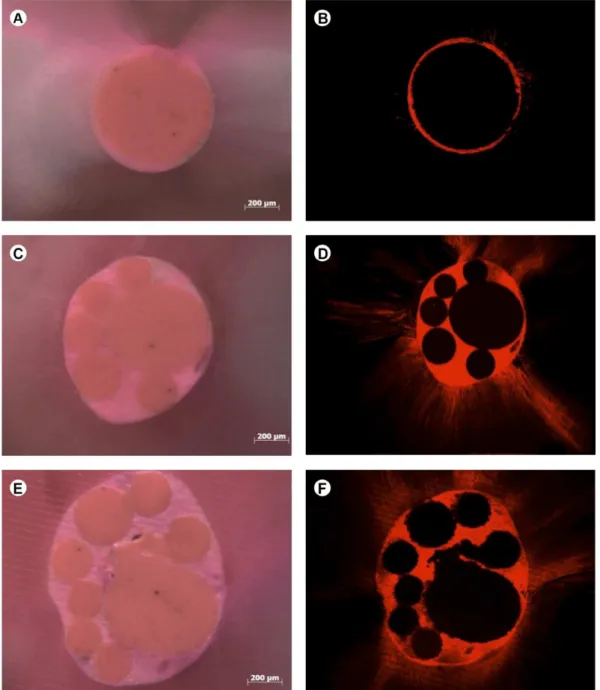
Figure 2. Representative stereomicroscopic and confocal microscopic images of the manual group. Different sections of the root canals are shown at 2 mm (A,B), 4 mm (C,D) and 6 mm (E,F).
298
D
.B
. Rossetto et al.
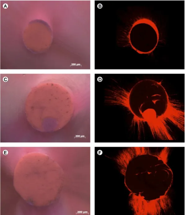
Figure 3. Representative stereomicroscopic and confocal microscopic images of the mechanical group. Different sections of the root canals are shown at 2 mm (A,B), 4 mm (C,D) and 6 mm (E,F).
Table 1.Median, minimal and maximal values of the percentages of gutta-percha, sealer and voids in the root canal fillings in the 2, 4 and 6 mm levels of the tested groups
Level
Manual method Mechanical method Ultrasonic method
Gutta-percha Sealer Voids Gutta-percha Sealer Voids Gutta-percha Sealer Voids
2 mm 80.00
(47.64-98.95)a
19.35 (1.05-52.06)a
0 (0-8.79)a
84.27 (23.33-96.44)a
15.73 (3.56-75.71)a
0 (0-1.68)a
83.52 (0-98.17)a
16.49 (1.83-100)a
0 (0-9.66)a
4 mm 69.46
(58.39-95.27)a
29.17 (4.73-37.15)a
0.81 (0-5.21)a
83.05 (71.87-99.59)b
16.73 (0.41-28.13)b
0 (0-3.00)a
73.35 (29.35-95.69)ab
26.57 (4.31-70.65)ab
0 (0-2.19)a
6 mm 75.98
(57.43-93.10)a
21.75 (6.9-35.60)a
0.68 (0-6.97)a
80.95 (56.83-91.21)a
18.88 (8.79-41.95)a
0.87 (0-6.78)a
77.37 (32.58-87.01)a
21.32 (11.12-58.36)a
0 (0-9.06)a
299
Obturation by active lateral compaction
the ultrasonic method was similar statistically the manual and mechanical methods (p>0.05). The results of the time spent (in s) for root canal filling are shown in Table 2.
Discussion
In this study, the influence of the method to perform the lateral compaction and the necessary time for this procedure was assessed. To determine the quality of the root canal fillings, the GP, sealer and void areas were measured, as well as the perimeter of penetration of the sealer into the dentinal tubules. The mechanical method was faster and favored the formation of a larger GP area in the 4 mm section.
Stereomicroscopy was used to evaluate the quality of the root canal filling (7,20) and the GP, sealer and voids area. Confocal laser microscopy was used to assess sealer
Figure 4. Representative stereomicroscopic and confocal microscopic images of the ultrasonic group. Different sections of the root canals are shown at 2 mm (A,B), 4 mm (C,D) and 6 mm (E,F).
Table 2.Mean and standard deviation of the time spent (seconds) for root canal filling in the groups.
Root filling technique Mean (S.D.)
Manual 338.6 (98.77)
Mechanical 222.5 (56.13)
Ultrasonic 286.7 (48.30)
300
D.B
. Rossetto et al.
distribution. This methodology was used in a previous study (14) where the filling ability established by the different filling techniques for the complex root canal anatomy was evaluated.
With regards to the presence of voids, no differences were found among the three methods for the different sections analyzed. None of the studied methods produced fillings without voids, which is in agreement with another study that used the lateral compaction technique in root canal filling of maxillary incisors (21). The lateral compaction technique presented an increase of the area of the root canal filled in this type of anatomy, but obvious voids and spreader tracts were evident in some cross-sections (Fig. 2), as previously reported (7). Probably a fine spreader creates a larger space than is required for the accessory cones used.
The ultrasound method, however, favored the softening of GP and did not eliminate the presence of voids. This result does not agree with the better sealing ability of the ultrasound compaction technique compared with the manual compaction technique observed in other studies (16-18). In another study that compared the cold lateral compaction technique and another technique using heated GP, no significant differences were verified regarding the sealing capacity (22), although the use of heated GP presented a greater amount of GP in the filling mass. This result demonstrated that even with a larger GP area, voids can still exist, as verified in the present study. However, this study (22) used AH 26 sealer, which presented a good sealing capacity.
GP and sealer areas showed significant differences for the manual method in comparison to the mechanical method at the 4 mm level, in which the mechanical method favored the formation of a larger GP area and a smaller area of sealer. The sealer is the component of root filling that could present greater solubility. With a thinner sealer film, there is a lesser probability of the formation of voids caused by contraction and the solubility of the sealer in the case of fluid infiltration.
A higher percentage of GP was verified in the 2-mm-thick sections in relation to the 4- and 6-mm-2-mm-thick sections, independent of the method. This result is contrary to that of another study (23) that found a higher percentage of GP in the cervical sections. This divergence is probably due to the type of accessory cone used. In this study, the XF cone was utilized, which is thinner and capable of better reaching the apical section. Another reason could be related to the method of instrumentation. The present study used ProTaper instrumentation up to the F5 instrument at the WL, which presented a taper variation of 0.05 mm, although with a lesser taper variation compared with another study that used manual instrumentation up to a size 50 K-File
with a 0.02 taper (23). This smaller taper variation could possibly contribute to the greater percentage of GP area and a thinner sealer film. Overall, using master cones with greater taper seem to be a more predictable and efficient technique (6).
The sealer penetrability in the dentinal tubules showed no significant differences among the three analyzed methods. In addition, the confocal images revealed a circumferentially continuous sealer layer at all levels. The results of the present study are consistent with those of others studies (6,24). The analysis of sealer penetrability is important because it relates to the higher or lower antiseptic action of the material through the root filling. Some authors consider that persistent intraradicular infection inside dentinal tubules can be responsible for the resurgence of apical periodontitis (25).
In relation to the time necessary for the execution of the lateral compaction technique, a faster root filling procedure was achieved when the accessory cone space was opened mechanically, possibly the mechanical movement of the spreader favored a greater opening of the space for the insertion of the accessory GP cones.
None of the lateral compaction methods prevented the occurrence of voids in the root canal filling. The mechanical method promoted a faster canal filling and a larger GP area than the manual method at the 4 mm level, probably due to GP plasticization with the heat spreader, which was evident in the cross-sections. This method seemed conducive to an efficient root canal filling due to GP flowability and shorter filling time.
Resumo
301
Obturation by active lateral compaction
entre os grupos. Foi possível concluir que o método mecânico foi mais rápido para a técnica de condensação lateral, mas todos os métodos de obturação estudados apresentaram espaços vazios na massa obturadora
Acknowledgements
This work was supported by the State of São Paulo Research Foundation (FAPESP) process 2011/03790-7 and 2011/18272-1. The authors deny any conflict of interest.
References
1. Schilder H. Cleaning and shaping the root canal. Dent Clin N Am 1974;18:269-296.
2. Ozok AR, Van Der Sluis LW, Wu MK, Wesselink PR. Sealing ability of a new polydimethylsiloxane-based root canal filling material. J Endod 2008;34:204-207.
3. Segura-Egea JJ, Jiménez-Pinzón A, Poyato-Ferrera M, Velasco-Ortega E, Ríos-Santos. Periapical status and quality of root fillings and coronal restorations in an adult Spanish population. Int Endod J 2004;37:525-530.
4. Tsesis I, Goldberger T, Taschieri S, Seifan M, Tamse A, Rosen E. The dynamics of periapical lesions in endodontically treated teeth that are left without intervention: a longitudinal study. J Endod 2013;39:1510-1515.
5. Oliver CM, Abbott PV. Correlation between clinical success and periapical dye penetration. Int Endod J 2001;34:637-644.
6. Weis MV, Parashos P, Messer HH. Effect of obturation technique on sealer cement thickness and dentinal tubule penetration. Int Endod J 2004;37:653-663.
7. Jarret IS, Marx D, Covey D, Karmazin M, Lavin M, Gound T. Percentage of canals filled in apical cross sections - an in vitro study of seven obturation techniques. Int Endod J 2004;37:392-398.
8. Elayouti A, Achleithner C, Lost C, Weiger R. Homogeneity and adaptation of a new gutta-percha paste to root canal walls. J Endod 2005;31:687-690.
9. Patel DV, Sherriff M, Ford TRP, Watson TF, Mannocci F. The penetration of Realseal primer and Tubliseal into root canal dentinal tubules: a confocal microscopic study. Int Endod J 2007;40:67-71.
10. Cavenago BC, Duarte MAH, Ordinola-Zapata R, Marciano MA, del Carpio-Perochena AE, Bramante CM. Interfacial adaptation of an epoxy-resin sealer and a self-etch sealer to root canal dentin using the System B or the single cone technique. Braz Dent J 2012;23:205-211. 11. Peak JD, Hayes SJ, Bryant ST, Dummer PM. The outcome of root canal
treatment. A retrospective study within the armed forces (Royal Air Force). Br Dent J 1991;190:140-144.
12. Levitan ME, Himel VT, Luckey JB. The effect of insertion rates on fill
length and adaptation of a thermoplasticized gutta-percha technique. J Endod 2003;29:505-508.
13. Chu CH, Lo EC, Cheung GSP. Outcome of root canal treatment using Thermafil and cold lateral condensation filling techniques. Int Endod J 2005;38:179-185.
14. Marciano MA, Ordinola-Zapata R, Cunha TVRN, Duarte MAH, Cavenago BC, Garcia RB, et al.. Analysis of four gutta-percha techniques used to fill mesial root canals of mandibular molars. Int Endod J 2011;44:321-329.
15. Plotinno G, Pameijer CH, Grande NM, Somma F. Ultrasonics in Endodontics: A review of the Literature. J Endod 2007;33:81-95. 16. Moreno A. Thermomechanical softened gutta-percha root canal filling.
J Endod 1977;5:186-188.
17. Baumgardner KR, Krell KV. Ultrasonic condensation of gutta-percha: an in vitro dye penetration and scanning electron microscopy study. J Endod 1990;16:253-259.
18. Deitch AK, Liewhr FR, West LA, William R, Patton WR. A comparison of fill density obtained by supplementing cold lateral condensation with ultrasonic condensation. J Endod 2002;28:665-667.
19. Bailey GC, Cunnington SA, Ng YL, Gulabivala K, Setchell DJ. Ultrasonic condensation of gutta-percha: the effect of power setting and activation time on temperature at the root surface – an in vitro study. Int Endod J 2004;37:447-454.
20. Romania C, Beltes P, Boutsioukis C, Dandakis C. Ex-vivo area-metric analysis of root canal obturation using gutta-percha cones of different taper. Int Endod J 2009;42:491–498.
21. Marciano MA, Bramante CM, Duarte MAH, Delgado RJR, Ordinola-Zapata R, Garcia RB. Evaluation of single root canals filled using the lateral compaction, Tagger’s Hybrid, Microseal and Guttaflow techniques. Braz Dent J 2010;21:411-415.
22. Wu MK, Kast’áková A, Wesselink PR. Quality of cold and warm gutta-percha fillings in oval canals in mandibular premolars. Int Endod J 2001;34:485-491.
23. Souza EM, Wu MK, van der Sluis LW, Leonardo RT, Bonetti-Filho I, Wesselink PR. Effect of filling technique and root canal area on the percentage of gutta-percha in laterally compacted root fillings. Int Endod J 2009;42:719-726.
24. Kok D, Duarte MAH, Da Rosa RA, Wagner MH, Pereira JR, Só MVR. Evaluation of epoxy resin sealer after three root canal filling techniques by confocal laser scanning microscopy. Microsc Res Tech 2012;75:1277-1280
25. Vieira AR, Siqueira Jr JF, Ricucci D, Lopes WSP. Dentinal tubule infection as the cause of recurrent disease and late endodontic treatment failure: a case report. J Endod 2012;38:250-254.