O
ABSTRACT
www.fob.usp.br/jaos or www.scielo.br/jaos
J Appl Oral Sci.2009;17(6):596-9
CO
2, ER:YAG AND ND:YAG LASERS IN ENDODONTIC
SURGERY
Daniel Humberto POZZA1, Patrícia Wehmeyer FREGAPANI2, Cristina Braga XAVIER3,
João Batista Blessmann WEBER4, Marília Gerhardt de OLIVEIRA5
1- PhD, Post Doctorate Program, Institute of Histology and Embryology, School of Medicine, University of Porto, City of Porto, Portugal. 2- DDS, Lutheran University of Brazil, Canoas, RS, Brazil.
3- PhD, Adjunct Professor, Department of Maxillofacial Trauma and Surgery, Federal University of Pelotas, Pelotas, RS, Brazil. 4- PhD, Department of Dentistry, Adjunct Professor, Pontifical Catholic University of Rio Grande do Sul, Porto Alegre, RS, Brazil.
5- PhD, Dentistry, Full Professor, Pontifical Catholic University of Rio Grande do Sul, Porto Alegre, RS, Brazil; ; Researcher, Brazilian Council for Scientific and Technological Development (CNPq).
Corresponding address: Marília Gerhardt de Oliveira Av. Cel. Lucas de Oliveira 1841/203 Petrópolis 90460001 Porto Alegre, RS Brasil -Phone/Fax: +55 51 3330 9545 - e-mail: [email protected]
Received: November 10, 2008 - Accepted: July 19, 2009
bjectives: CO2, Er:YAG and Nd:YAG lasers have been used in endodontic surgery. This in vitro study evaluated 1% Rhodamine B dye penetration using computer-assisted morphometry (ImageTool Software®) of 108 endodontically treated human permanent
canines. Material and methods: Teeth were divided into 9 groups according to the technique used: A: 90-degree apicoectomy with bur, root-end cavity preparation with ultrasound and filled with MTA; B: 90-degree apicoectomy with bur, root-end cavity prepared with ultrasound and filled with MTA, and treatment of apical surface with CO2 laser (1 W, CW/CW); C: 90-degree apicoectomy with bur, and treatment of apical surface with Nd:YAG laser (150 mJ, 10 Hz); D: 90-degree apicoectomy with bur, and treatment of apical surface with CO2 laser,(1 W, CW/CW); E: apicoectomy with Er:YAG laser (400 mJ, 10 Hz), root-end cavity prepared with ultrasound and filled with MTA; F: apicoectomy with Er:YAG laser (400 mJ, 10 Hz) and treatment of apical surface with Nd:YAG laser (150 mJ, 10Hz); G: apicoectomy with CO2 laser (5W, CW/SP), root-end cavity prepared with ultrasound and filled with MTA; H: irradiation of apical end with CO2 laser (1 W, CW/CW); I: irradiation of apical end with Nd:YAG laser (150 mJ, 10 Hz). Results:
Dye penetration was found in all specimens at different rates, the lowest penetration occurring in groups C (16.20%), B (17.24%) and F (17.84%). Conclusions: Groups B, C and F represent the best technical sequences to perform endodontic surgery.
Key words: Apicoectomy. Lasers. Ultrasonics. Tooth apex. Dental leakage. Seepage.
INTRODUCTION
The permeability of dentin exposed by apicoectomy is one of the causes of endodontic surgery failure because microleakage and bacterial contamination trigger inflammation6. Pécora, et al.15 (2001) reported that dentin
permeability decreased when smear layer was found close to the apical third of the root canal. In contrast, the removal of smear layer after apicoectomy using rotary instruments is beneficial because it promotes cementum deposition on the exposed dental surface and favors tissue repair.
Dye penetration has been used to investigate apical permeability in studies with different endodontic surgery techniques. According to Gagliani, et al.1(1998), apical
microleakage increases at increased resection angles because a larger number of dentinal tubules are sectioned and exposed. Less dye penetration is found when apicoectomy is performed at 90 degrees because the apical delta is more fully removed in perpendicular resections.
Gouw-Soares, et al.3 (2003) conducted a study with
human teeth with apicoectomies performed with burs, Er:YAG laser or CO2 laser. The use of lasers resulted in smoother surfaces and more homogeneous dentin fusion and recrystallization, which occluded tubules and decreased permeability. Paghdiwala13 (1991) found that thermal
ablation with Er:YAG laser can cause the dissolution of mineral components and fusion of amorphous particles, without crystallization, which results in a clean and smooth surface. The advantages of this type of laser over burs are: better visibility; accurate apical resection; no contact; removal of lesion in a shorter time by vaporization; hemostasis; no vibration or discomfort and minimal pain; and less bacterial risk of trauma to adjacent tissues10.
Grgurevic, et al.4 (2005)tested different Er:YAG laser
parameters for apicoectomy, and found that 380 mJ and 20 Hz had an ablation speed four times greater than 200 mJ and 8 Hz. They concluded that even high-energy lasers are safe when used under water-air refrigeration.
Irradiation with Nd:YAG laser is absorbed by mineral structures, such as phosphates and carbonate hydroxyapatite, and disorganizes crystal structures by thermochemical ablation. In this process, dentin turns into an ionizing gas free of debris, and has no porosity or cracks, which reduces dentinal permeability because it occludes tubules6.
CO2 laser, however, is absorbed by water in tissues and leads to a rapid increase in temperature and intracellular pressure. The heated particles become incandescent and, when deposited on the irradiated tissue again, form a carbonized layer. In mineralized tissues, this irradiation is absorbed by calcium carbonate and phosphates, which causes molecular vibration and generalized heat as a consequence of dental tissue removal. Tubules are occluded by conversion of hydroxyapatite into calcium orthophosphate (apatite) and by transfer of thermal energy to the organic portion17. Miserendino9 (1988) recommended
the use of CO2 laser at 10 W and continuous wave for endodontic surgery. Those authors found that irradiated root surfaces had craters with a superficial carbonized layer, formation of a matrix similar to cementum, and no dentinal tubules.
To define the ideal parameters of energy for the use of CO2 laser, Pashley, et al.14 (1992) used 11, 113 or 556 J/
cm2. The two lowest laser energy levels increased
permeability, whereas the highest produced a fully glazed surface that occluded dentinal tubules. Such findings were confirmed by Kimura, et al.5 (2000), who visualized areas
of melted dentin under scanning electron microscopy (SEM), with partial occlusion of dentinal tubules and no cracks, fractures or thermal damage to adjacent structures or the pulp.
The apical cavity should be at least 2 mm deep, and should reach the largest possible number of accessory canals. Its walls should be parallel and follow the root canal to produce a safe and effective sealing11,24. The preparation of
root-end cavities with ultrasound facilitates the distribution of root-end filling material and provides a more efficient apical sealing because it results in more parallel walls and a
deeper cavity (mean = 2.5 mm), which provides better retention2,11. Root-end cavities prepared with ultrasound,
combined or not with burs, result in less debris than those prepared with rotary instruments only22.
Torabinejad, et al.20 (1995) conducted an in vitro study
to evaluate the apical sealing ability of MTA, whose resistance to compression is similar to that of amalgam used in root-end obturations. Since then, several authors found good results with the use of MTA because of its greater biocompatibility, less apical microleakage when used in endodontic surgeries, less bacterial contamination, better marginal adaptation to the cavity walls, and effective antibacterial action2,12,16,23.
The present in vitro study evaluated dye penetration using computer-assisted morphometry to evaluate different techniques, indicate the best technique, and compare the results of the different groups under study.
MATERIAL AND METHODS
This study used 108 human permanent canines. The teeth were radiographed, sectioned with an abrasive disk under irrigation, stored in 0.9% sodium chloride, and later underwent conventional endodontic treatment.
Specimens were divided into 9 groups of 12 teeth each: A: 90-degree apicoectomy with bur, root-end cavity prepared with ultrasound and filled with MTA; B: 90-degree apicoectomy with bur, root-end cavity prepared with ultrasound and filled with MTA and treatment of apical surface with CO2 laser(1 W, CW/CW); C: 90-degree apicoectomy with bur, and treatment of apical surface with Nd:YAG laser (150 mJ, 10 Hz); D: 90-degree apicoectomy with bur, and treatment of apical surface with CO2 laser (1 W, CW/CW); E: apicoectomy with Er:YAG laser (400 mJ, 10 Hz), root-end cavity prepared with ultrasound and filled with MTA; F: apicoectomy with Er:YAG laser (400 mJ, 10 Hz) and treatment of apical surface with Nd:YAG laser (150 mJ, 10Hz); G: apicoectomy with CO2 laser (5W, CW/SP),
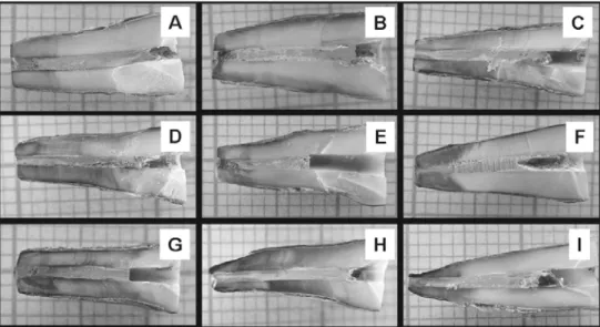
FIGURE 1- Photographs of teeth in groups A, B, C, D, E, F, G, H and I to be evaluated using computer-assisted morphometry (x4)
597
COroot-end cavity prepared with ultrasound and filled with MTA; H: irradiation of apical end with CO2 laser (1 W, CW/ CW); I: irradiation of apical end with Nd:YAG laser (150 mJ, 10 Hz).
Two layers of a sealing material were applied to the specimens, which were then immersed in 1% Rhodamine B for 24 h.
Specimens were photographed, and the photographs were digitized to undergo computer-assisted morphometry using the ImageToolä software (UTHSCSA, San Antonio, TX, USA) (Figure 1). The following measurements were made: total dentin area and total area of dentin with dye penetration. The area of dye penetration was then divided by the value of total dentin area and the result corresponded to the percentage of dentin with dye penetration.
Kruskal-Wallis and Dunn’s post-hoc test were used to evaluate the differences in dye penetration between groups at 5% significance level.
RESULTS
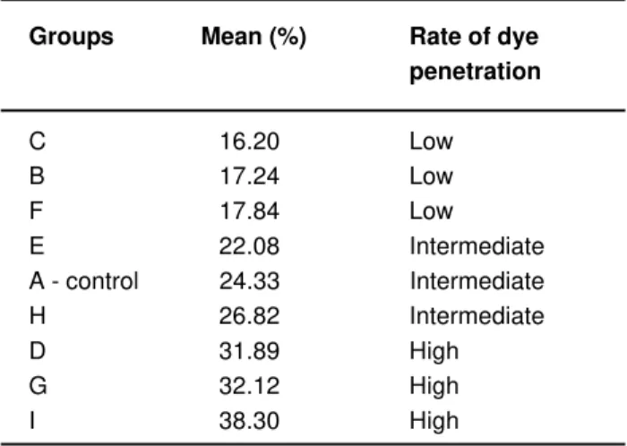
According to the different rates of penetration and statistical analysis, the groups in this study were classified into three subgroups: lower (below 20%); intermediate (between 20 and 30%); and higher (above 30%) rates of
dye penetration (Table 1). Statistically significant differences were found between group C and group D (p<0.05); group C and group G (p<0.05); and group C and group I (p<0.01).
DISCUSSION
Human permanent canines were selected for this study because their roots are straight in 88.2% of mandibular and 100% of maxillary canines. Apicoectomies were performed at 90 degrees and at 3 mm from the apical end because such angle and distance result in less microleakage than when apical resection is beveled1,16,21.
The use of ultrasound tips provides a more adequate access to the apical end of the root canal. Therefore, resections may be performed perpendicular to the long axis of the tooth, which preserves structure and decreases the number of sectioned dentinal tubules11,16,22.
MTA was used in this study because it does not require a dry field, is easy to handle, apply and remove excess, has good biocompatibility, results in less apical microleakage in endodontic surgeries, has excellent marginal adaptation to the walls of the cavity, and requires little force for condensation16,19.
Groups C, B and F had the lowest rates of dye penetration. In group C, this seemed to be associated with the smear layer that resulted from apical resection with a bur. This layer served as the substrate for fusion when treated with Nd:YAG laser and, consequently, it occluded dentinal tubules, which is in agreement with results found in other studies that investigated fusion using this type of laser6-8.
In group B, the low rate of dye penetration may have been associated with fusion and recrystallization promoted by CO2 laser on the smear layer, which resulted from the apicoectomy performed with a bur and served as a substrate for dentinal tubule occlusion5,9,10,14.
Group F was statistically very similar to group C (p = 1.0000). The variation between these two groups was assigned only to the difference in instruments used for apicoectomy: Er:YAG laser in group F, and bur in group C. Er:YAG laser removes the smear layer during ablation, and leaves the dentinal tubules open. This removal may be responsible for the greater dye penetration in this group compared to group C13,15,18.
Group E had an intermediate rate of dye penetration, lower than that of the control group, which suggests that Er:YAG laser may cause some degree of dentin fusion and reduce the lumen of tubules, as reported by Paghdiwala13.
In group H, the intermediate rate of dye penetration may be explained by the fact that CO2 laser affects the adaptation of the filling material to the root canal walls due to the rapid increase of temperature generated by laser applied to the gutta-percha and root canal sealer. Therefore, a variation in volume during and after irradiation may result in a loss of adaptation of the endodontic obturation. It is assumed that such thermal damage did not occur in dentin, which was found to be glazed after removal of superficial carbonization, in agreement with the findings of Pashley, et al.14 (1992).
Groups Mean (%) Rate of dye
penetration
C 16.20 Low
B 17.24 Low
F 17.84 Low
E 22.08 Intermediate
A - control 24.33 Intermediate
H 26.82 Intermediate
D 31.89 High
G 32.12 High
I 38.30 High
TABLE 1- Classification of the groups according to severity of dye penetration
A: 90° apicoectomy with bur, ultrasonic root-end cavity preparation and filling with MTA; B: A + apical surface
treatment with CO2 laser; C: 90° apicoectomy with bur and
apical surface treatment with Nd:YAG laser; D: 90°
apicoectomy with bur and apical surface treatment with CO2
laser; E: apicoectomy with Er:YAG laser, ultrasonic root-end cavity preparation and filling with MTA; F: apicoectomy with Er:YAG laser and apical surface treatment with Nd:YAG
laser; G: apicoectomy with CO2 laser, ultrasonic root-end
cavity preparation and filling with MTA; H: apical end
irradiation with CO2 laser; I: apical end irradiation with
Nd:YAG laser. Source: Study data, Instat 3.0 for Windows.
598
In group D, the high rate of dye penetration may be explained by the same process as in group H. CO2 laser may have promoted a loss of adaptation of the endodontic obturation in the root canal by the rapid increase in temperature16.
Group G had the poorest result for CO2 laser in the present study. In this group, the highest power level (5 W) was used, which was justified by the need to perform ablation and dentinal resection for apicoectomy. Of the groups with an apicoectomy at 3 mm, this was the group that had the most irregular resected surface, which confirms similar findings of other studies1,16.
Apical penetration of 1% Rhodamine B dye at different rates was found in all specimens evaluated in this study using computer-assisted morphometry and the ImageToolä software.
CONCLUSIONS
The present in vitro results indicate that the following techniques had the lowest rates of dye penetration, and are, therefore, the best technical sequences among the ones evaluated in this study to perform endodontic surgeries: apicoectomy with bur and treatment of apical surface with Nd:YAG laser; apicoectomy with bur, root-end cavity preparation with ultrasound, filling with MTA, and treatment of apical surface with CO2 laser; and apicoectomy with Er:YAG laser and treatment of apical surface with Nd:YAG laser.
ACKNOWLEDGMENTS
The authors are grateful to CAPES for financial support.
REFERENCES
1- Gagliani M, Taschieri S, Molinari R. Ultrasonic root-end preparation: influence of cutting angle on the apical seal. J Endod. 1998;24(11):726-30.
2- Gondim E, Zaia AA, Gomes BP, Ferraz CC, Teixeira FB, Souza-Filho FJ. Investigation of the marginal adaptation of root-end filling materials in root-end cavities prepared with ultrasonic tips. Int Endod J. 2003;36(7):491-9.
3- Gouw-Soares S, Stabholz A, Lage-Marques JL, Zezell DM, Groth EB, Eduardo CP. Comparative study of dentine permeability after apicectomy and surface treatment with 9.6 microm TEA CO2 and Er:YAG laser
irradiation. J Clin Laser Med Surg. 2004;22(2):129-39.
4- Grgurevic J, Grgurevic L, Miletic I, Karlovic Z, Krmek SJ, Anic I. In vitro study of the variable square pulse Er:YAG laser cutting efficacy for apicectomy. Lasers Surg Med. 2005;36(5):347-50.
5- Kimura Y, Wilder-Smith P, Matsumoto K. Lasers in endodontics: a review. Int Endod J. 2000;33(3):173-85.
6- Lee BS, Lin CP, Lin FH, Lan WH. Ultrastructural changes of human dentin after irradiation by Nd:YAG laser. Lasers Surg Med. 2002;30(3):246-52.
7- Lin CP, Lee BS, Lin FH, Kok SH, Lan WH. Phase, compositional, and morphological changes of human dentin after Nd:YAG laser treatment. J Endod. 2001;27(6):389-93.
8- Liu HC, Lin CP, Lan WH. Sealing depth of Nd:YAG laser on human dentinal tubules. J Endod. 1997;23(11):691-3.
9- Miserendino LJ. The Laser apicoectomy: endodontic application of the CO2 laser for periapical surgery. Oral Surg Oral Med Oral Pathol.
1988;66(5):615-9.
10- Moritz A, Gutknecht N, Goharkhay K, Schoop U, Wernisch J, Pöhn C, et al. The carbon dioxide laser as an aid in apicoectomy: an in vitro study.J Clin Laser Med Surg. 1997;15(4):185-8.
11- Navarre SW, Steiman HR. Root-end fracture during retropreparation: a comparison between zirconium nitride-coated and stainless steel microsurgical ultrasonic instruments. J Endod. 2002;28(4):330-2.
12- Oliveira MG, Xavier CB, Demarco FF, Pinheiro AL, Costa AT, Pozza DH. Comparative chemical study of MTA and Portland cements. Braz Dent J. 2007;18(1):3-7.
13- Paghdiwala AF. Root resection of endodontically treated teeth by erbium: YAG laser irradiation.J Endod. 1991;19:91-4.
14- Pashley EL, Horner JA, Liu M, Kim S, Pashley DH. Effects of CO2
laser energy on dentin permeability. J Endod. 1992;18(6):257-62.
15- Pécora JD, Cussioli AL, Guerisoli DM, Marchesan MA, Sousa-Neto MD, Brugnera A Jr. Evaluation of Er:YAG laser and EDTAC on dentin adhesion of six endodontic sealers. Braz Dent J. 2001;12(1):27-30.
16- Peters CI, Peters OA, Barbakow F. An in vitro study comparing root-end cavities prepared by diamond-coated and stainless steel ultrasonic retrotips. Int Endod J. 2001;34(2):142-8.
17- Sasaki KM, Aoki A, Ichinose S, Ishikawa I. Ultrastructural analysis of bone tissue irradiated by Er:YAG laser. Lasers Surg Med. 2002;31(5):322-32.
18- Takeda FH, Harashima T, Kimura Y, Matsumoto K. A comparative study of the removal of smear layer by three endodontic irrigants and two types of laser. Int Endod J. 1999;32(1):32-9.
19- Torabinejad M, Chivian N. Clinical applications of mineral trioxide aggregate. J Endod. 1999;25(3):197-205.
20- Torabinejad M, Hong CU, Lee SJ, Monsef M, Pitt Ford TR. Investigation of mineral trioxide aggregate for root-end filling in dogs. J Endod. 1995;21(12):603-8.
21- von Arx T, Kurt B. Root-end cavity preparation after apicoectomy using a new type of sonic and diamond-surfaced retrotip: a 1-year follow-up study. J Oral Maxillofac Surg. 1999;57(6):656-61.
22- von Arx T, Walker WA 3rd. Microsurgical instruments for root-end cavity preparation following apicoectomy: a literature review. Endod Dent Traumatol. 2000;16(2):47-62.
23- Xavier CB, Weismann R, Oliveira MG, Demarco FF, Pozza DH. Root-end filling materials: apical microleakage and marginal adaptation. J Endod. 2005;31(7):539-42.
24- Zuolo ML, Perin FR, Ferreira MO, Faria FP. Ultrasonic root-end preparation with smooth and diamond-coated tips. Endod Dent Traumatol. 1999;15(6):265-8.