http://doi.org/10.1590/2317-6431-2017-1850 ISSN 2317-6431
Otological findings in patients following infection with
Zika virus: case report
Achados otológicos em pacientes pós-infecção pelo zika vírus:
estudos de caso
Odailza Rosa Martins1, Priscila de Araújo Lucas Rodrigues2, Andréia Cristina Munzlinger dos Santos3, Erica
Zattar Ribeiro4, Andreia Ferreira Nery5, Jazon Baracat Lima4, Cynthia Castilho Moreno4, Angela Rúbia Oliveira
Silveira4
ABSTRACT
The Zika virus is transmitted between humans by the Aedes aegypti
mosquito and has become a public health emergency, as declared by the World Health Organization. Studies have reported the virus to be associated with hearing alterations in patients, following infection. This study was conceived in view of the importance of hearing for communication, associated with the limited number of reports on the impact of Zika virus on the hearing system of adults. The aim of the study was to characterize the otological findings in two adult patients, following infection with the Zika virus. The otological findings observed in two outpatients of an otorhinolaryngology service were described, both of whom reported hearing complaints immediately following the clinical symptoms of Zika virus infection. Both adult patients reported the following symptoms: discomfort, skin rash, joint pain, auricular fullness, and hearing loss. Serologic testing (IgG) for Zika virus was performed. The audiological findings were unilateral peripheral hearing loss in one of the patients, alterations in contralateral acoustic reflexes in both patients, and alteration in the brainstem auditory-evoked potential in one patient. The audiological findings demonstrate possible neuronal involvement in the manifested complaints, whether or not associated with the peripheral component, in patients infected with Zika virus.
Keywords: Zika virus; Hearing loss; Diagnostic techniques, Otological; Hearing; Zika virus infection
RESUMO
O Zika vírus em humanos é transmitido pelo mosquito Aedes aegypti
e se tornou uma emergência de saúde pública, conforme declaração da Organização Mundial de Saúde. Estudos relataram a associação do vírus com alterações auditivas em pacientes, pós-infecção. Este estudo foi concebido em função da importância da audição para a comunicação, associado aos poucos relatos sobre o impacto que o Zika vírus pode causar no sistema auditivo de indivíduos adultos. O objetivo do estudo foi caracterizar os achados otológicos em dois pacientes adultos, pós-infecção pelo Zika vírus. Foram descritos os achados otológicos de pacientes de um ambulatório de otorrinolaringologia, que apresentaram queixas auditivas logo após os sintomas clínicos de infecção pelo Zika vírus. Os dois pacientes adultos relataram os seguintes sintomas: mal-estar, erupção cutânea, artralgia, plenitude auricular e perda auditiva. Exame sorológico reagente para Zika vírus (IgG). Os achados audiológicos foram: perda auditiva periférica unilateral em um paciente, alterações de reflexos contralaterais em ambos e do potencial evocado de tronco encefálico em um paciente. Os achados audiológicos demonstram possível envolvimento neuronal nas queixas apresentadas, associado, ou não, ao componente periférico, nos pacientes infectados pelo Zika vírus.
Palavras-chave: Zika vírus; Perda auditiva; Técnicas de diagnóstico otológico; Audição; Infecção pelo Zika vírus
Research performed at the Júlio Müller University Hospital, Cuiabá (MT), Brazil.
(1) Speech-Language Pathology and Audiology Course, Centro Universitário de Várzea Grande – UNIVAG – Várzea Grande (MT), Brazil.
(2) Department of Surgery Clinical, Head and Neck Unit, Júlio Müller University Hospital, Cuiabá (MT), Brazil, and Speech-Language Pathology and Audiology Course, Centro Universitário de Várzea Grande – UNIVAG – Várzea Grande (MT), Brazil.
(3) Speech-Language Pathology and Audiology Course, Centro Universitário de Várzea Grande – UNIVAG – Várzea Grande (MT), Brazil. (4) Department of Surgery Clinical, Head and Neck Unit, Júlio Müller University Hospital, Cuiabá (MT), Brazil.
(5) Department of Clinical Medicine, Faculty of Medicine, Júlio Müller University Hospital, Cuiabá (MT), Brazil.
Conflict of interests: No
Authors’ contribution: ORMliterature review, auditory evaluations and article writing; PLAR conception and design of the study, auditory evaluations and article writing; ACMS literature review and article writing; EZR otorhinolaryngological evaluation and article writing; AFN infectious diseases evaluation and article writing; JBL otorhinolaryngological evaluation and article writing; CCM otorhinolaryngological evaluation and article writing; AROS otorhinolaryngological evaluation and article writing.
Corresponding author: Odailza Rosa Martins. E-mail: [email protected]
INTRODUCTION
The Zika virus (ZIKV) is a flavivirus similar to dengue, chikungunya, yellow fever, and West Nile viruses. Transmission occurs via mosquitoes of the Aedes genus, specifically the urban species, namely Aedes aegypti.
Currently, records of occupational transmission in research laboratories, perinatal transmission, and sexual and transfusion-related transmission have been the subject of scientific studies. Patients infected with ZIKV show symptoms similar to those of patients infected with dengue virus, such as low-grade fever, arthralgia, myalgia, migraine, retro-orbital pain, skin rashes with itchiness, sometimes accompanied by abdominal pain, diarrhea, constipation, and small ulcers in the oral mucous membranes(1).
In a historical context, ZIKV was named after the place in which it was first identified in 1947, in monkeys that were being used as sentinels for the control of yellow fever, in the Zika forest in Uganda, Africa(2). From 1951 until 1981, ZIKV was rarely described in Africa and Southeast Asia. Despite having been identified in 1947, the first large outbreak of ZIKV was registered in 2007, in Micronesia (Yap Island), where the virus was responsible for an widespread epidemic. Additional outbreaks occurred in October 2013 and April 2014, in several Pacific islands, including French Polynesia, which experienced a large-scale outbreak(2).
In Brazil, the first identified and confirmed case of ZIKV occurred in April 2015. Since then, several hypotheses have been proposed to explain how the virus entered the country. One hypothesis concerns the large influx of foreigners received during the football World Cup period, in 2014(2). Since the confirmation of ZIKV in Brazil, it is estimated that at least one million Brazilians, if not more, have already been infected, due to the ability of the virus to be transmitted through the bite of the Aedes aegypti mosquito, which promotes large-scale outbreaks. In Brazil, cases of this disease have been recorded in all states, and confirmed in laboratories, with the highest incidence occurring in the Northeast states(2).
A number of viral infections are known to cause hearing loss. Viral-induced hearing loss can be congenital or acquired and unilateral or bilateral. As such, certain viral infections can directly damage structures in the inner ear, and others can induce inflammatory responses which in turn cause hypoacusis(3).
Many studies have focused on the impact of ZIKV on newborns, showing that congenital viral infection can affect hearing, and as such should be considered a risk factor for hearing loss in hearing screening programs. Therefore, babies with evidence of congenital ZIKV infection should receive regular follow-up, taking into account the possibility of late occurrence and/or progressive hearing loss(4). In view of the ZIKV outbreak in Brazil, it is important to highlight that any decline in hearing function directly interferes with the
individual’s capacity to communicate, be it a child or an adult, since hearing is fundamental for socializing, the use of speech, and the development of language.
The aim of this study, therefore, was to characterize the otological findings in adolescent and adult patients infected with ZIKV.
CLINICAL CASE PRESENTATION
A series of studies of patient cases was performed, under the hypothesis that ZIKV virus can cause hearing impairment. The subjects selected for the research signed an Informed Consent form (ICF), in order to participate in the study. The research was approved by the Research Ethics Committee of the Júlio Müller University Hospital, and sent to the
Plataforma Brasil database, according to protocol number
050964/2016.
The inclusion criteria for the research were as follows: patients infected with ZIKV, of both sexes, aged between 13 and 59 years, with no recorded history of otological and/or hearing impairment, and who were able to be present for all the examinations proposed. Patients outside the age bracket mentioned, patients with some symptoms or signs indicative of previous hearing impairment, and those who were not present for an examination proposed in the protocol were excluded from the study. The patients selected came spontaneously to the otorhinolaryngology outpatient service, with otological complaints following possible ZIKV infection.
The assessments were performed in the Júlio Müller University Hospital, and the Integrated Clinic of the University Centre of Várzea Grande on the same day. As such, the sample was initially composed of five patients, though two of those patients declined to participate in the study, and one was not present for the audiology tests, thus the final sample was composed of the two remaining patients.
First, an otorhinolaryngological evaluation was performed through anamnesis, in order to collect otological information such as the presence of tinnitus, dizziness, otitis, otorrhea, otalgia, and previous records (regarding family, illnesses, drug use, ear or skull trauma). Subsequently, a clinical examination was performed by an infectious diseases specialist, in order to collect serological samples of the patients interviewed, and to confirm the information on the clinical signs and symptoms related to the ZIKV.
After these assessments, the patients performed the following audiology examinations:
- Tone Threshold Audiometry: the frequencies of 250 Hz to 8000 Hz were evaluated via air conduction, and from 500 Hz to 4000 Hz via bone conduction.
- Impedance tests: tympanograms were performed, and the contralateral stapedial reflexes were established, at the frequencies of 500 Hz, 1000 Hz, 2000 Hz, and 4000 Hz, using a 226 Hz probe.
- Transient Otoacoustic Emissions (TOAEs): the cochlear responses were analyzed at the 2000 Hz, 3000 Hz, and 4000 Hz frequency bands, using non-linear click-evoked stimuli, with an intensity of 83 dB Sound Pressure Level (dB SPL). Signal-to-noise ratio (SNR) values higher than or equal to 3 dB were considered as otoacoustic emission. - Distortion-product otoacoustic emissions (DP-OAEs): the
cochlear responses at the frequencies of 2000 Hz, 3000 Hz, 4000 Hz, and 5000 Hz were analyzed, using two pure tones, with an f1 intensity of 65 dB SPL, and f2 of 55 dB SPL, with 2 f1-f2 ratios. Signal-to-noise ratio (SNR) values higher than or equal to 6 dB were considered as otoacoustic emission.
- Auditory Brainstem Response (ABR): the integrity of the neural transmission and the electrophysiological threshold were investigated, using a click stimulus, rarefied polarity, and speed of 27.1 kHz, 100-3000 Hz filter, and maximum impedance limit allowed between the electrodes equal to or lower than 5 kΩ. The reference values of the absolute and inter-peak latencies adopted in the analysis were as follows: I: 1.3-1.8 ms; III: 3.3-3.9 ms; V: 5.1-5.9 ms; I-III: 1.6-2.5 ms; III-V: 1.4-2.2 ms; I-V: 3.5-4.4 ms. It is worth noting that the search for the electrophysiological threshold was performed due to the possibility of obtaining retro--cochlear findings in this study, whereby it was necessary to evaluate the compatibility between the behavioral and the electrophysiological responses. Two measurements were performed for each intensity evaluated, in order to verify the reproducibility of the waves.
The equipment used for the auditory evaluation was: - Audiometer: Resonance model, from Audi tech®. - Supra-aural headphone: TDH39 model, from Telephonics®. - Impedance Audiometer: AT235 model, from Interacoustic®,
226 Hz probe.
- Otoacoustic Emission analyzer: Otoread model, from Interacoustic®.
- Auditory Evoked Potential Analyzer: MB9400 model, from Nihon Kohden®.
The results from the examinations mentioned and the case studies will be presented.
Case presentation will be presented in the following order: anamnesis data, otorhinolaryngological evaluation, clinical evaluation performed by an infectious diseases specialist, and audiology exams described in the tables. The audiometry, logoaudiometry, Impedance test, and BAEP of both cases are included in full in Appendices 1 and 2. Each clinical case was described separately, due to distinct presenting otological results.
Case 1
Otorhinolaryngological evaluation
A 41-year-old woman reported that, starting in June 2016, she experienced body discomfort, with presence of red blemishes on her thorax, arms, and left side of the face, fever, wrist edema, as well as vertigo. Following otorhinolaryngological evaluation, she was medicated; however, her condition worsened after seven days, with symptoms such as dizziness and loss of hearing in the left ear, accompanied by constant tinnitus. She denied any family history of hearing impairment, previous conditions, traumas, exposure to noise, and the use of drugs and ototoxic medication. During the evaluation, the integrity of the tympanic membrane was verified bilaterally, via otoscopy.
Clinical evaluation of the infection
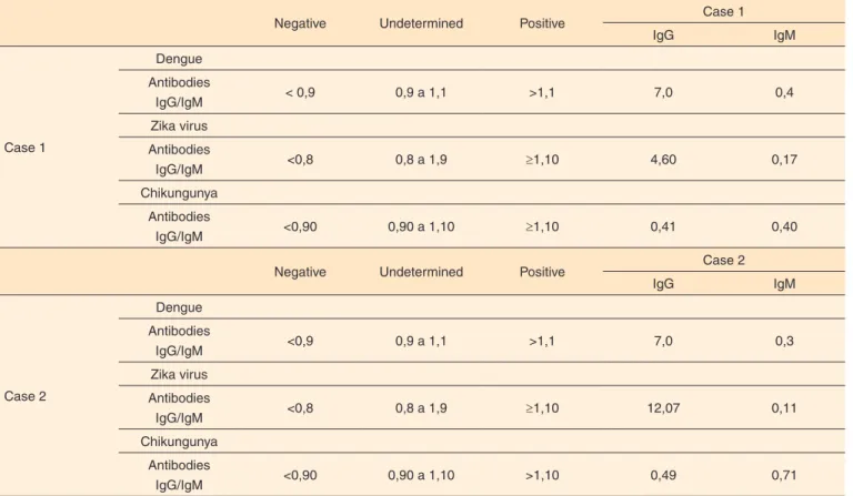
Considering that the patient was clinically assessed after the acute stage of the disease, blood samples were collected for the detection of immunoglobulins M (IgM) and G (IgG), using the technique of enzyme-linked immunosorbent assay (ELISA). Due to the simultaneous circulation of different arboviruses in the current virological profile in the Americas, and the extensive cross-reactivity with other flaviviruses, the serological tests were performed for dengue (DENV), chikungunya (CHIKV), and zika (ZIKV) viruses. The results showed ZIKV IgG with a titer of 4.6. The values obtained, including the reference standard used, are shown in Table 1.
Audiological evaluation
It was observed, through analysis of the results of the contralateral stapedial reflexes, that there was apparently a neuronal impairment associated with the peripheral impairment. The results, for the majority of frequencies tested, showed an absent reflex, even with an intact afferent pathway and with sufficient auditory thresholds to elicit the acoustic reflex. The WRS of the left ear revealed speech recognition incompatible with the tone thresholds (Table 2).
Case 2
Otorhinolaryngological evaluation
or cranial traumas, noise exposure, allergies, or the use of drugs or ototoxic medication. During the evaluation, the integrity of the tympanic membrane was verified bilaterally, via otoscopy.
Clinical evaluation of the infection
Considering that the patient was clinically assessed after the acute stage of the disease, blood samples were collected for the detection of immunoglobulins M (IgM) and G (IgG), using the technique of enzyme-linked immunosorbent assay (ELISA). Due to the simultaneous circulation of different arboviruses in the current virological profile in the Americas, and the extensive cross-reactivity with other flaviviruses, the serological tests were performed for DENV, CHIKV, and ZIKV. The results showed ZIKV IgG with a titer of 12.0. The values obtained, including the reference standard used, are shown in Table 1.
Audiological evaluation
Through analysis of the results of the contralateral stapedial reflexes, it was observed that there was apparently a neuronal impairment. The results for some of the frequencies tested showed absent reflexes, even with an intact afferent pathway, bilaterally. The BAEP also indicated neuronal alterations, due to the incompatibility of the electrophysiological threshold with the psychoacoustic threshold (Table 3).
DISCUSSION
Although there have been several outbreaks of disease associated with ZIKV in Brazil, namely microcephaly, there are few reports of hearing impairment in adults proven to be infected with ZIKV. It is believed that the present study pioneers the characterization of audiology findings in adult individuals following infection with ZIKV.
The results from this research point towards an important aspect regarding hearing in adult individuals. ZIKV may be able to alter hearing function in distinct ways between different individuals, according to this research. Both patients had a history of dengue and ZIKV infection, as shown in Table 1. However, both presented with otological and audiological alterations following ZIKV infection, with the first case study showing permanent hearing impairment in the left ear, while the audiology exams for the second case study were within normal ranges. It is worth noting that the second case had previously undergone examinations at a different institution before being asked to participate in this study, which showed a low degree of sensorineural alteration in the left ear (LE).
Regarding the peripheral evaluation performed through tone threshold audiometry, it was observed that the first patient presented with an impairment of the sensorineural type in the left ear. Concerning this finding, there are few studies relating ZIKV to this type of impairment in adult patients. However, it
Table 1. Serology results of cases 1 and 2
Negative Undetermined Positive Case 1
IgG IgM
Case 1
Dengue
Antibodies
IgG/IgM < 0,9 0,9 a 1,1 >1,1 7,0 0,4
Zika virus
Antibodies
IgG/IgM <0,8 0,8 a 1,9 ≥1,10 4,60 0,17
Chikungunya
Antibodies
IgG/IgM <0,90 0,90 a 1,10 ≥1,10 0,41 0,40
Negative Undetermined Positive Case 2
IgG IgM
Case 2
Dengue
Antibodies
IgG/IgM <0,9 0,9 a 1,1 >1,1 7,0 0,3
Zika virus
Antibodies
IgG/IgM <0,8 0,8 a 1,9 ≥1,10 12,07 0,11
Chikungunya
Antibodies
IgG/IgM <0,90 0,90 a 1,10 >1,10 0,49 0,71
Table 2. Results of the audiology exams for case 1
Procedures Right ear Left ear
Tone audiometry
Type Normal auditory
thresholds
Sensorineural
Degree Moderate
Configuration - Descending
accentuated
Logoaudiometry
SRT 25dB 35dB
WRS 100% 48% Mon / 56%
Dis
Impedance audiometry
Tympanometry CURVE A CURVE A
Acoustic reflexes
CLAR (500 Hz) 110 Absent
CLAR (1000 Hz) Absent Absent
CLAR (2000 Hz) Absent Absent
CLAR (4000 Hz) Absent Absent
TEOAE
SNR 2 kHz Present Present
SNR 3 kHz Present Present
SNR 4 kHz Present Present
DP-OAE
SNR 2 kHz Present Present
SNR 3 kHz Present Absent
SNR 4 kHz Present Absent
BAEP
Absolute latency I (ms) 1.6 1.6
Absolute latency III (ms) 3.8 3.6
Absolute latency V (ms) 5.3 5.6
Relative latency I-III (ms) 2.1 2.0
Relative latency III-V (ms) 1.5 1.9
Relative latency I-V (ms) 3.7 3.9
Interaural wave V difference (ms)
0.3
Interaural wave I-V difference (ms)
0.2
Electrophysiological threshold
30 dBnHL 40 dBnHL
Subtitle: SRT = Speech recognition threshold; WRS = word recognition score; Mon = Monosyllables; Dis = Disyllables; CLAR = contra lateral acoustic reflex; TEOAE = Transient evoked otoacoustic emissions; DP-OAE = Distortion product otoacoustic emissions; ABR = Auditory Brainstem Response; kHz = Kilohertz; ms = milliseconds; dBnHL = normal hearing level decibel
Table 3. Results of the audiology exams for case 2
Procedures Right ear Left ear
Tone audiometry
Type Normal auditory
thresholds
Normal auditory thresholds
Degree -
-Configuration -
-Logoaudiometry
SRT 10 dB 20 dB
WRS 100% Mon 100% Mon
Impedance audiometry
Tympanometry Curve A Curve A
Acoustic reflexes
CLAR (500 Hz) 100 110
CLAR (1000 Hz) 100 Absent
CLAR (2000 Hz) 100 110
CLAR (4000 Hz) 110 Absent
TEOAE
SNR 2 kHz Present Absent
SNR 3 kHz Present Present
SNR 4 kHz Absent Present
DP-OAE
SNR 2 kHz Present Present
SNR 3 kHz Present Present
SNR 4 kHz Present Present
BAEP
Absolute latency I (ms) 1.8 1.7
Absolute latency III (ms) 3.7 3.6
Absolute latency V (ms) 5.9 5.9
Relative latency I-III (ms) 1.8 1.8
Relative latency III-V (ms) 2.2 2.2
Relative latency I-V (ms) 4.1 4.1
Interaural wave V difference (ms)
0.0
Interaural wave I-V difference (ms)
0.0
Electrophysiological threshold
30 dBnHL 50 dBnHL
is known that viral infections can cause auditory alterations, which are in most cases the sensorineural type of hearing loss(3).
A study performed in the Agamenon Magalhães Hospital, Recife, Brazil, between November 2015 and May 2016, has confirmed the possible relationship between congenital ZIKV infection and hearing loss. In that study, the authors reported that among 69 children with microcephaly and laboratory tests proving ZIKV congenital infection, four (5.8%) presented with sensorineural hearing loss, with no other potential causes(4). Another study reported the case of a newborn with microcephaly, from a twin pregnancy, whose auditory exams indicated transient absent otoacoustic emissions, unresponsive brainstem evoked auditory potential in the left ear, and a 99-dB response to click stimulus in the right ear, thus confirming a profound hearing loss bilaterally. The behavioral hearing evaluation showed no response, even for high intensity stimuli(5). Even though those studies were performed on a pediatric population, the inner ear impairments found in children as a result of ZIKV infection are compatible with the type of hearing loss described in the first case of the present study.
The auditory thresholds in the second case were normal. A case study from 2014, performed in an outpatient service in Heidelberg, Germany, verified the association between Zika and hearing loss. The study reported the case of a 45-year-old woman returning from a holiday on a peninsula on the east of Malaysia, who presented with hearing loss in the left ear, with a short delay between the sound and its perception(6). The hearing difficulty lasted ten days and gradually improved; however, there are no reports indicating which examinations and audiological tests were performed or the audiological findings observed. According to the study report, it may be assumed that the hearing loss fluctuated. In the present study, it was possible to observe the relationship between the auditory characteristics of the second case with the study performed in 2014, regarding the loss of auditory perception.
During the Logoaudiometry evaluation, a poor performance in the WRS was observed in the first case in the left ear only, which was incompatible with the tone thresholds. Studies have reported that the incompatibility of results, such as a low word recognition score in tone audiometry, is characteristic of altered neural synchrony(7,8).
In the evaluation of the middle ear and central auditory pathways up to the brainstem, using impedance audiometry, it was perceived that the acoustic reflexes of both were absent, with the contralateral reflex absent in the first case for the majority of frequencies tested, and in the second case, absent at 1000 Hz and 4000 Hz in the left ear. Both observations confirm the suggestion of impairment of the central auditory system, as the integrity of the middle ear was confirmed during tympanometry. Studies have confirmed that the absence of the acoustic reflex, and the increase of its thresholds or latency without apparent justification, can be due to the superior olivary
complex being unable to execute the command to the facial nerve, to contract the stapedius muscle(9).
Cochlear function, in both cases, was compatible with the psychoacoustic evaluation, as demonstrated by the transient evoked otoacoustic emissions and the DP-OAE. Some authors have reported that, when normal OAEs are observed, with some alteration in the central auditory pathways, it can be due to retro-cochlear alterations. That is, hearing compatible with normal cochlear function presenting with neural synchrony changes, can be classified as a neuropathy(7). As such, it is verified that this definition is compatible with the characteristics observed in both cases in this study.
The role of the BAEP is to verify the integrity of the auditory pathway up to the brainstem, and to determine the electrophysiological threshold. In the first case, the BAEP showed altered electrophysiological thresholds in the left ear, with the right ear within normal range, demonstrating compatibility with the cochlear impairment observed. In the second case, the BAEP showed altered electrophysiological thresholds in the left ear, although in this case, the result was incompatible with the tone audiometry. Therefore, the diagnosis suggests a possible alteration in the auditory system up to the level of the brainstem. The BAEP can give electrophysiological evidence on the auditory processing of linguistic information(10). As such, the alteration observed with the BAEP in the second case, associated with normal auditory thresholds, suggests that speech discrimination in the presence of noise may be impaired. Regarding the second case, it is worth emphasizing that the data indicating retro-cochlear impairment is atypical, as in general, patients with neuronal alterations have hearing loss, as well as poor performance on speech comprehension in silence(11), which did not occur. However, it is noteworthy that in adult patients suffering from retro-cochlear pathology, a variety of clinical findings has been discussed in the literature, where it is claimed that the audiological findings vary from normal hearing thresholds to profound hearing loss, which can be of the fluctuating type, and the acoustic reflexes can be either absent or increased(11,12).
As ZIKV is now recognized as a factor that can lead to neurological complications in adults following infection, such as Guillaine-Barré syndrome, facial paralysis, myelitis, and others that are still being analyzed, the importance of studying the damage caused by ZIKV action is further emphasized(13,14). From this perspective, a study was performed investigating the risks of ZIKV for neuronal cells during the embryonic stage, as well as in childhood and adulthood. The authors reported that the virus has the potential to destroy neuronal cells at any stage of life(15).
Finally, it is critically important to highlight the limitations of the present study, as due to the small size of the sample obtained, it would not be prudent to generalize the results. However, the findings indicate that ZIKV infection in adult individuals merits follow up and monitoring, due to its manifestations having fluctuating and central characteristics. The central auditory behavioral evaluation and the long latency potentials can be investigated in the future, as a way to verify the impact of the neuronal impairment on sound processing.
In view of the rapid spread of ZIKV in Brazil, it is suggested that patients should monitor their auditory health following ZIKV infection, since even though those patients may not report any alterations in their hearing accuracy, it is possible that the central auditory system could be affected.
Finally, in addition to the importance of thoroughly investigating the various risks that ZIKV pose for human health, it is fundamental to develop policies that aim to improve vector control strategies and to make the population aware of the diseases and health damages caused by the virus, including its impact auditory health.
FINAL COMMENTS
Both patients presented with possible hearing impairment following ZIKV infection, although each patient presented in a unique way. The audiology findings demonstrate that the complaints have a neuronal involvement, whether or not associated with the peripheral component.
REFERENCES
1. Ventura CV, Maia M, Ventura BV, Linden VVD, Araújo EB, Ramos RC et al. Ophthalmological findings in infants with microcephaly and presumable intra-uterus Zika virus infection. Arq Bras Oftalmol. 2016;79(1):1-3. https://doi.org/10.5935/0004-2749.20160002 2. Vasconcelos CPF. Doença pelo vírus Zika: um novo problema
emergente nas Américas? Rev Pan-Amaz Saúde. 2015;6(2):9-10. https://doi.org/10.5123/S2176-62232015000200001
3. Cheloni VAB, Mancini P, Gonçalves DU. Doenças infecciosas e perda auditiva. Rev Med Minas Gerais. 2010;20(1):102-6.
4. Leal MC, Muniz LF, Ferrera TSA, Santos CM, Almeida LC, Linden
VVD et al. Hearing loss in infants with microcephaly and evidence of congenital zika virus infection — Brazil, November 2015–May 2016. MMWR Rep. 2016;65(34):917-9. https://doi.org/10.15585/ mmwr.mm6534e3
5. Leal MC, Muniz LF, Caldas Neto SS, Linden V, Ramos RCF. Sensorineural hearingloss in a case of congenital Zika Vírus. Braz J Otorhinolaryngol. Forthcoming 2016. https://doi.org/10.1016/j. bjorl.2016.06.001
6. Tappe D, Nachtigall S, Kapaun A, Schnitzler P, Stephan G, Jonas S.Acute Zika virus infection after travel to Malaysian Borneo. Emerg Infect Dis. 2015;21(5):911-2. https://doi.org/10.3201/ eid2105.141960
7. Parra VM, Matas CG, Neves IF. Estudo de caso: neuropatia auditiva.Rev Bras Otorrinolaringol. 2003;69(2):283-8. https://doi. org/10.1590/S0034-72992003000200022
8. Yellin MW, Jerger J, Fifer RC. Norms for disproportionate loss in speech intelligility. Ear Hear. 1989;10(4):231-4.
9. Linares AE. Emissões otoacústicas. In: Bevilacqua M, organizer. Tratado de audiologia. São Paulo: Santos; 2011. p. 137.
10. Matas CG, Hataiama NM, Goncalves IC. Estabilidade dos potenciais evocados auditivos em indivíduos adultos com audição normal. Rev Soc Bras Fonoaudiol. 2011;16(1):37-41. https://doi.org/10.1590/ S1516-80342011000100008
11. Hood LJ. Auditory neuropathy/dys-synchrony disorder: diagnosis and management. Otolaryngol Clin North Am. 2015;48(6):1027-40. https://doi.org/10.1016/j.otc.2015.06.006
12. Kaga K. Auditory nerve disease and auditory neuropathy spectrum disorders. Auris Nasus Larynx. 2016;43(1):10-20. https://doi. org/10.1016/j.anl.2015.06.008
13. Silva RC, Souza AM. Zika vírus: what do we know about the viral structure, mechanisms of transmission, and neurological outcomes? Rev Soc Bras Med Trop. 2016;49(3):267-73. https://doi. org/10.1590/0037-8682-0150-2016
14. Araujo LM, Ferreira MLB, Nascimento OJM. Guillain-Barré syndrome associated with the Zika virus outbreak in Brazil. Arq Neuropsiquiatr. 2016;74(3):253-5. https://doi.org/10.1590/0004-282X20160035
Appendix 2. Audiometry, logoaudiometry, impedance audiometry, and BAEP for case 2
ERRATUM
On page 1, wich read:
“Priscila Lucas de Araújo Rodrigues”
Read:
“Priscila de Araújo Lucas Rodrigues”