http://doi.org/10.1590/2317-6431-2016-1723 ISSN 2317-6431
Bite force in children with posterior crossbite
Força de mordida em crianças com mordida cruzada posterior
Monize Vilela1, Melissa Nara de Carvalho Picinato-Pirola2, Lúcia Dantas Giglio3, Wilma Terezinha
Anselmo-Lima3, Fabiana Cardoso Pereira Valera3, Luciana Vitaliano Voi Trawitzki3, Tais Helena Grechi1
ABSTRACT
Introduction: The bite force is influenced by the occlusal condition. In children with posterior crossbite the results are controversial. Purpose:
To investigate the influence of posterior crossbite in maximal isometric bite force (MIBF) in children with mixed dentition. Methods: In this cross-sectional study, 32 children participated, 21 of them belonging to the posterior cross-bite group (10 girls and 11 boys, mean age 9.2 years) and 11 to the control group (6 girls, 5 boys, mean age 9.3 years). The children were evaluated by an orthodontist for occlusal diagnosis and characterization of the groups, by otorhinolaryngologists for evaluation of respiratory symptoms and by a speech therapist to identify the clinical and MIBF myofunctional orofacial condition. The dynamometer was placed in the molar region and the children were instructed to bite it as hard as possible three times alternately. For data analysis, Student’s t-test for independent samples was used. The level of significance was set at 5%. Results: While comparing the groups crossbite vs. control, there was no significantly difference; also, among only children belonging to the crossbite group, there was no difference between the sides (crossed bite vs. Noncrossed one). Conclusion: The presence of posterior crossbite did not influence the maximal isometric bite force in children with mixed dentition.
Keywords: Bite force; Malocclusion; Child; Dentition, Mixed
RESUMO
Introdução: A força de mordida é influenciada pela condição oclusal. Em crianças com mordida cruzada posterior, os resultados são controversos. Objetivo: Investigar a influência da mordida cruzada posterior na força isométrica máxima de mordida, em crianças na fase de dentição mista. Métodos: Participaram deste estudo transversal 32 crianças, sendo 21 do grupo mordida cruzada posterior (10 meninas e 11 meninos, média de idade 9,2 anos) e 11 do grupo controle, sem alterações oclusais (seis meninas, cinco meninos, média de idade 9,3 anos). As crianças foram avaliadas por um ortodontista, para diagnóstico oclusal e caracterização dos grupos, pela equipe de otorrinolaringologia, para avaliação do quadro respiratório, e por uma fonoaudióloga. O dinamômetro foi posicionado na região dos molares e as crianças foram instruídas a mordê-lo o mais forte possível, por três vezes, alternadamente. Para análise dos dados foi utilizado o teste t de Student para amostras independentes e dependentes. O nível de significância estabelecido foi de 5%. Resultados: Na comparação entre os grupos mordida cruzada e controle não foi encontrada diferença significativa e no grupo mordida cruzada, não houve diferença entre o lado cruzado e o não cruzado. Conclusão: A presença de mordida cruzada posterior não esteve relacionada à força de mordida em crianças na fase de dentição mista.
Palavras-chave: Força de mordida; Má oclusão; Criança; Dentição mista
Research conducted at Department of Ophthalmology, Otorhinolaryngology and Head and Neck Surgery, School of Medicine of Ribeirão Preto – Universidade de São Paulo – USP – Ribeirão Preto (SP), Brazil.
(1) Clinical Hospital, School of Medicine of Ribeirão Preto – Universidade de São Paulo – USP – Ribeirão Preto (SP), Brazil. (2) School of Ceilândia, Universidade de Brasília – UnB - Brasília (DF), Brazil.
(3) School of Medicine of Ribeirão Preto – Universidade de São Paulo – USP – Ribeirão Preto (SP), Brazil.
Conflict of interests: No
Authors’ contribution: MV main author, it’s responible for data organization, tabulation of results, analysis and writing; MNCPP writing and critically revising the manuscript; LDG writing and critically revising the manuscript; WTAL contribution to assess respiratory condition and critically revising the manuscript; FCPV
contribution to assess respiratory condition and critically revising the manuscript; LVVT contribution to analysis of the results and writing; THG data collection, writing and critically revising the manuscript.
Corresponding author: Monize Vilela. E-mail: [email protected]
INTRODUCTION
Posterior crossbite is a malocclusion in the canine, premolar and molar regions, where the buccal cusps of the upper teeth lingually occlude the vestibular cusps of the corresponding lower teeth(1). It can occur unilaterally or bilaterally and be
present in the different stages of the dentition.
Some studies have related posterior crossbite with the presence of deleterious oral habits, orofacial myofunctional disorders and oral breathing(2,3). Posterior crossbite,
consi-dered one of the most frequent types of malocclusion in the deciduous and mixed dentition phase, presents a prevalence of 7.2% to 23%(4). It can produce changes in mandibular
sym-metry(5), in the electromyographic activity of the muscles of
mastication(6), in the coordination and masticatory pattern(7),
in the swallowing(3) and in the bite force(8,9). The bite force
is understood as the exertion between the upper and lower teeth when the jaw is raised by the muscles of mastication(10).
It is an important tool to evaluate the functional status of the masticatory system(11) and was used to evaluate the oral
function in different malocclusions, in the oral surgeries, temporomandibular disorders and in the neuromuscular diseases(12).
The bite force was previously investigated in children with posterior crossbite, in the deciduous and mixed dentition phase. The main objective of the studies was to evaluate the effect of orthodontic treatment on the correction of maloc-clusion(8,13,14,15). The results of these studies differ from each
other, but generally point to similar forces in the orthodontic pre-treatment phase, as well as after the restraint, and to different forces in the phase immediately after orthodontic treatment.
It is worth remembering that the bite force can be influenced according to the craniofacial morphology(16), sex(17), age(18),
pre-sence of signs and symptoms of temporomandibular disorder(19)
and number of teeth(16).
Although some studies have focused on the myofunctional orofacial condition and bite force in children with posterior crossbite, in different age groups, the results are still contro-versial, which makes it difficult to understand the occlusal relationship and myofunctional orofacial condition.
The purpose of this study was to investigate the influence of the posterior crossbite on the maximal isometric bite force (MIBF) in children in the mixed dentition phase.
METHODS
Sample
The present project was approved by the Research Ethics Committee of Clinical Hospital of School of Medicine of Ribeirão Preto, Universidade de São Paulo (HC/FMRP- USP), under no. 6443/2007. Thirty-two children in the mixed dentition phase, aged between 7 and 10 years, participated in this cross-sectional study. The children were divided into two groups: crossbite group, 21 children with posterior crossbite, of whom 14 had unilateral posterior crossbite, 3 with unilateral posterior crossbite associated with anterior open bite, and 4 with bilateral crossbite; control group, with 11 children without occlusal alterations. The children were selected by an orthodontist at Preventive and Interceptive Orthodontics Clinic of School of Dentistry of Ribeirão Preto - USP and at Center of The Mouth Respirator (CERB) of the Otorhinolaryngology Division of HC/FMRP-USP.
Descriptive data regarding the sex and age of the studied groups are shown in Table 1.
In the crossbite group, children in the mixed dentition phase, with unilateral or bilateral crossbite, involving canines and deciduous molars and first permanent molars, with indi-cation of orthodontic treatment, without restriction regarding the respiratory condition and other associated malocclusions were included.
In the control group, children in the mixed dentition phase, without occlusal alterations, who had never used orthodontic and/or orthopedic appliances, with age close to the crossbite group and without restrictions regarding the respiratory con-dition were included.
Children with genetic syndrome, congenital and acquired dentofacial deformities, the ones with extensive tooth cavities, history of neurological treatment, history of gastroesophageal reflux, history of orthodontic and/or functional orthopedic treatment and previous orofacial myofunctional speech therapy were excluded.
Dental (oral) evaluation
For occlusal diagnosis of the children, clinical evaluation was performed and it was asked complete orthodontic docu-mentation (lateral and occlusal cephalometric x-rays, study
Table 1. Sex and age of the researched groups
Groups n Sex Age (mean) Standard
deviation p-value
F M
Crossbite 21 10 11 9.2 1.04 0.74
Control 11 6 5 9.3 0.92
Student’s t-test (p<0.05)
models, intraoral photographs and extraoral photographs, from front and profile). For the posterior crossbite group, children who presented the region of canines, premolars and molars in an abnormal position, in the vestibulolingual direction (vestibular cusps of the upper teeth lingually occluding the vestibular cusps of the corresponding lower teeth) were selected. The children of the control group should present Class I (Angle Classification) of the deciduous canines (mesial surface of the superior canine cusp occluding the distal surface of the lower canine) and verti-cal and horizontal trespass (overbite and overjet, respectively) of the normal incisors(1).
Otorhinolaryngological evaluation
The evaluation of the upper airways was performed at the otorhinolaryngology department of a school hospital, including anterior rhinoscopy and nasofibroscopy (Pentax® FNL – 10RP2, 3.4 mm flexible fibroscope for children) for measurement of adenoid size.
Of the 21 children in the crossbite group: 10 (47.62%) had from 10 to 50% of cavum obstruction; 6 (28.58%) from 50 to 70% of obstruction; and 5 (23.80%) from 70 to 100% of obs-truction. Of the 11 children in the control group: 7 (63.64%) presented from 10 to 50% of cavum obstruction; 2 (18.18%) from 50 to 70% of obstruction; and 2 (18.18%) from 70 to 100% of obstruction.
Bite force evaluation
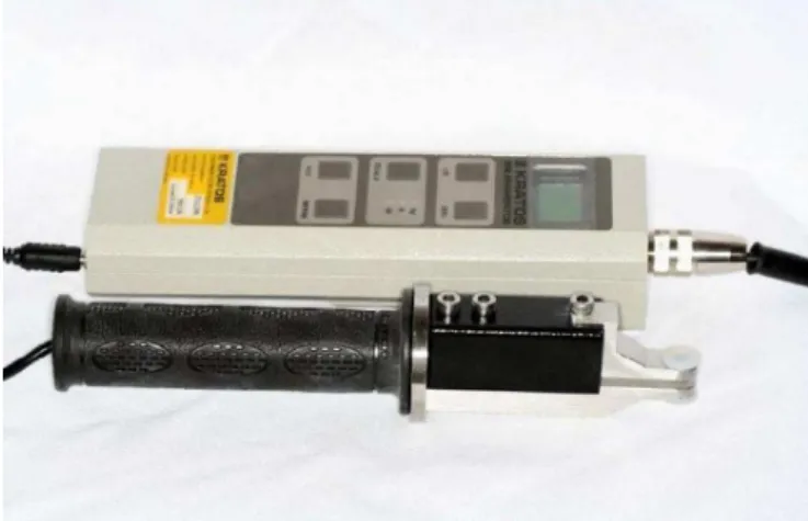
The bite force measurements were performed using a digital dynamometer, IDDK model (Kratos®, Cotia, São Paulo, Brazil), with capacity up to 100 kilograms-force (Kgf), adapted to oral conditions. The apparatus has a scale in Kgf and Newton (N), set zero key, which allows exact control of the obtained values and also a peak register that, during the obtainment of the values, facilitates the reading of the maxi-mum force (Figure 1).
During the examination, the children remained seated in a comfortable chair with their feet flat on the floor and their head parallel to the horizontal plane. To measure the bite force, the device was positioned in the region of the molar teeth, on both sides of the dental arcade, alternately, and the children were instructed to bite it as hard as possible. Three records were made for each side, with a non-standard rest between the records. The maximum bite force was recorded in Kgf by
recording the force peak indicated on the screen and the values were noted in the protocol of each child for further analysis.
Data analysis
For the analysis of the bite force, the average of the three measurements obtained on each side was considered. The statistical package SPSS (version 17.0) was used and the value of p<0.05 was adopted as a level of significance. The Kolmogorov-Smirnov test was applied to check the normality of data distribution.
The Student’s t parametric test was then performed, being unpaired when the crossbite group and the control group were compared, and paired when the crossed and noncrossed sides were compared in children with unilateral crossbite.
RESULTS
There was no significant difference (p<0.05) between the posterior crossbite and the control groups for MIBF, on both sides (Table 2).
There was no significant difference (p>0.05) between the crossed and noncrossed sides for MIBF in the posterior cros-sbite group (Table 3).
DISCUSSION
The bite force makes it possible to verify the functional state of the masticatory system. Thus, it results from the action
Table 2. Mean, standard deviation and comparison between the crossbite and control groups in relation to the maximal isometric bite force
Group n Right side Left side
Mean SD P Mean SD P
Crossbite 21 23.81 10.32 0.94 23.94 9.61 0.67
Controle 11 24.08 7.40 22.35 9.43
Subtitle: SD = standard deviation; P = probability in Student’s t-test for independent samples. Values in Kgf.
of the mandibular elevator muscles and can be modified by craniofacial biomechanics(20).
In this study, a sample of children with posterior crossbite was selected, in order to verify its influence on the bite force. The hypothesis was that the altered morphological condition of the children with this malocclusion could influence on the bite force.
Authors reported that children with unilateral posterior crossbite have a tendency to irregular and contralateral masti-catory cycles to the crossed side(21,22,23). Other studies(7,24,25)
hi-ghlighted the presence of asymmetry of the electromyographic activity of the muscles of mastication between the crossed and noncrossed sides.
In the present study, no significant difference was found in the comparison between the groups (crossbite and control) and in the intragroup analysis (crossed and noncrossed sides) in children in the mixed dentition phase. One study(8) verified
the bite force in children with posterior crossbite, with no res-trictions on respiratory conditions and at a similar age to that of the children in this study. The results also showed no significant difference between children with and without malocclusion.
The bite force in children with malocclusion was studied by some authors(15) who compared them with children without
malocclusion, but in the deciduous dentition phase, differing from this research regarding the teething phase. However, the authors verified that the type of occlusion did not affect the bite force values, confirming the findings of the present study.
Another study(14) analyzed the bite force at different stages
of orthodontic treatment and found similar forces between the right and left sides in children with unilateral crossbite. The level of the bite force was lower immediately after the orthodontic treatment, and higher after the restraint, with approximate values of children without malocclusion. These results are in agreement with the findings of the present study, although the objective was not to analyze the bite force after the orthodontic treatment.
Some studies(8,13) compared children in the mixed
denti-tion phase, with and without posterior crossbite and found a significant difference, with higher strength values in children without malocclusion. This divergence from the results of this study can be attributed to the number of participants, since the number was lower in this sample.
Another factor that may influence bite force results is the positioning of the assessment tool. Authors(26) have pointed out
that may occur variations in bite forces, associated with the
instrumentation and the position of the transducer in relation to the dental arch. A transducer positioned more posteriorly produces a greater bite force, which may be attributed probably to the mechanical lever system of the jaws.
It is also worth noting that the bite force can be influenced by the eruption stage of the teeth, the number of teeth in occlusal contact, the presence of malocclusion and the degree of axial inclination of the teeth in crossbite(8).
The differences between the studied age range(15) and the
sample size(8,13) made it difficult the direct comparison of the
studies found with the results of this research, in which children with different respiratory conditions were included in both groups and the presence of nasal obstruction may have influen-ced the bite force values. The relationship of the respiratory condition and the bite force in children is little investigated in the literature. It is known that oral breathing can influence the growth pattern and the craniofacial morphology, variables that are important in determining the bite force(27).
New studies, with a greater number of participants, are needed to better elucidate the effects of posterior crossbite on the orofacial musculature, thus contributing to the basis of orofacial myofunctional work.
CONCLUSION
The presence of posterior crossbite did not influence the bite force in children in the mixed dentition phase.
REFERENCES
1. Björk A, Krebs A, Solow B. A method for epidemiological registration of malocclusion. Acta Odontol Scand. 1964;22:27-41. http://dx.doi.org/10.1016/0002-9416(66)90175-8
2. Bresolin D, Shapiro PA, Shapiro GG, Chapko MK, Dassel S. Mouth breathing in allergic children: Its relationship to dentofacial development. Am J Orthod. 1983;83(4):334-40. http://dx.doi. org/10.1016/0002-9416(83)90229-4
3. Stahl F, Grabowski R, Gaebel M, Kundt G. Relationship between occlusal findings and orofacial myofunctional status in primary and mixed dentition. Part II: Prevalence or orofacial dysfunctions. J Orofac Orthop. 2007;68(2):74-90. http://dx.doi.org/10.1007/s00056-007-2606-9
4. Grabowski R, Stahl F, Gaebel M, Kundt G. Relationship between occlusal findings and orofacial myofunctional status in primary and mixed dentition. Part I: Prevalence of malocclusions. J Orofac Orthop. 2007;68(1):26-37. http://dx.doi.org/10.1007/s00056-007-1606-0
5. Pinto AS, Buschang PH, Throckmorton GS, Chen P. Morphological and positional asymmetries of yang children with functional unilateral posterior crossbite. Am J Orthod Dentofacial Orthop. 2001;120(5):513-20. http://dx.doi.org/10.1067/mod.2001.118627a 6. Iodice G, Danzi G, Cimino R, Paduano S, Michelotti A. Association
between posterior crossbite, skeletal, and muscle asymmetry: a Table 3. Mean, standard deviation and intragroup comparison between
crossed and noncrossed sides in relation to maximal isometric bite force
Mean SD P
Crossed side 26 9.3 0.6
Noncrossed side 26.7 8.5
systematic review. Eur J Orthod. 2016;38(6):638-51. http://dx.doi. org/10.1093/ejo/cjw003
7. Andrade AS, Gavião MB, Gameiro GH, De Rossi M. Characteristics of masticatory muscles in children with unilateral posterior crossbite. Braz Oral Res. 2010;24(2):204-10. http://dx.doi.org/10.1590/S1806-83242010000200013
8. Sonnesen L, Bakke M, Solow B. Bite force in pre-orthodontic children with unilateral crossbite. Eur J Orthod. 2001;23(6):741-9. 9. Andrade AS, Gameiro GH, Derossi M, Gavião MB. Posterior
crossbite and functional changes: a systematic review. Angle Orthod. 2009;79(2):380-6. http://dx.doi.org/10.2319/030708-137.1 10. Ahlberg J, Könönen M, Nissinen M, Rantala M, Sarna S, Lindholm
H. Self-reported stress among multiprofissional media personnel. Occup Med (Lond). 2003;53(6):403-5. http://dx.doi.org/10.1093/ occmed/kqg074
11. Van der Bilt A. Assessment of mastication with implications for oral rehabilitation: a review. J Oral Rehabil. 2011;38(10):754-80. http:// dx.doi.org/10.1111/j.1365-2842.2010.02197.x
12. Van der Bilt A, Tekamp A, van der Glas H, Abbink J. Bite force and electromyography during maximum unilateral and bilateral clenching. Eur J Oral Sci. 2008;116:217-22.
13. Castelo PM, Gavião MBD, Pereira LJ, Bonjardim LR. Masticatory muscle thickness, bite force, and occlusal contacts in young children with unilateral posterior crossbite. Eur J Orthod. 2007;29(2):149-56. http://dx.doi.org/10.1093/ejo/cjl089
14. Sonnesen L, Bakke M. Bite force in children with unilateral crossbite before and after orthodontic treatment: a prospective longitudinal study. Eur J Orthod. 2007;29(3):310-3. http://dx.doi.org/10.1093/ ejo/cjl082
15. Rentes AM, Gavião MBD, Amaral JR. Bite force determination in children with primary dentition. J Oral Rehabil. 2002;29(12):1174-80.
16. Moriya Y, Tuchida K, Moriya Y, Sawada T, Koga J, Sato J et al. The influence of craniofacial form on bite force and EMG activity of masticatory muscles.VIII-1. Bite force of complete denture wearers. J Oral Sci. 1999;4(1)1:19-27.
17. Tate GS, Throckmorton GS, Ellis E 3rd, Sinn DP. Masticatory performance, muscle activity, and occlusal force in preorthognathic surgery patients. J Oral Maxillofac Surg. 1994;52(5):476-81.
18. Kiliaridis S, Kjellberg H, Wenneberg B, Engstrom C. The relationship between maximal bite force, bite force endurance, and facial morphology during growth: a cross-sectional study. Acta Odontol Scand. 1993;51(5):323-31. http://dx.doi. org/10.3109/00016359309040583
19. Pizolato RA, Gaviao MB, Berretin-Felix G, Sampaio AC, Trindade AS Jr. Maximal bite force in young adults with temporomandibular disorders and bruxism. Braz Oral Res. 2007;21(3):278-83. http:// dx.doi.org/10.1590/S1806-83242007000300015
20. Bakke M. Bite force and occlusion. Semin Orthod. 2006;12(2):120-6. http://dx.doi.org/10.1053/j.sodo.2006.01.005
21. Ben-Bassat Y, Yaffe A, Brin I, Freeman J, Ehrlich Y. Functional and morphological-occlusal aspects in children treated for unilateral posterior cross-bite. Eur J Orthod. 1993;15(1):57-63. http://dx.doi. org/10.1093/ejo/15.1.57
22. Brin I, Ben-Bassat Y, Blustein Y, Ehrlich J, Hochman N, Marmary Y et al. Skeletal and functional effects of treatment for unilateral posterior crossbite. Am J Orthod Dentofacial Orthop. 1996;109(2):173-9. http://dx.doi.org/10.1016/S0889-5406(96)70178-6
23. Throckmorton GS, Buschang PH, Hayasaki H, Pinto AS. Changes in the masticatory cycle following treatment of posterior unilateral crossbite in children. Am J Orthod Dentofacial Orthop. 2001;120(5):521-9. http://dx.doi.org/10.1067/mod.2001.118626 24. Kecik D, Kocadereli I, Saatci I. Evaluation of the treatment changes
of functional posterior crossbite in the mixed dentition. Am J Orthod Dentofacial Orthop. 2007;131(2):202-15. http://dx.doi.org/10.1016/j. ajodo.2005.03.030
25. De Rossi M, De Rossi A, Hallak JE, Vitti M, Regalo SC. Electromyographic evaluation in children having rapid maxillary expansion. Am J Orthod Dentofacial Orthop. 2009;136(3):355-60. http://dx.doi.org/10.1016/j.ajodo.2007.08.027
26. Braun S, Hnat WP, Freudenthaler JW, Marcotte MR, Honigle K, Johnson BE. A study of maximum bite force during growth and development. Angle Orthod. 1996;66(4):261-4. http://dx.doi. org/10.1043/0003-3219(1996)066<0261:ASOMBF>2.3.CO;2 27. García-Morales P, Buschang PH, Throckmorton GS, English JD.