Article
Printed in Brazil - ©2015 Sociedade Brasileira de Química0103 - 5053 $6.00+0.00A
*e-mail: [email protected]
Can Ergosterol Be an Indicator of
Fusarium
Fungi and Mycotoxins in Cereal
Products?
Ewa Stanisz,a Agnieszka Zgoła-Grześkowiak,*,a Agnieszka Waśkiewicz,b Łukasz Stępieńc
and Monika Beszterdab
aInstitute of Chemistry and Technical Electrochemistry, Poznan University of Technology, 60-965 Poznań, Poland
bDepartment of Chemistry, Poznań University of Life Sciences, 60-625 Poznań, Poland
cInstitute of Plant Genetics, Polish Academy of Sciences, 60-479 Poznań, Poland
Presence of fungi in food and feed products is a major problem. Fungi synthesize a large number of secondary metabolites including particularly harmful mycotoxins. They can be produced in plant tissues and are commonly found all over the world in many products including cereals. A total of 44 samples were taken for identification of ergosterol - the potential marker of fungal presence. Fourteen of these samples were chosen for further studies that included the evaluation
of the relationship between ergosterol content and three major mycotoxins produced by Fusarium
spp.: fumonisin B1, zearalenone and deoxynivalenol. Fungal strains were also isolated and identified
by molecular means in those samples. The results of the studies give a further and more detailed insight into the relationship between contents of ergosterol and mycotoxins in different cereal products. It was found that there was no correlation between content of ergosterol and mycotoxins in the tested food products. Also, the presence of mycotoxins was not correlated with occurrence of species able to produce these toxins.
Keywords: ergosterol, mycotoxins, Fusarium, flour, groat, flakes
Introduction
Fungi are very common all over the globe. They produce a great number of metabolites with many chemical structures and biological activities. Certain fungal metabolites are highly desired components in some foods such as cheese, whereas other metabolites, such as
penicillin and cephalosporin, are important antibiotics.1
However, some genera, for example Aspergillus,
Penicillium, and Fusarium, produce metabolites called
mycotoxins that can have adverse effects, such as estrogenic effects, carcinogenicity, mutagenicity and teratogenicity in
humans and animals.1,2
Fungal toxins such as aflatoxins and ochratoxins, fumonisins, trichothecenes, zearalenone, patulin and ergot alkaloids receive the most attention due to their frequent occurrence and their severe effects on animal
and human health.3 Toxic properties of mycotoxins found
in the cereal products (like fumonisins, zearalenone
and deoxynivalenol) are widely known.4-8 Severe
contaminations of cereals with mycotoxins could be
linked to bad storage conditions in individual places9-12
and harvesting process.3,13
Consequently, food and feed should be monitored for the presence of mycotoxins and fungi. Microbiological methods used to determine fungi usually require several days for analysis and are not able to detect dead fungi. Analytical methods for determination and identification of mycotoxins are faster. These include mainly chromatographic techniques
(thin layer chromatography (TLC),14-16 gas chromatography
(GC)15-17 and high-performance liquid chromatography
(HPLC)15,16,18-20) as well as immunochemistry based
methods.15-17,18-23
Apart of these tests various chemical indicators are used as an indirect measure of the total fungal biomass in samples, e.g., ergosterol (ERG) has been proposed as
a fungal contamination marker24,25 as it is present in most
while being absent or only a minor constituent in most
vascular (higher) plants and insects.26,27 As in the case of
mycotoxins, ergosterol is usually determined in food and
feed by HPLC.26,28-33
Recently, the discussion on the usefulness of ERG as an indicator of the fungi presence as well as on the correlations between its content and levels of different mycotoxins in
food has been initiated, e.g., in rye,34 corn,32,35,36 wheat,37
rice27,38 and asparagus.39 Some research provided the
evidence showing that the presence of various mycotoxins
was correlated with the presence of ergosterol.31 Several
studies were presented concerning significant correlations between fungal biomasses, having been estimated by
ergosterol concentrations.40
On the other hand in some cases no simple relationship was noted between ergosterol content and the biomass of
some fungal genera.29 Some authors suggested caution,
especially when dealing with samples that have been exposed to sunlight, since the exposure might alter the
ergosterol amounts severely.28
This publication is one of the voices in the discussion and gives further data on the relationship between ergosterol and mycotoxins contents in different cereal products. Three mycotoxins widely found in these products were taken into
account: fumonisin B1, zearalenone and deoxynivalenol.
Experimental
Characteristics of cereal products
A total of 44 materials were tested, including 15 flours (13 wheat and 2 corn), 8 groats (3 barley, 1 buckwheat, 2 corn and 2 wheat), 13 flakes (1 barley, 2 wheat, 2 rye, 5 oat and 3 corn), 4 oat bran and 4 rice (3 white and 1 brown) samples, were randomly collected from local
retail markets in Poznań, Poland. Each sample (except
flours) was ground in the A-11 IKA laboratory analytical mill (Staufen, Germany). Humidity was determined directly after milling and the rest of the samples was put in sealed bags and stored at 4 °C until analysis.
Isolation of fungal strains
Materials used in the study were ground to fine powder which was later used for isolation of fungi. Each sample was distributed onto 90 mm sterile potato dextrose agar (PDA)
plates with streptomycin (at concentration 300 mg L-1) and
incubated at room temperature for 3 days. Then, individual fungal strains were sub-cultured via several passages on PDA medium. Pure strains were harvested to Eppendorf tubes and used for genomic deoxyribonucleic acid (DNA) extraction.
DNA extraction and species identification
Genomic DNAs of all strains were extracted using a modified hexadecyltrimethylammonium bromide
method.41,42 Mycelia of pure cultures, grown on solid
potato dextrose agar (PDA) medium, were harvested and homogenized using liquid nitrogen. Subsequently, 800 µL of cetyl trimethylammonium bromide (CTAB)
buffer buffer with 0.4% of β-mercaptoethanol were added,
followed by the addition of 150 µL of chloroform:octanol mixture (24:1,v/v). The samples were incubated at 65 °C for 25 min. After addition of 150 µL of chloroform-isoamyl alcohol mixture (24:1, v/v), samples were shaken vigorously and left at room temperature for 10 min and then centrifuged for 15 min at 12 000 rpm. The aqueous upper phase was recovered and the DNA precipitated with 1 mL of ice-cold ethanol at –20 °C for 20 min. The precipitate was centrifuged at 12 000 rpm for 15 min, the DNA was washed with ethanol (70%) and centrifuged for 5 min at 12 000 rpm. Samples were air-dried and re-dissolved in 200 µL of tris ethylenediaminetetraacetic acid buffer (pH 8.0).
PCR primers, cycling profiles
The ITS4: 5’-TCCTCCGCTTATTGATATGC-3’ and ITS5: 5’-GGAAGTAAAAGTCGTAACAAGG-3’ were chosen to amplify the genomic region encoding the rRNA
subunits.43 The primers have been validated previously.44
The PCR was done in 20 µL volume using C-1000 Bio-Rad thermal cycler (Hercules, USA). Each sample contained 1 unit of Phire II HotStart Taq DNA polymerase purchased by Thermo Scientific (Miami, USA), 4 µL of 5x PCR buffer,
12.5 pmol of forward/reverse primers, 2.5 mmol L-1 of each
dNTP and about 10-20 ng of fungal DNA. PCR conditions were as follows: 30 s at 98 °C, 35 cycles of (5 s at 98 °C, 5 s at 58 °C, 10 s at 72 °C) and 1 min at 72 °C. Amplicons were electrophoresed in 1.5% Invitrogen agarose gels (Carlsbad, USA) with GelView staining.
DNA sequencing and analysis
For sequence analysis PCR-amplified DNA fragments were purified with an Epicentre exonuclease I (Madison, USA) and a Promega shrimp alkaline phosphatase (Madison, USA) using the following program: 30 min at 37 °C, followed by 15 min at 80 °C. Both DNA strands were labeled using an Applied Biosystems BigDyeTerminator
3.1 kit (Foster City, USA), according to Stępieńet al.44 and
of the reagents. Sequence reading was performed using Applied Biosystems equipment (Foster City, USA). The sequences of PCR products were compared to the reference accessions obtained from NCBI GenBank database using BLASTn algorithm.
Reagents and chemicals
Ergosterol, fumonisin B1, deoxynivalenol and
zearalenone analytical standard were purchased from Sigma-Aldrich (St. Louis, USA). MS grade methanol, HPLC grade acetonitrile and pentane were from POCh (Gliwice, Poland). HPLC grade methanol was from Sigma-Aldrich (St. Louis, USA). Water for the HPLC mobile phase was purified using a Millipore Milli-Q system (Bedford, USA) or prepared by reverse osmosis in a Demiwa system from Watek (Ledec nad Sazavou, The Czech Republic), followed by double distillation from a Heraeus Bi18 quartz apparatus (Hanau, Germany).
Analytical grade NaOH, KOH, sodium dihydrophosphate,
acetic acid, n-hexane and o-phosphoric acid were from
POCh (Gliwice, Poland). Analytical grade disodium
tetraborate, o-phthalaldehyde, 2-mercaptoethanol, t
-butyl-ammonium hydroxide, sodium acetate and all the other chemicals were purchased from Sigma-Aldrich (Steinheim, Germany).
Determination of ergosterol
The determination of ergosterol was conducted
according to previously described procedure.45 Briefly,
200 mg of the sample was placed in 12 mL glass culture
tube and 2 mL of methanol followed by 0.5 mL of 2 mol L-1
NaOH were added. The tube was sealed with a rubber lined screw cap and placed in a Bel-Art Products 200 mL screw-capped high-density polyethylene bottle (Wayne, USA). A Moulinex Microchef 460 microwave oven (Caen, France) operating at 2450 MHz and 300 W through 20 s was used for irradiation of the samples. The cooled down sample was extracted four times with 1 mL portions of pentane. Pentane extracts were separated by one minute centrifugation at 4 000 rpm. The combined extracts were evaporated with a gentle stream of nitrogen, reconstituted in 0.5 mL of methanol and filtered through the 0.2 µm PTFE syringe filter from Agilent Technologies (Santa Clara, CA, USA). The samples were analyzed using the chromatographic system UltiMate 3000 RSLC from Dionex (Sunnyvale, USA) connected with the API 4000 QTRAP triple quadrupole mass spectrometer from AB Sciex (Foster City, USA) using the atmospheric pressure chemical ionization (APCI) interface. 10 µL of samples were injected
into a Gemini-NX C18 column (100 mm × 2.0 mm; 3 µm)
from Phenomenex (Torrance, USA) maintained at 35 °C. The isocratic mobile phase employed in the analysis consisted of methanol:water (95:5, v/v) at a flow rate of
0.4 mL min-1. The APCI source operated in positive ion
mode. The following settings for the ion source and mass spectrometer were used: curtain gas 10 psi, nebulizer
gas 20 psi, temperature 400°C, nebulizing current 3 µA
and collision gas 10 psi. Declustering potential was 65 V and the dwell time was set to 200 ms. The quantitative
transition was from 379.3 to 69.1 m/z at collision energy set
to 45 V and the confirmatory transition was from 379.3 to
145.1 m/z at collision energy set to 22 V. Abundance for the
confirmatory transition for ergosterol standard was equal to 30% of the quantitative transition. Therefore, according
to the EU guidelines46 the maximum permitted tolerance
for relative ion intensity was set to ± 25%.
Determination of fumonisin B1
The determination of fumonisin B1 was conducted
according to previously described procedure.47 Briefly,
5 g of the sample were homogenized for 3 min in 10 mL of methanol:water (3:1, v/v) and filtered through Sigma-Aldrich Whatman Grade 4 filter paper (Steinheim, Germany). The extract was adjusted to pH 5.8-6.3 with
the use of 0.1 mol L-1 KOH. A strong anion exchange
cartridge(SAX, 500 mg, 6 mL) from Supelco (Bellefonte, PA, USA) was attached to the Supelco SPE manifold
unit and conditioned at a flow rate of 2 mL min-1 with
5 mL of methanol followed by 5 mL of methanol:water (3:1, v/v). Next, the filtered extract was applied at a flow
rate of 2 mL min-1, the cartridge was washed with 8 mL
methanol:water (3:1, v/v) followed by 3 mL of methanol.
Fumonisin B1 was eluted with 10 mL of 1% acetic acid in
methanol. The eluate was evaporated to dryness at 40 °C under the stream of nitrogen. Dry residue was stored at –20 °C until HPLC analysis.
The o-phthalaldehyde (OPA) reagent was prepared by
dissolving 20 mg OPA in 0.5 mL methanol and dilution with
2.5 mL of 0.1 mmol L-1 disodium tetraborate and addition
of 25 µL of 2-mercaptoethanol. 20 µL of the extract was derivatized with 80 µL of the OPA reagent for 3 min. The samples were analyzed using the Waters 2695 apparatus (Milford, USA) equipped with the Waters 2475 fluorescence
detector (λEx = 335 nm; λEm = 440 nm). 10 µL of the reaction
mixture was injected into a Waters C18 Nova-Pak column
(150 mm × 3.9 mm; 4 µm) (Milford, USA). Methanol:sodium
dihydrophosphate (0.1 mol L-1 in water) solution (77:23, v/v)
adjusted to pH 3.35 with o-phosphoric acid was used as the
Determination of deoxynivalenol and zearalenone
The determination of deoxynivalenol and zearalenone was conducted according to previously described
procedure.48 Briefly, the sample was extracted with
acetonitrile:methanol:water (16:3:1, v/v/v) using 5 mL of
solvent per 1 g of sample. The extracts were defatted with
n-hexane (3 × 50 mL) and then concentrated. The extract
was purified by filtration on a column (Celite 545-charcoal Darco G-60–neutral alumina 3:9:5, v/v/v) conditioned with acetonitrile:water (82:18, v/v) according to the method
described in Chełkowski et al.49 Deoxynivalenol was
quantified by high performance liquid chromatography using the Waters 2695 apparatus with a C18 Nova Pak
column (300 mm × 3.9 mm; 4 µm) and the Waters
2996 photodiode array detector (λmax = 224 nm).
Deoxynivalenol was eluted from the column with a
25% methanol at a flow rate 0.7 mL min-1. Zearalenone
was determined using the Waters 2695 apparatus with
the Waters 2475 fluorescence detector (λEx = 274 nm;
λEm = 440 nm). Acetonitrile:water:methanol (46:46:8,
v/v/v) was used as the mobile phase with a flow rate of
0.5 mL min-1.
Results and Discussion
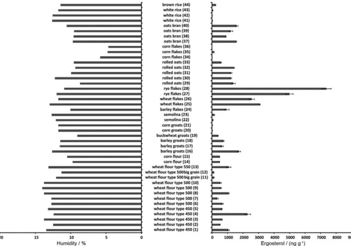
Cereal products are widely consumed all over the world. Different types of flours, groats and flakes are available to the consumers. They can be found in the basis of the food pyramid of many peoples. Therefore, safety of these products is of the highest importance. Toxins that are produced by different fungi are among the most important impurities found in the cereal products and, therefore, should be controlled. They can be determined directly or with the use of ergosterol as a marker of fungal presence. In this study the presence of ergosterol was analysed in different cereal products including 15 samples of flour, 13 samples of flakes, 8 samples of groats, 4 samples of oats and 4 samples of rice. The number of samples was selected in such a way that they reflect availability of these products to people. All accessible samples from different producers/suppliers were taken from the selected markets. ERG was present in all of the samples tested. However, the results presented in Figure 1 showed high diversity of ergosterol content in the particular samples. Generally, higher concentrations of ergosterol
(i.e., above 1000 ng g-1) were found in flakes than in flours
(except for sample No. 4) and groats (except for sample No.
16). The corn flakes are an exception here, however, as all three samples of this product contained modest amounts of ergosterol, which can be attributed to the specific procedure utilized to the preparation of these flakes in comparison to the other types of flakes.
The wheat flour samples (except of big grain wheat flours - samples No. 11 and No. 12) contained similar amounts of ERG independently of their type. The flours type 450 (samples No. 1 to 5) containing 0.45% mineral substances), with one exception (sample No. 4), contained ergosterol at similar concentrations like these found in the flours type 500 (samples No. 6 to 10) and 550
(sample No. 13), i.e., about 400 to 1100 ng g-1. There
were only two samples type 500 (samples No. 11 and 12) with considerably lower concentration of ergosterol (i.e.,
about 100 ng g-1). These samples are the special flours
with bigger grain size. Similar phenomenon can be found among the groats. The corn groats (samples No. 20 and 21) and semolina groats (samples No. 22 and 23) have bigger grains than the other groats (samples No. 16 to 19) and also very low ergosterol content. Apparently, bigger grain size and, thus, lower surface area make fungal growth difficult. Therefore, low ergosterol content was also found in the samples of rice having relatively big grain size.
The ergosterol content found in the samples was also compared with their humidity. The aim of this experiment
was to check the influence of humidity on the fungi growth and thus on amount of ergosterol found in the samples. It is widely known, that fungal growth is dependent on several factors including humidity. Therefore, it should be relatively low during storage of the cereal products. For flours air humidity should be lower than 70% and flour
humidity should not exceed 14%.50,51 The samples in this
study were subjected to the analysis and their humidity was determined. The results obtained (Figure 1) proved proper storage conditions – the humidity was lower than 14%. Then, the influence of humidity on the ergosterol content was checked. However, no correlation was found between these parameters.
Seven samples of flour (No. 2-5,7,14,15) and seven samples of flakes (No. 27-29,32,33,35,36) were chosen for mycotoxin content analysis and subjected to fungal strains isolation and identification. The samples from both groups were chosen to contain both higher and lower ERG content. Three mycotoxins characteristic to cereal products:
fumonisin B1 (FB1), zearalenone (ZON) and deoxynivalenol
(DON) (Figure S1 of the Supplementary Information) were determined in these samples. Moreover, fungi present in the selected products were also identified molecularly using rDNA-coding sequence analysis. The results obtained during the determination of mycotoxins are presented in Table 1. None of the determined mycotoxins was detected
Table 1. Content of ergosterol and three major mycotoxins and fungal species identified in the cereal products tested
Sample name (number) a Ergosterol / (ng g-1) ± SD
Fumonisin B1 / (ng g-1) ± SD
Zearalenone / (ng g-1) ± SD
Deoxynivalenol /
(ng g-1) ± SD Identified fungi strains Wheat flour type 450 (2) 529 ± 18 nd 4.1 ± 0.4 789 ± 31 Stemphylium sp.
Alternaria sp., Penicillium commune
Wheat flour type 450 (3) 644 ± 24 nd nd nd Alternaria alternata,
Penicillium chrysogenum
Wheat flour type 450 (4) 2295 ± 198 nd nd nd Penicillium caseifulvum,
Penicillium aurantiogriseum
Wheat flour type 450 (5) 657 ± 19 nd 2.6 ± 0.7 368 ± 24 Penicillium commune
Wheat flour type 500 (7) 349 ± 70 nd nd nd Aspergillus oryzae
Corn flour (14) 495 ± 1 87.5 ± 7.9 16.9 ± 1.1 1011 ± 88 Fusarium verticillioides, Fusarium subglutinans
Corn flour (15) 495 ± 19 112.0 ± 9.3 21.5 ± 3.7 1050 ± 98 Mucor sp.
Rye flakes (27) 4995 ± 238 nd nd nd Penicillium fuscoglaucum,
Microdochium nivale, Fusarium proliferatum
Rye flakes (28) 7380 ± 298 nd 3.7 ± 0.8 nd Fusarium oxysporum,
Penicillium aurantiogriseum
Rolled oats (29) 1381 ± 123 nd 8.1 ± 1.9 799 ± 40 Penicillium expansum
Rolled oats (32) 1421 ± 26 nd 12.6 ± 1.8 1224 ± 101 Fusarium oxysporum
Rolled oats (33) 590 ± 32 nd 4.5 ± 0.5 nd nd
Corn flakes (35) 150 ± 11 158.2 ± 12.5 14.3 ± 2.6 nd nd
Corn flakes (36) 17.5 ± 0.5 nd nd nd Cladosporium cladosporioides
anumbers of samples according to Figure 1; nd: not detected (detection limits for mycotoxins are: 0.1 ng g-1 for fumonisin B
in four samples out of 14 samples tested. Other samples contained from a few to more than a thousand nanograms of
mycotoxin (particularly for DON) per gram. No correlation
was found between ERG content and mycotoxin levels in samples tested (Figure 2).
Furthermore, fungi present in these samples were isolated and identified (Table 1). Twenty-two strains were isolated and purified out of 12 samples. The remaining two samples failed to display living fungi presence. Using molecular tools, all of the strains were identified in order to analyse their mycotoxigenic potential. Most
of the strains belonged to Penicillium and Fusarium
genera. Members of both groups are able to produce
a range of toxic metabolites.48,52 As it is obvious that
they produce different mycotoxins (if any) and that amount of mycotoxins produced by these fungi can be diversified, lack of correlation can be explained easily. However, only in the case of sample No. 14 (corn flour),
the presence of fumonisin B1 can be linked directly to
Fusarium verticillioides, a species known to be massive
fumonisin producer, identified in the sample. Interestingly, in all of the samples containing ZON and/or DON, none of the species able to produce these toxins (particularly
F. culmorum or F. graminearum) were detected. Similar
results have been obtained for pineapple53 and maize
(authors’ studies, unpublished), and, most likely, can be explained by low viability of those species in plant tissues. Thus, the presence of those toxins can be regarded as a “trace” of the producer species obviously infecting the plant in the past, but not at the stage of sampling. Another possible explanation is the transport of the mycotoxins within the plant to the tissues that normally do not contain pathogen, although, in the case of cereal grain products, this seems to be very unlikely. Another possibility is the competition of other fungal species colonizing the sample (e.g., the
fast-growing Mucor sp. or several Penicillium species), as it
was proven by ergosterol and fungi identification (Table 1), which actually are not able to produce the mycotoxins studied, but capable of synthesizing different metabolites. It
is likely to be the case of F. oxysporum identified in samples
No. 28 (rye flakes) and 32 (rolled oats) and F. proliferatum
detected in sample No. 27 (rye flakes), both species being able to produce beauvericin (BEA) in various plant
tissues.53-55 The analysis of BEA content in the respective
samples could provide the evidence to clarify this issue. Finally, the method used for mycotoxin quantification is much more sensitive than any of the methods available for fungal species identification in the respective plant material (perhaps with the exception of RealTime-PCR-based methods, which were, unfortunately, not used here).
The obtained results show that the determination of ergosterol for estimation of mycotoxin content in the cereal products is not possible when a wide variety of samples is used. Although high correlations exist in
different products,56 the use of ergosterol as a marker for
mycotoxin content must always be considered and validated individually.
Conclusions
The results obtained in this study show that the use of ergosterol for estimation of the mycotoxins amount present in the cereal products is sometimes of little value. Even the sample with ergosterol concentration
over 7000 ng g-1 contained only insignificant amounts of
mycotoxins. Low ergosterol level did not indicate low concentrations of mycotoxins, too. At least two different factors can play role here. It must be taken into account that mycotoxins are not produced by every fungi. Also, death of fungi leads to slow decrease of ergosterol content in the tested products while the amount of mycotoxins usually stays at constant level.
Supplementary information
Supplementary information is available free of charge at http://jbcs.sbq.org.br/ as PDF file.
Acknowledgements
This work was supported by the 03/31/DSPB/0276 grant from Polish Ministry of Science and Higher Education.
References
1. Shibamoto, T.; Bjeldanes, L. F.; Introduction to Food Toxicology,
2nd ed.; Academic Press Elsevier Inc.: San Diego, 2009. 2. Omaye, S. T.; Food and Nutritional Toxicology; CRC Press:
Boca Raton, 2004.
3. Tangni, E. K.; Pussemier, L.; Food Addit. Contam. 2006,23, 181.
4. Delgado, J. E.; Wolt, J. D.; Int. J. Environ. Res. Public Health
2011,8, 3179.
5. European Commission, Health & Consumer Protection Directorate-General, Scientific Committee on Food; Opinion of the Scientific Committee on Food on Fusarium Toxins, Part 3: Fumonisin B1 (FB1); EC: Brussels, 2000.
6. Pestka, J. J.; Anim. Feed Sci. Tech. 2007,137, 283.
7. Tardieu, D.; Bailly, J. D.; Benard, G.; Tran, T. S.; Guerre, P.;
Poult. Sci. 2004,83, 1287.
8. Zinedine, A.; Soriano, J. M.; Moltó, J. C.; Mañes, J.; Food Chem. Toxicol. 2007,45, 1.
9. Czerwiecki, L.; Czajkowska, D.; Witkowska-Gwiazdowska, A.;
Food Addit. Contam. 2002,19, 470.
10. Czerwiecki, L.; Czajkowska, D.; Witkowska-Gwiazdowska, A.;
Food Addit. Contam. 2002,19, 1051.
11. Pitt, J. I.; Taniwaki, M. H.; Cole, M. B.; Food Control 2013,
32, 205.
12. Pussemier, L.; Larondelle, Y.; Van Peteghen, C.; Huyghebaert, A.;
Food Control 2006, 17, 14.
13. Magan, N.; Hope, R.; Cairns, V.; Aldred, D.; Eur. J. Plant Pathol.
2003, 109, 723.
14. Berny, P. J. In Encyclopedia of Chromatography, 3rd ed.; Cazes, J., ed.; CRC Press: Boca Raton, 2011, vol. 2.
15. Cigić, I. K.; Prosen, H.; Int. J. Mol. Sci. 2009,10, 62. 16. Shephard, G. S.; Berthiller, F.; Burdaspal, P. A.; Crews, C.;
Jonker, M. A.; Krska, R.; Mac Donald, S.; Malone, R. J.; Maragos, C.; Sabino, M.; Solfrizzo, M.; Van Egmond, H. P.; Whitaker, T. B.; World Mycotoxin J. 2012,5, 3.
17. Kuzdraliński, A.; Solarska, E.; Mazurkiewicz, J.; Food Control 2013,33, 68.
18. Manova, R.; Mladenova, R.; Food Control 2009,20,362.
19. Nicholas, W.; Turner, N.; Subrahmanyam, S.; Piletsky, S. A.;
Anal. Chim. Acta 2009,632, 168.
20. Pappa, E.; Otta, K. H.; Záray, G.; Mincsovics, E.; Microchem. J.
2002,73, 39.
21. Kos, J.; Mastilović, J.; Hajnal, E. J.; Šarić, B.; Food Control
2013,34, 31.
22. Pleadin, J.; Vahčić, N.; Peši, N.; Ševelj, D.; Markov, K.; Frece, J.; Food Control 2013,32, 49.
23. Urusov, A. E.; Zherdev, A. V.; Dzantiev, B. B.; Appl. Biochem. Microbiol. 2010,46, 253.
24. Perkowski, J.; Buśko, M.; Stuper, K.; Kostecki, M.; Matysiak, A.; Szwajkowska-Michałek, L.; Biologia 2008,63,
542 (ISSN: 0006-3088).
25. Zhang, H.; Wolf-Hall, C.; Hall, C.; J. Agric. Food Chem. 2008,
56,11077.
26. Karaca, H.; Nas, S.; Food Addit. Contam. 2006, 23,502.
27. Saxena, J.; Munimbazi, C.; Bullerman, L. B.; Int. J. Food Microbiol. 2001,71, 29.
28. Mille-Lindblom, C.; Von Wachenfeldt, E.; Tranvik, L. J.;
J. Microbiol. Methods 2004, 59, 253.
29. Bermingham, S.; Maltby, L.; Cooke, R. C.; Mycol. Res. 1995,
99, 479.
30. Kadakal, C.; Nas, S.; Ekinci, R.; Food Chemistry 2005,90, 95.
31. Neuhof, T.; Koch, M.; Rasenko, T.; Nehls, I.; J. Agric. Food Chem. 2008,56, 7566.
32. Pietri, A.; Bertuzzi, T.; Pallaroni, L.; Piva, G.; Food Addit. Contam. 2004,21, 479.
33. Schwadorf. K.; Müller, H. M.; Arch. Anim. Nutr. 1990,40, 385.
35. Bakan, B.; Melcion, D.; Richard-Molard, D.; Cahagnier, B.;
J. Agric. Food Chem. 2002,50, 728.
36. Waśkiewicz, A.; Wit, M.; Goliński, P.; Chełkowski, J.; Warzecha, R.; Ochodzki, P.; Wakuliński, W.;.Food Addit. Contam.2012,29, 1752.
37. Tothill, I. E.; Harris, D.; Magan, N.; Mycol. Res. 1992,96, 965.
38. Gourama, H.; Bullerman, L. B.; LWT - Food Sci. Technol. 1995,
28, 185.
39. Waśkiewicz, A.; Irzykowska, L.; Bocianowski, J.; Karolewski, Z.; Weber, Z.; Goliński, P.; Food Addit. Contam.
2013,30, 1332.
40. Dong, Y.; Steffenson, B. Y.; Mirocha, C. J.; J. Agric. Food Chem.
2006, 54, 4121.
41. Doohan, F. M.; Parry, D. W.; Jenkinson, P.; Nicholson, P.; Plant Pathol. 1998,47, 197.
42. Stępień, Ł.; Gromadzka, K.; Chełkowski, J.; J. Appl. Genet.
2012,53, 227.
43. White, T. J.; Bruns, T.; Lee, S.; Taylor, J. In PCR Protocols: A Guide to Methods and Applications; Innis M. A.; Gelfand D. H.; Shinsky J. J.; White T. J., eds.; Academic Press: San Diego, 1990, ch. 3.
44. Stępień, Ł.; Koczyk, G.; Waśkiewicz, A.; Fungal Biol. 2011, 115, 112.
45. Horbik, D.; Łowińska-Kluge, A.; Górski, Z.; Stanisz, E.; Zgoła-Grześkowiak, A.; J. Braz. Chem. Soc. 2013,24, 1478. 46. The Commission of the European Communities; Commission
Regulation No. 2002/657/EC, implementing Council Directive
96/23/EC; Official Journal of the European Union: Luxembourg, 2002.
47. Waśkiewicz, A.; Irzykowska, L.; Drzewiecka, K.; Bocianowski, J.; Dobosz, B.; Weber, Z.; Karolewski, Z.; Krzyminiewski, R.; Goliński, P.; Cent. Eur. J. Biol. 2013,8, 1065.
48. Wiśniewska, H.; Stępień, Ł.; Waśkiewicz, A.; Beszterda, M.; Góral T.; Belter, J.; Cent. Eur. J. Biol. 2014,9, 163.
49. Chełkowski, J.; Gromadzka, K.; Stępień, Ł.; Lenc, L.; Kostecki, M.; Berthiller F.; World Mycotox. J.2012,5,133.
50. Belitz H. D.; Grosch, W.; Schieberle, P.; Food Chemistry,
4thed.;Springer: Berlin, 2009.
51. Robertson, G.L. Food Packaging. Principles and Practice,
3rded.;CRC Press: Boca Raton, 2013.
52. Wawrzyniak J.; Waśkiewicz A.; Food Addit. Contam. 2014,31, 138.
53. Stępień, Ł.; Koczyk, G.; Waśkiewicz, A.; J. Appl. Genet. 2013,
54, 367.
54. Stępień, Ł.; Waśkiewicz, A.; Toxins 2013,5, 537.
55. Waśkiewicz, A.; Stępień, Ł.; Arh. Hig. Rada. Toksikol. 2012,
63, 437 (ISSN 0004-1254).
56. Waśkiewicz, A.; Beszterda, M.; Bocianowski, J.; Goliński, P.;
Food Microbiol. 2013,36, 426.