© 2013 Sociedade Brasileira de Hemodinâmica e Cardiologia Intervencionista. Published by Elsevier Editora Ltda. All rights reserved.
Feasibility, Safety, and Efficacy of Percutaneous
Atrial Septal Defect Closure in Infants
Marcelo Silva Ribeiro
1, Fabricio Leite Pereira
2, Wanda Teixeira do Nascimento
3,
Rodrigo Nieckel da Costa
4, Daniela Lago Kreuzig
5, Simone Rolim Fernandes Fontes Pedra
6,
Patricia Figueiredo Elias
7, Cristiane Pessoti
8, Ieda Bosisio Jatene
9, Maria Aparecida Paula Silva
10,
Ricardo Fonseca Martins
11, Maria Virginia Tavares Santana
12, Valmir Fernandes Fontes
13,
Carlos Augusto Cardoso Pedra
14RESUMO
Factibilidade, Segurança e Eficácia do Fechamento Percutâneo da Comunicação
Interatrial em Crianças Pequenas
Introdução: A experiência com o fechamento percutâneo
da comunicação interatrial (CIA) em crianças pequenas é limitada. Avaliamos a factibilidade, a segurança e a eicácia desse procedimento em crianças com peso < 20 kg. Métodos: Estudo descritivo observacional de uma coorte de crianças < 20 kg submetidas a tratamento percutâneo. Pacientes com dilatação ventricular direita e sintomas evidentes foram in cluídos. Implantamos próteses aprovadas pela ANVISA, sob monitorização ecocardiográica transesofágica. Os pacientes foram avaliados 1 mês, 3 meses, 6 meses e 12 meses após.
Resultados: Entre outubro de 1997 e maio de 2012, 80 pacien
tes foram tratados. As medianas de idade e peso foram de 4 anos (112) e 13,5 kg (520), respectivamente, 20 pacientes apresentavam alguma síndrome genética (25%) e 4 pacien tes (5%) apresentavam CIA adicional. Somente um paciente necessitou duas próteses. Dois pacientes tinham defeitos
ABSTRACT
Background: The experience with percutaneous closure of
atrial septal defect (ASD) in infants is limited. We sought to determine the feasibility, safety and eficacy of this procedure in children weighing < 20 kg. Methods: Observational study of a cohort of children weighing < 20 kg undergoing percutane ous closure. Patients with right ventricular enlargement and evident symptoms were included. ANVISA approved devices were implanted under transesophageal echocardiography moni toring. Patients were evaluated 1, 3, 6 and 12 months after the procedure. Results: Eighty patients were treated between October 1997 and May 2012. Median age and weight were 4 years (112) and 13.5 kg (520), respectively, 20 patients had a genetic syndrome (25%) and 4 patients (5%) had an additional ASD. Only one patient required 2 devices. Two patients had associated defects that were treated in the same procedure (pulmonary valve stenosis and arteriovenous istula). One patient developed total atrioventricular block during device implantation, solved spontaneously 36 hours after de vice removal, with no need for pacemaker implantation. This patient was successfully treated percutaneously 6 months later
Original Article
1 Physician of the Department of Interventional Cardiology for Conge
nital Heart Defects of the Instituto Dante Pazzanese de Cardiologia. São Paulo, SP, Brazil.
2 Physician. Resident at the Interventional Cardiology for Congenital
Heart Defects Service of the Instituto Dante Pazzanese de Cardiologia. São Paulo, SP, Brazil.
3 Physician. Resident at the Interventional Cardiology for Congenital
Heart Defects Service of the Instituto Dante Pazzanese de Cardiologia. São Paulo, SP, Brazil.
4 Ph.D. student. Interventional Cardiologist in Congenital Heart Defects
of the Instituto Dante Pazzanese de Cardiologia. São Paulo, SP, Brazil.
5 Physician at the Department of Echocardiography in Congenital Heart
Defects and Structural of the Instituto Dante Pazzanese de Cardiologia. São Paulo, SP, Brazil.
6 Ph.D. Director of the Department of Echocardiography in Congenital
Heart Defects and Structural of the Hospital do Coração da Associação Sanatório Sírio. São Paulo, SP, Brazil.
7 Physician at the Department of Pediatric Cardiology of the Hospital
do Coração da Associação Sanatório Sírio. São Paulo, SP, Brazil.
8 Physician at the Department of Pediatric Cardiology of the Coração
da Associação Sanatório Sírio. São Paulo, SP, Brazil.
9 Ph.D. Director of the Pediatric Cardiology Department of the Coração
da Associação Sanatório Sírio. São Paulo, SP, Brazil.
10 Physician at the Department of Pediatric Cardiology of the Instituto
Dante Pazzanese de Cardiologia. São Paulo, SP, Brazil.
11 Physician at the Department of Pediatric Cardiology of the Instituto
Dante Pazzanese de Cardiologia. São Paulo, SP, Brazil.
12 Ph.D. Director of the Pediatric Cardiology Department of the Instituto
Dante Pazzanese de Cardiologia. São Paulo, SP, Brazil.
13 Ph.D. Director of the Interventional Cardiology for Congenital Heart
Defects Service of the Hospital do Coração da Associação Sanatório Sírio. São Paulo, SP, Brazil.
14 Ph.D. Director of the Interventional Cardiology for Congenital Heart
Defects Service of the Instituto Dante Pazzanese de Cardiologia. São Paulo, SP, Brazil.
Correspondence to: Carlos A. C. Pedra. Av. Dr. Dante Pazzanese, 500 – 14o andar – São Paulo, SP, Brazil – CEP 04012180
Email: [email protected]
without complications. Seventynine patients were discharged within 24 hours after the procedure. A mild residual shunt (12 mm) was observed in 5% of the cases before discharge. There was no residual shunt 6 months after the procedure. There were no complications in the late followup.
Conclu-sions: Percutaneous ASD closure in selected symptomatic
infants is a feasible, safe and effective alternative and should be the irst option therapy.
DESCRIPTORS: Heart septal defects, atrial. Prostheses and
implants. Child.
T
he irst description of interatrial septal defects dates back to 1875 and was performed by Rokitansky, but their physiopathology and clinical picture started to be revealed only after 1941.1 Ostium secundumatrial septal defect (ASD) has the fourth or ifth highest incidence of congenital heart diseases, corresponding to approximately 5% to 10% of cases.24
With the gradual decrease in pulmonary vascular resistance, and improved hypertrophy and right ven tricular compliance within the irst year of life, ASD begins to have hemodynamic consequences with right chamber volume overload. However, most of these patients remain asymptomatic during the irst years of life. The defect is usually electively corrected before the child reaches school age, at around 5 years old. Occasionally, patients younger than 5 years with ASD develop isolated symptoms generated by pulmonary hyperlow, including recurrent respiratory infections, bronchospasm, congestive heart failure, or failure to thrive.5,6 This scenario is aggravated when the ASD is
associated with prematurity, chronic pulmonary diseases (asthma, bronchodysplasia), genetic syndromes, or other systemic diseases (renal and liver failure, among others). These patients require early treatment.
The surgical approach was considered the method of choice for ASD management for over four decades. Although it has very good results,7,8 it requires cardio
pulmonary bypass, hemotherapy, and longer hospital stay, in addition to some postoperative morbidity, such as pain, infections, pericardial effusion, arrhythmias, and sternotomy scars.911 The irst percutaneous ASD closure
was described in 1976.12 With the technical advances
and surgeons’ greater experience, percutaneous treatment has become the modality of choice for the management of most patients with ASD.1319 Surgery is reserved for
cases with unfavorable anatomy for the percutaneous approach, associated cardiac anomalies, or other types
associados, os quais foram tratados no mesmo procedimento (estenose pulmonar valvar e fístula arteriovenosa). Um pa ciente desenvolveu bloqueio atrioventricular total durante o implante da prótese, resolvido espontaneamente 36 horas após a remoção da prótese, sem necessidade de implante de marcapasso. Esse paciente foi tratado percutaneamente 6 meses após com sucesso, sem complicações. Setenta e nove pacientes receberam alta hospitalar em até 24 horas após o procedimento. Fluxo residual discreto (12 mm) foi observado em 5% dos casos antes da alta. Após 6 meses de seguimento, não foi detectado luxo residual. Não houve complicações tardias no seguimento. Conclusões: O fechamento percutâneo da CIA em crianças pequenas selecionadas e sintomáticas é uma alternativa terapêutica factível, segura e eicaz, devendo ser a primeira opção para seu tratamento.
DESCRITORES: Comunicação interatrial. Próteses e implantes.
Criança.
of ASD (e.g. ostium primum ASD, sinus venosus, and coronary sinus).19 Although the percutaneous treatment
is well established for older children, adolescents, and adults, there are few reports on the use of this method in small children.2024
This article describes an experience with the per cutaneous treatment of ASD in children weighing < 20
kg, assessing its feasibility, eficacy, and safety.
METHODS
Study design
Observational, longitudinal descriptive study of a cohort of children weighing < 20 kg submitted to
percutaneous closure of ostium secundum ASD in two cardiology referral centers in Brazil. Data collection was performed retrospectively, through medical record analysis. Demographic, clinical, echocardiographic, and hemodynamic data were collected. Parents or guard ians of patients were informed of the procedure and signed an informed consent approved by the ethics and research committee of both institutions.
Inclusion criteria
Children weighing < 20 kg with clinical and echo
cardiographic diagnosis of ostium secundum ASD with hemodynamic consequences, characterized by right ventricular volume overload associated with one or more of the following diagnoses:
– congestive heart failure;
– recurrent respiratory infections (six or more epi sodes in 12 months);
– failure to thrive;
– genetic syndromes (e.g. Down syndrome, etc.); – severe systemic disease (e.g. renal or liver failu re, etc.).
Exclusion criteria
Exclusion criteria included weight < 5 kg, ASD
classiied as nonostium secundum, ixed pulmonary artery hypertension, presence of associated intracardiac defects with surgical indication, and unfavorable anatomy for percutaneous closure of the defect.1925 The absolute
diameter of the ASD was not considered an exclusion factor for the procedure, nor was a deicient anterior superior border, provided that the contralateral border was adequate.25 Patients with large ASDs that required
devices that were too large for the size of the heart, with risk of interference with atrioventricular valve func tion and pulmonary venous or coronary sinus drainage were excluded. Patients with more than one deicient border (usually contralateral) around the defect, usu ally accompanied by thin and distensible atrial septum, were also excluded. Patients with two adjacent ASDs that could be occluded with only one prosthesis, with two large and distant ASDs, but with the possibility of occlusion with two devices; with two distant ASDs, but in which the smallest defect was < 34 mm (therefore,
with no clinical signiicance); with cribriform ASD with multiple small defects that could be occluded with only one prosthesis; and with simple additional heart defects that could be treated by catheterisation, such as patent ductus arteriosus (PDA), pulmonary valve stenosis, or vascular malformation were not excluded.
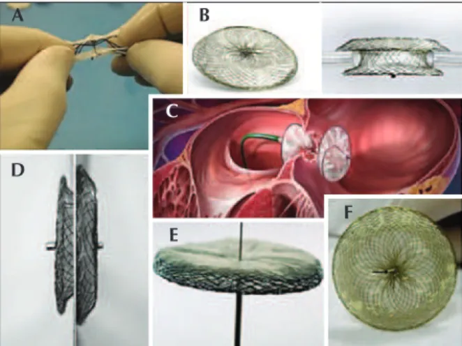
Prostheses used in the trial
The prostheses used in this trial were approved by the Brazilian Health Surveillance Agency (Agência Nacional de Vigilância Sanitária – ANVISA), with well established safety and eficacy in previous experiments in Brazil, the United States, and Europe.17,18,2628 These
were: Amplatzer
Atrial Septal Occluder and Amplatzer
Cribriform Occluder (St. Jude Medical Inc. – St. Paul, United States), Gore Helex
Atrial Septal Occluder (W.L. Gore & Associate – Newark, United States), Lifetech Cera
ASD Occluder (Lifetech Scientiic Corporation – Shenzhen, China), Occlutech Figulla
(Occlutech AB – Helsingborg, Sweden), Cardia
Atriassept (CardioLogic Ltd.), and NitOcclud ASDR
(PFM Medical Ag – Koln, Germany) (Figure 1).
The spatial conformation of these prostheses consists of two discs of varying sizes with a central connecting waist.17,18,27,28 With the exception of the
Helex
prosthesis, all others use nitinol – an alloy of nickel and titanium – as the main component of their selfexpanding mesh. Occlusion is performed by the illing of the septal oriice by the selfcentring waist, which exerts radial force on the ASD edges, as well
as physical barrier to blood low exerted by the discs positioned in each of the atria after implantation.
Nitinol is also present in the Helex
prosthesis, but in the form of a helical strut that provides support to a dense mesh of polytetraluoroethylene (PTFE), which has occluding power after it is fully exposed and po sitioned.26 In this prosthesis, the waist only connects
the two disks and is not selfcentring.
Procedure
The procedure was performed under general anaes thesia, controlled by luoroscopy and transesophageal echocardiography (TEE) using a twodimensional pae diatric probe suitable for children weighing < 20 kg
with a Philips (Healthcare, DA – Best, the Netherlands) or General Electric (GE – United States) equipment. Femoral venous access was used in all patients, except in two, in whom the transhepatic route was necessary due to the absence of the hepatic portion of the inferior vena cava, caused by left atrial isomerism. Heparin at a dose of 100 IU/kg was administered through the lateral route of the venous sheath, immediately after its insertion. The right cardiac chambers were catheter ized and measures of pulmonary pressure were directly acquired. Calculations of pulmonary/systemic low and resistance made using Fick’s method were restricted to cases with severe pulmonary hypertension.29
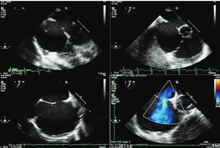
ASD measures assessed at baseline were evalu ated, as well as the appearance of its borders, through TEE25 (Figure 2). The total atrial septum length in the
anteriorposterior and superiorinferior views was deter mined using the fourchamber projection and the short axis at the level of the aortic valve, respectively. This measurement was performed to assess the adequacy of prosthesis size selected for the size of the patient’s heart. The stretched ASD diameter measurement using
Figure 1 – Prostheses used: A) Cardia
Atriassept (CardioLogic Ltd.) B) Figulla
ASD (Occlutech International AB), C) Helex
ASD (Gore Medical Inc.), D) Amplatzer
ASO (St. Jude Medical, Inc.), E) Nit Occlud
ASD – R (PFM Medical Ag.), and F) Cera
(Lifetech Scientiic).
A B
D
E
a lowpressure NuMed balloon (Hopkinton, United States) or AGA balloon (Golden Valley, United States), with the stoppedlow technique30 was performed when
there were thin, lexible, or aneurysmal borders or in the presence of additional ASD in addition to the
main defect (Figure 3). Balloon measurement was also performed in all cases in which the Helex
prosthesis was chosen; the selected prosthesis was twofold larger than the measured value. Selfcentring prostheses from 0 mm to 2 mm larger than the central waist diameter
Figure 2 – Twodimensional echocardiographic assessment of atrial septal defect, showing its favorable aspect for percutaneous treatment. In the lower panel, to the right, analysis of low through the defect (left atriumright atrium) at the color low mapping.
of the balloon were chosen. In patients with single ASD with irm borders, the prosthesis of choice for implantation had a waist diameter that was 20% to 30% greater than the largest diameter of the defect at baseline. The selfcentring prostheses with central waist (Amplatzer
ASO, Figulla
ASD, Nit Occlud
ASD and Cera
) were used in most cases (Figure 4). The Helex
, Figulla
PFO, or Amplatzer
Cribriform prostheses, not selfcentring, were selected in the presence multi fenestrated atrial septum, and were implanted through the most central oriice. The Helex
prosthesis was also occasionally employed for defects < 1012 mm, both
single and central. When there were two ASDs, self centring prostheses were used, implanted in the larger defect (usually anteriorsuperior), with closure of the additional ASD (usually posteriorinferior) resulting from the coverage of the disks around the central waist. Two prostheses were necessary in only one case, one self centring and the other cribriform, in order to occlude a more anterior ASD and a cribriform posteriorinferior septum (Figure 5).
In some patients, especially those whose weight was
< 15 kg and who had smaller dimensions of the left
atrium (LA), in order to prevent extrinsic LA compres sion, the opening of the left disc was performed after the TEE probe was pulled. To avoid possible prolapse of left disc through the anterior portion (retroaortic) of the atrial septum, some technical manoeuvres were employed, including clockwise rotation of the sheath (which is almost in parallel with the column in anteriorposterior projection at the fluoroscopy) and opening of the left disk in the left or right superior pulmonary vein.
Prophylactic antibiotic therapy with secondgeneration cephalosporin was administered during prosthesis im plantation and maintained for 24 hours. Acetylsalicylic acid was introduced ive days before the procedure at a dose of 3 mg/kg/day to 5 mg/kg/day and maintained
for six months. Patients were hospitalized for at least 24 hours for clinical observation, and were discharged from the hospital after undergoing a transthoracic echo cardiography (TTE) and electrocardiography (ECG), and after ruling out acute complications of the treatment. The presence of residual shunt was determined through the analysis of the Doppler color low through the atrial septum. Any immediate lowspeed residual shunt through the prosthesis mesh was disregarded. Residual shunt was considered as adjacent to the prosthesis border and classiied as minimum (< 1 mm in diameter), small
(12 mm), medium (24 mm), and large (> 4 mm).31
Follow-up
Outpatient followup was performed at one month, three months, six months, and 12 months after the percutaneous treatment, accompanied by chest radio graphy, ECG, and TTE. After 12 months, followup was performed annually. Prophylaxis for infective endocar ditis was recommended for up to six months after the procedure in cases with total defect occlusion (Figure 6), and indeinitely if any residual shunt was noticed through the atrial septum after six months.
Definitions and studied variables for outcome assessment
The realisation of the procedure was deined as successful implantation with prosthesis release and ap propriate positioning in the atrial septum. Safety was assessed by complication rates.31 Higher incidence of
death, brain embolism, cardiac perforation with tampon ade, endocarditis, need for reintervention (surgical or percutaneous), arrhythmias requiring permanent cardiac pacemaker or antiarrhythmic drugs for prolonged periods, pleural or pericardial effusion requiring surgical drain age, and need for surgery due to device embolisation were considered complications.
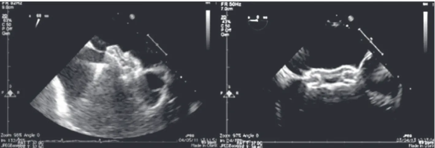
Figure 5 – Fluoroscopy demonstrating the simultaneous positioning of two Amplatzer prostheses (ASO and Cribriform) in the interatrial septum. At the top, both prostheses positioned and still connected to the delivery system; at the bottom, after disconnecting the delivery cable.
Figure 6 – Transthoracic echocardiography showing good positioning of the two prostheses implanted in the interatrial septum, with no residual shunt six months after percutaneous treatment, as shown by the color low mapping. RA, right atrium, LA, left atrium, RV, right ventricle; LV, left ventricle.
RA RA
LA
RV RV
LV LV
Minor complications included prosthesis embolisa tion with percutaneous removal, cardiac arrhythmias with no need for prolonged treatment, hematoma at the venous access site, retroperitoneal hematoma with no need for intervention, anaemia requiring blood products, noncardiac infection, and several respiratory conditions (pulmonary edema, atelectasis, laryngitis). Eficacy was assessed by the total occlusion rates or by the presence of residual shunt with no hemodynamics consequences (with normal right ventricular dimensions) during followup.
Statistical Analysis
Statistical analysis was performed using the Sigmastat software, version 2011. The data are shown as absolute values and frequencies, means and standard deviations, or medians and ranges, according to sample distribution.
RESULTS
Between October of 1997 and May of 2012, 80 patients (49 females) with a median age of 4 years (1 12 years) and median weight of 13.6 kg (520 kg) were submitted to percutaneous ASD closure in two Brazil ian cardiology referral centers. Of these 80 patients, 18 (22.5%) were younger than 2 years and 9 (11%) weighed < 10 kg. Twenty patients (25%) had genetic
syndromes (Down syndrome, HoltOram syndrome, and other unknown syndromes) or multiple malforma tions (gastrointestinal, renal, neurological, bone). Ten (12.5%) had a history of prematurity. During routine catheterisation, none of the patients showed severe pulmonary artery hypertension, and all patients had mean pressure < 30 mmHg in the pulmonary artery.
Seventysix patients (95%) had a single ASD, with a mean diameter of 12 ± 4 mm at the TEE. Four patients
had ASD with one or more additional oriices (5%). The balloonstretched diameter assessment was performed in 40 patients (50%), with a mean value of 16 ± 5 mm.
The mean diameter of the chosen prosthesis was 16.9 ±
5 mm and the mean proile of the long delivery sheath was 8 ± 1.5 F. The mean duration of procedures was
80 ± 15 minutes. The distribution of prostheses used
in the procedures is shown in Table 1.
Two patients had associated heart defects, treated percutaneously in the same procedure. One had mo derate pulmonary valve stenosis and was submitted to balloon pulmonary valvuloplasty prior to ASD occlusion. The other patient had pulmonary arteriovenous istula presented in right lung base that resulted in signiicant cyanosis, which was successfully occluded with the Amplatzer® Duct Occluder II (Figure 7).
Technical success was achieved in all 80 patients (100%). However, a patient submitted to Helex
prosthe sis implantation had total atrioventricular block (TAVB) during prosthesis positioning, with no improvement after its full opening and locking into the inal position in
the atrial septum. The prosthesis was removed according to the usual technique. Since the patient had adequate junctional escape rhythm with no signs of low output, he was taken to the intensive care unit for monitoring, receiving intravenous corticosteroids, with no need for pacemaker stimulation during evolution.
The TAVB was solved in approximately 36 hours and the patient was discharged within 72 hours af ter conirmation of sustained sinus rhythm in Holter monitoring. This patient returned after – six months, was successfully submitted to ASD occlusion with Amplatzer
without complications. Only one patient with multiple distant ASDs required a second prosthesis, both successfully implanted. In the other patients with multiple ASDs, additional defects were occluded after device implantation in the main ASD. Except for one patient who had TAVB, and another with a congenital syndrome that awaited discharge to return to home care, all others were discharged within 24 hours after the procedure. There were no hemodynamic, vascular, or pulmonary complications.
Slight/mild residual shunt (12 mm) was observed in four patients (5%) at the TTE prior to hospital discharge. The outpatient followup comprised 95% of the patients, lasting 4 ± 3 years. The rate of ASD occlusion in the
irst clinical evaluation at one month and six months after the procedure was 100%. The echocardiography showed no signs of interference with atrioventricular valve function or blood low in the right superior pul monary vein or coronary sinus. There were no clinical complications detected during followup.
DISCUSSION
This prospective observational longitudinal study demonstrated that in children weighing < 20 kg with
ASD with hemodynamic consequences and signiicant clinical impact, percutaneous closure is feasible, safe, and effective.
TABLE
Distribution of prostheses used
Prosthesis Number %
Amplatzer
Septal Occluder 40 50
Lifetech Cera
ASD 13 16.25
Helex
ASD 10 12.5
Cardia Atriassept
10 12.5
Occlutech Figulla
ASD 5 6.25
Nit Occlud
ASDR 1 1.25
Amplatzer
Cribriform Occluder
Clinical indications for early percutaneous closure
The decision to treat an ASD in children weigh ing < 20 kg was reserved for those patients in whom
pulmonary blood low resulting from the defect brought signiicant clinical consequences, characterized by congestive heart failure, recurrent respiratory conditions (six or more episodes in 12 months) and low weight gain. In addition, patients who beneit the most from early percutaneous approach are those with chronic pulmonary disease, several genetic syndromes, and severe systemic diseases.
In this clinical practice, when these conditions are not present, percutaneous treatment of ASD with hemo dynamic consequences (with enlarged right ventricle) is usually scheduled to occur when the child is aged between 4 and 5 years.
Procedure feasibility in young children
Until recently, there were some dogmas and uncertainties regarding the feasibility of performing percutaneous closure in small children. The catheter proile, the size of the devices, and the possibility of intracardiac trauma, among others, were reasons to justify the impossibility of percutaneous treatment. In this study, although such problems were not observed, considerable care was taken regarding the procedure. The femoral vein, although of a smaller calibre in young children, was not a limiting factor to the procedure in this series. The sheaths were advanced without tech nical dificulties and without vascular complications in all patients. The mean proile of 8F sheaths was suitable for the median weight of the sample (13 kg). The procedure was feasible even in a patient weigh ing 5 kg, with a genetic syndrome, in whom a long
7F sheath was used to implant a 9mm Amplatzer®
prosthesis. Even in patients who do not have normal femoral venous access, the transhepatic route, used in two patients in this sample, allows the procedure to be performed. In these two cases, long 9F or 10F sheaths (the largest sheaths used in the present study) were employed uneventfully.
The smaller size of the left atrium can cause some dificulty to open the left disk of the commercially avail able prostheses, and thus should be performed more carefully and slowly. Pulling the TEE probe helped to prevent a possible extrinsic compression, which would further reduce the size of the left atrium. In patients with an appropriate window (common in young children), another possibility to properly guide the procedure would be the use of TTE.32 It was chosen not to use
intracardiac echocardiography in this population due to the need for a second venous access. In this experience, the manoeuvres used to avoid disc prolapse on the left through the anterior portion (retroaortic) of the septum were effective. However, greater care when performing these manoeuvres was required due to the increased fragility of intracardiac structures. Despite the small number of patients with multiple ASDs in this study, the ASDs were occluded in all ive patients with one or even two prostheses.
Percutaneous treatment of associated heart diseases using the same anaesthesia time of the ASD occlusion added no technical dificulty, nor did it inluence the success of the procedure. The only additional care that was planned for the catheterisation was treating the associated disease irst, thus preventing additional intra cardiac manipulations after the prosthesis implantation and release in the atrial septum, which could theoreti cally cause prosthesis displacement or embolisation. Thus, both the balloon pulmonary valvuloplasty and
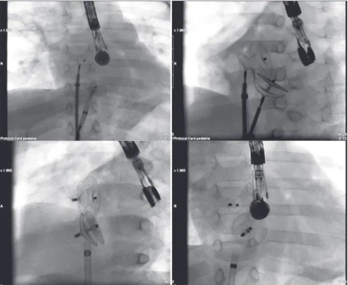
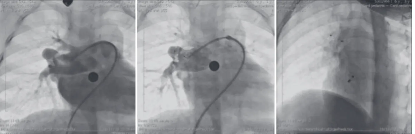
Figure 7 – To the left, coronary angiography demonstrating the presence of large pulmonary arteriovenous istula originating from the middle lobar branch of the right pulmonary artery. In the center, control coronary angiography showing good prosthesis positioning, with istula occlusion. To the right, chest luoroscopy in the left oblique projection with cranial angulation showing the spatial association between the implanted prosthesis in the istula and in the atrial septal defect (Amplatzer
the occlusion of the pulmonary atrioventricular istula were performed before the ASD occlusion.
Taking into consideration all the aforementioned aspects, technical success was achieved in 100% of cases. The only unsuccessful case was caused by an electrical complication requiring device removal. This high success rate reflects the group’s experience with the techniques and prostheses used and the manage ment of young children with severe congenital heart defects.
Although weight < 5 kg was considered an exclu
sion criterion in this study, the percutaneous closure of ASD in preterm neonates weighing < 23 kg with
bronchodysplasia and on mechanical ventilation has been performed in some centers, resulting in early extubation and optimal outcomes (Dr. John Cheatham, personal communication). This will probably be the next challenge in future experiments.
Safety in this population
In this series, percutaneous ASD closure in young children was shown to be very safe. The only severe complication was the development of TAVB in one patient. This TAVB probably resulted from the lack of the selfcentring mechanism in the device used (Helex®) causing the device to fall into the coronary
sinus, temporarily injuring the region of the atrioven tricular node, which required device removal and a subsequent new procedure. The implantation of a selfcentring prosthesis a few months later minimized this lower migration and prevented the occurrence of TAVB. The small size of the atrial cavities, typical of a small child, may also have influenced the emergence of this complication during the attempted implanta tion. The transitory nature of TAVB and the successful implantation of a new prosthesis – the Amplatzer
– confirm the hypothesis that there was no permanent injury to atrioventricular node or electrical conduction bundles. In one study, percutaneous closure of ASD in young children resulted in rates of approximately 4% of TAVB.33 However, large prostheses were used
in that experiment.
This observation, together with those obtained from this and other trials, show the importance of proper patient selection for the percutaneous procedure, espe cially regarding the stringent selection of the size and type of device for the underlying anatomy.34
The use of balloons to assess the stretched ASD diameter did not involve complications resulting from the technique in this selected group of young children, demonstrating safety when well indicated. As for the cases in which it was decided not to use them (50%), there were no complications secondary to prosthesis under – or overestimation. The use of the balloon was restricted to the ASDs with features that complicated
the choice of prosthesis (e.g. thin and redundant bor ders, etc.) or when the option was the use of prosthesis without selfcentring mechanism (e.g., Helex
). Previ ously published works corroborate the nonmandatory use of this resource when choosing the prosthesis, with no impairment regarding the safety or the eficacy of the procedure in selected cases.30
One of the authors’ concerns was to avoid, as much as possible, the overestimation of the prosthesis chosen for implantation, which could interfere with the atrioventricular valve function or the blood low in the left superior pulmonary vein or coronary sinus. None of the patients had such complications. The strategy of measuring the total length of the interatrial septum at the echocardiography (TEE and TTE) and avoiding pros thetic discs greater than those measures was probably effective in preventing these complications. Therefore, some authors recommend performing the angiography with the catheter placed in the left superior pulmonary vein in hepatoclavicular projection to obtain the inter atrial septum length measurement (Dr. Ziyad M. Hijazi, personal communication).
In this study, no vascular complications were observed due to the adequacy of the profile of the prostheses used (mean 8F) to the size of patients (median weight of 13 kg). In this sense, the use of the Amplatzer
in this population has been preferred, due to the lower profile of their implant sheaths when compared to similar prostheses, such as the Cera
or Figulla
. The launch of the Figulla Flex
II is expected, which will reduce the calibre of the sheath by 2F to 3F, allowing for a safer use of this device in this population.
Efficacy
The occlusion rate of 100% observed from one month to six months in all followed patients (95%) is consistent with previous experiences reported in the literature, and demonstrates the high eficacy of this procedure. The progressive endothelialisation of the pros theses explains the spontaneous occlusion of immediate residual shunt.35 Although this aspect was not assessed
in this study, the concept of clinical eficacy must not be forgotten. Previous studies have demonstrated that young patients with ASD and who are highly symptom atic greatly beneit from the percutaneous procedure, with fast weight gain and signiicant improvement of recurrent pulmonary conditions.23
Study limitations
CONCLUSIONS
Percutaneous closure of ASD in very symptomatic selected young children is a feasible, safe, and effective therapeutic alternative. As it is less invasive than surgery, especially considering that this subgroup of patients is at highsurgical risk, this therapeutic modality should be regarded as the option of choice in the management of these patients, and should be offered early.
CONFLICTS OF INTEREST
Carlos Pedra is a consultant for the following re presentation and manufacturer companies: Bioassist and St. Jude (Amplatzer
), Boynton and Lifetech (Cera
), CMS (PFM), and TecMedic (Figulla
). The other authors declare to have no conlicts of interest.
CLARIFICATIONS
After completion of data collection and drafting of this manuscript, the percutaneous closure of ASD was performed in seven more patients weighing < 20 kg
between May of 2012 and May of 2013. Although similar results were obtained, a severe complication was observed in a patient undergoing occlusion by transhepatic route, due to the impossibility of peripheral venous access due to multiple previous hospitalisations. This patient with genetic syndrome, weighing 7 kg, with severe gastroesophageal relux and chronic pulmonary disease, developed right hemothorax secondary to liver puncture. After drainage, there was satisfactory progress without additional complications. This complication, which was not related to the occlusion procedure itself, but rather to the process of obtaining an access route, indicates the frailty of some of these patients and does not invalidate the aforementioned observations.
REFERENCES
1. Bedford DE, Papp C, Parkinson J. Atrial septal defect. Br Heart J. 1941;3(1):3768.
2. Fyler DC, Bucley LP, Hellenbrand WE. Report of the New England Regional Infant Cardiac Program. Pediatrics. 1980; 65(2 Pt 2):375461.
3. Dickinson DF, Arnold R, Wilkinson JL. Congenital heart disease among 160,480 liveborn children in Liverpool 1960 to 1969: implications of surgical treatment. Br Heart J. 1981;46(1): 5562.
4. Carlgren LE. The incidence of congenital heart disease in children born in Gothenburg 19411950. Br Heart J. 1959;21(1):4050. 5. Bull C, Deanield J, de Leval M, Stark J, Taylor JF, Macartney
FJ. Correction of isolated secundum atrial septal defect in infancy. Arch Dis Child. 1981;56(10):7846.
6. Dimich I, Steinield L, Park SC. Symptomatic atrial septal defect in infants. Am Heart J. 1973;85(5):6014.
7. Horvath KA, Burke RP, Collins JJ Jr, Cohn LH. Surgical treat ment of atrial septal defect: early and longterm results. J Am Coll Cardiol. 1992;20(5):11569.
8. Kirklin JW, BarrattBoyes BG. Cardiac surgery, 2nd ed. New York: Churchill Livingstone; 1993. p. 60944.
9. Galal MO, Wobst A, Halees Z, Hatle L, Schmaltz AA, Khougeer F, et al. Perioperative complications following surgical closure of atrial septal defect type II in 232 patients: a baseline study. Eur Heart J. 1994;15(10):13814.
10. Butera G, Carminati M, Chessa M, Youssef R, Drago M, Giamberti A, et al. Percutaneous versus surgical closure of secundum atrial septal defect: Comparison of early results and complications. Am Heart J. 2006;151(1):22834. 11. Hopkins R, Bert A, Buchholz B. Surgical patch closure of
atrial septal defects. Ann Thorac Surg. 2004;77(6):214450. 12. King TD, Mills NL. Secundum atrial septal defects: nonoperative
closure during cardiac catheterization. JAMA. 1976;235(23): 25069.
13. Rome JJ, Keane JF, Perry SB, Spevak PJ, Lock JE. Doubleumbrella closure of atrial septal defects: initial clinical applications. Circulation. 1990;82(3):7518.
14. Justo RN, Nykanen DG, Boutin C, McCrindle BW, Freedom RM, Benson LN. Clinical impact of transcatheter closure of secundum atrial septal defects with the double umbrella device. Am J Cardiol. 1996;77(10):8892.
15. Masura J, Gavora P, Formanek A, Hijazi ZM. Transcatheter closure of secundum atrial septal defects using the new self centering Amplatzer septal occluder: initial human experience. Cathet Cardiovasc Diagn. 1997;42(4):38893.
16. Thanopoulos BD, Laskari CV, Tsaousis GS, Zarayelyan A, Vekiou A, Papadopoulos GS. Closure of atrial septal defects with the Amplatzer occlusion device: Preliminary results. J Am Coll Cardiol. 1998;31(5):11106.
17. Chessa M, Carminati M, Butera G, Bini RM, Drago M, Rosti L, et al. Early and late complications associated with transcatheter occlusion of secundum atrial septal defect. J Am Coll Cardiol. 2002;39(6):10615.
18. Du ZD, Hijazi ZM, Kleinman CS, Silverman NH, Larntz K; Amplatzer Investigators. Comparison between transcatheter and surgical closure of secundum atrial septal defect in children and adults. J Am Coll Cardiol. 2002;39(11):183644. 19. Feltes TF, Bacha E, Beekman RH 3rd, Cheatham JP, Feinstein
JA, Gomes AS, et al.; American Heart Association Congenital Cardiac Defects Committee of the Council on Cardiovascular Disease in the Young; Council on Clinical Cardiology; Council on Cardiovascular Radiology and Intervention. Indications for cardiac catheterization and intervention in pediatric cardiac disease: a scientiic statement from the American Heart As sociation. Circulation. 2011;123(22):260752.
20. Vogel M, Berger F, Dahnert I, Ewert P, Lange PE. Treatment of atrial septal defects in symptomatic children aged less than 2 years of age using the Amplatzer septal occluder. Cardiol Young. 2000;10(5):5347.
21. Butera G, De Rosa G, Chessa M, Rosti L, Negura DG, Lu ciane P, et al. Transcatheter closure of atrial septal defect in young children results and followup. J Am Coll Cardiol. 2003;42(2):2415.
22. Cardenas L, Panzer J, Boshoff D, MalekzadehMilani S, Ovaert C. Transcatheter closure of secundum atrial defect in small children. Catheter Cardiovasc Interv. 2007;69(3):44752. 23. Fraisse A, Losay J, Bourlon F, Agnoletti G, Lusson J, Godart
F, et al. Eficiency of transcatheter closure of atrial septal defects in small and symptomatic children. Cardiol Young. 2008;18(3):3437.
25. Pedra SR, Pontes SC Jr, Cassar RS, Pedra CA, Braga SL, Esteves CA, et al. The role of echocardiography in the percutaneous treatment of septal defects. Arq Bras Cardiol.
2006;86(2):8796.
26. Jones TK, Latson LA, Zahn E, Fleishman CE, Jacobson J, Vin cent R, et al.; Multicenter Pivotal Multicenter Pivotal Study of the HELEX Septal Occluder Investigators. Results of the U.S. multicenter pivotal study of the HELEX septal occluder for percutaneous closure of secundum atrial septal defects. J Am Coll Cardiol. 2007;49(22):221521.
27. Pac A, Polat TB, Cetin I, Oflaz MB, Balli S. Figulla ASD occluder versus Amplatzer septal occluder: a comparative study on validation of a novel device for percutaneous closure of atrial septal defects. J Interv Cardiol. 2009;22(6): 48995.
28. Krizanic F, Sievert H, Pfeiffer D, Konorza T, Ferrari M, Hijazi Z, et al. The Occlutech Figulla PFO and ASD occluder: a new nitinol wire mesh device for closure of atrial septal defects. J Invasive Cardiol. 2010;22(4):1827.
29. Lopes AA, O’Leary PW. Measurement, interpretation and use of hemodynamic parameters. Cardiol Young. 2009;19 Suppl 1:812.
30. Gupta SK, Sivasankaran S, Bijulal S, Tharakan JM, Harikrishnan S, Ajit K. Transcatheter closure of atrial septal defect: Balloon sizing or no Balloon sizing: single centre experience. Ann Pediatr Cardiol. 2011;4(1):2833.
31. Butera G, Lucente M, Rosti M, Chessa M, Micheletti A, Giam berti A, et al. A comparison between the early and midterm results of surgical as opposed to percutaneous closure of defects in the oval fossa in children aged less than 6 years. Cardiol Young. 2007;17(1):3541.
32. Sahin M, Ozkutlu S, Yıldırım I, Karagöz T, Celiker A. Trans catheter closure of atrial septal defects with transthoracic echocardiography. Cardiol Young. 2011;21(2):2048.
33. Suda K, Raboisson MJ, Piette E, Dahdah NS, Miro J. Revers ible atrioventricular block associated with closure of atrial septal defects using the Amplatzer device. J Am Coll Cardiol. 2004;43(9):167782.
34. Costa RN, Ribeiro MS, Pereira FL, Pedra SR, Jatene MB, Jatene IB, et al. Percutaneous versus surgical closure of atrial septal defects in children and adolescents. Arq Bras Cardiol. 2013;100(4):34754. 35. Pedra CA, Pedra SR, Esteves CA, Cassar R, Pontes SC Jr, Braga