© 2014 Sociedade Brasileira de Hemodinâmica e Cardiologia Intervencionista. Published by Elsevier Editora Ltda. All rights reserved.
Percutaneous Closure of Small
Ostium
secundum
Atrial Septal Defects
Francisco Chamié
1, Daniel Chamié
2ABSTRACT
Background: Small atrial septal defects are usually well toler-ated in childhood but may present problems later in life. The percutaneous occlusion of such defects is safe and presents very low risk, but is questioned. This manuscript was aimed at evaluating the results of the percutaneous occlusion of small defects and patients’ mid-term follow-up. Methods: Retrospec-tive analysis of percutaneous closure procedures performed in patients with ostium secundum atrial septal defects whose static diameters were ≤ 6 mm. Results: From November 2003 to March 2014, procedures performed in 34 patients were evaluated. Of these, 22 were female, with ages ranging from 2 to 60 years. The static diameters ranged between 2 to 6 mm and a mean increase of 55% was observed after balloon sizing. Implants were feasible in all of the cases and each patient received one device, of different types and brands. There were no deaths and all patients had their defects com-pletely closed. There was only one minor complication during the procedure and a major complication 4 months after the procedure in another patient. Conclusions: The percutaneous closure of small ostium secundum atrial septal defects was safe and effective. The good results of occlusion procedures suggest that the use of devices may be considered the treatment of choice for ostium secundum atrial septal defects ≤ 6 mm.
DESCRIPTORS: Heart septal defects, atrial. Cardiac cathe-terization. Septal occluder device. Prostheses and implants.
1 Hospital Federal dos Servidores do Estado, Rio de Janeiro, RJ, Brazil. 2 Instituto Dante Pazzanese de Cardiologia, São Paulo, SP, Brazil.
Correspondence to: Francisco Chamié.Intercat – Cardiologia Interven-cionista – Rua Real Grandeza, 108, salas 223-224 – Botafogo – CEP: 22281-034 – Rio de Janeiro, RJ, Brazil
E-mail: [email protected]
Received on: 06/05/2014 • Accepted on: 08/23/2014
Original Article
RESUMO
Fechamento Percutâneo de Pequenas Comunicações Interatriais Tipo Ostium secundum
Introdução: As comunicações interatriais pequenas são ge-ralmente bem toleradas na infância, mas podem apresentar problemas na vida adulta. O procedimento de oclusão percutânea desses defeitos é seguro e apresenta baixíssimo risco, mas é questionado. Este manuscrito objetivou avaliar os resultados do fechamento de defeitos considerados pequenos e sua evolução a médio prazo. Métodos: Análise retrospectiva dos procedimentos de oclusão em pacientes portadores de co-municações interatriais tipo ostium secundum cujos diâmetros estáticos eram ≤ 6 mm. Resultados: De novembro de 2003 a março de 2014, foram analisados os procedimentos realiza-dos em 34 pacientes, realiza-dos quais 22 eram do sexo feminino, com idades variando de 2 a 60 anos. Os diâmetros estáticos variaram de 2 a 6 mm e apresentaram um aumento médio de 55% após o uso do balão medidor. Os implantes foram possíveis em todos os casos e foi utilizado um dispositivo por paciente, de diferentes tipos e marcas. Não ocorreram óbitos e todos os pacientes apresentaram oclusão completa de seus defeitos. Houve apenas uma complicação menor durante um procedimento e uma complicação maior, após 4 meses, em outro paciente. Conclusões: A oclusão percutânea de pequenas comunicações interatriais tipo ostium secundum foi segura e eicaz. Os bons resultados dos procedimentos de oclusão sugerem que o uso de dispositivos possa vir a ser considerado o tratamento de escolha das comunicações interatriais tipo ostium secundum ≤ 6 mm.
A
trial septal defects (ASD) are very prevalent, ranging from 7 to 11% of all congenital heart defects.1,2 These are generally well tolerated, and ASD diagnosis can be dificult in childhood, due to the low expression of clinical symptoms. Not infrequently, this defect is not discovered until adulthood, at a more advanced age, or haphazardly, during a survey for other heart conditions.3Small defects are known to have the possibility of spontaneous closure, but they can also become larger, to the point of precluding their occlusion through the use of devices.4 Some authors have pointed out that adult individuals with ostium secundum ASD (osASD) are at increased risk of adverse cardiovascular events if the defect is not closed, and emphasize the importance of an earlier correction, before the onset of complica-tions, such as atrial ibrillation (AF), pulmonary arterial hypertension, and right-ventricular failure.5-7 Although they are not candidates for surgical correction, small defects should not be excluded from transcatheter percutaneous closure which, in such cases, is a fairly simple procedure with high success rate and a very low risk of complications in qualiied hands.6
The aim of this study was to evaluate the results, in the medium and long term, in patients with small osASD undergoing transcatheter percutaneous closure. Indications for occlusion were discussed, and the com-plications encountered were evaluated.
METHODS Study design
This monocentric, single-arm study presents a retrospective review of patients with small osASD un-dergoing percutaneous closure with different prostheses.
Patient selection
This study retrospectively analyzed the clinical records of all patients with osASD referred for percutaneous closure, and whose defects had a static diameter ≤ 6 mm by transesophageal echocardiography (TEE). Patients with patent foramen ovale (PFO) or multifenestrated communications, who needed more than one device for occlusion, were excluded from this analysis.
Procedure
All patients underwent general anesthesia and orotracheal intubation, after at least 8 hours of fasting. Patients underwent right and left heart catheterization via femoral vein puncture and under monitoring by TEE (three-dimensional type, when available). Angiography was not performed.
Heparin was administered at doses of 100 IU/kg for children or 5,000 to 10,000 IU for adults, after obtaining venous access and following the introduction
of the transesophageal probe. A dose of cefazolin (50 mg/kg in children or 2 g in adults) was administered.
The stretched diameter (SD) measurement of the defect, with an appropriate balloon provided by the manufacturer, was performed in all cases. The choice of the prosthesis was based on the SD, using devices ranging from the exact size up to 2 mm above the SD. The prosthesis was loaded through the long sheath with a compatible size, according to manufacturer specii-cations, and released by usual techniques, previously detailed.8
After the procedure, patients were followed in the closed unit and were discharged after performing a control transthoracic echocardiogram (TTE).
FOLLOW-UP
All patients were advised to use acetylsalicylic acid (3-5 mg/kg/day in children and 200 mg/day in adults) for 6 months. Since 2007, the use of clopi-dogrel 75 mg for 3 months in adults has been as-sociated. It was recommended to observe procedures for infective endocarditis prophylaxis for 6 months, when necessary.
TTEs were performed after 1, 3, and 12 months, and then yearly. A TEE was performed at the 6th month.
Statistical analysis
Continuous variables were expressed as means and standard deviations, and categorical variables as numbers and percentages. The comparison between the values of the static diameters (measured by TEE) and of stretched diameters of osASDs was performed with the Wilcoxon’s signed-rank test for paired samples.
RESULTS
From November 2003 to March 2014, 514 patients underwent osASD occlusion. Of these, 34 (6.6%) had defects whose static diameter, measured by TEE, was ≤ 6 mm. Twenty-two patients were female, and 12 patients were male, with a ratio of 1.8:1. Their ages ranged from 2 to 60 years (22 ± 18.2 years), and their weights from 12 to 98 kg (60 ± 20.7 kg).
Six patients, mistakenly diagnosed with PFO, were diagnosed with osASD and referred for occlusion. Four patients had suffered ischemic stroke, and two professional divers were referred due to a decompression accident.
A 6-year-old patient showed high systolic pulmonary pressure (40 mmHg), which returned to normal level (25 mmHg) immediately after the occlusion of the defect.
An atrial septal aneurysm was present in three cases. Four patients had additional oriices with mul-tiple fenestrations, besides the main oriice; all defects were closed with a single device. Two patients had their osASD previously occluded with GORE HELEX
prostheses and showed signiicant residual shunts, requiring a new device. Both showed signiicant low passage through the lateral edge of the prosthesis, with oriices that measured 8 and 8.5 mm, respectively, with the measuring balloon.
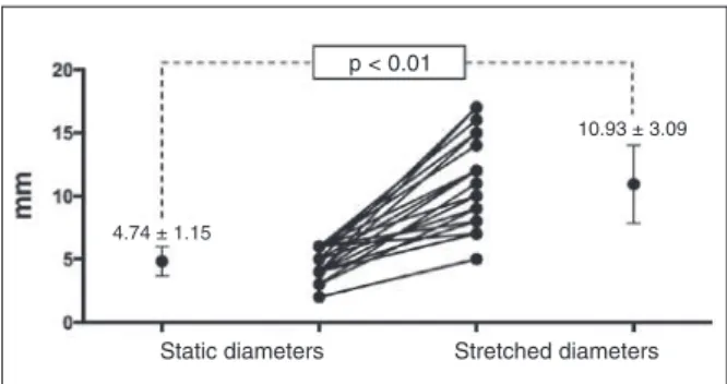
The diameters of the main static oriices ranged from 2 to 6 mm (4.5 ± 1.1 mm), and the stretched diameters from 5 to 16 mm (10 ± 3.0 mm). The use of the measuring balloon caused a mean increase of 55% compared to the static diameter of the defects (Figure 1).
It was possible to perform the implantation in all patients; different types of devices were used, in appropriate sizes: Amplatzer
ASD Occluder (St. Jude Medical Inc., St. Paul, United States) in eight patients; Cardia
ASD Occluder (PFM Medical Ag, Koln,
Ger-many) in seven; OcclutechASD Occluder (Occlutech
AB, Helsingborg, Sweden) in ive; Lifetech Cera
ASD Occluder (Lifetech Scientiic Corporation, Shenzhen, China) in ive; Cardia
PFO Occluder (PFM Medical
Ag, Koln, Germany) in three; AmplatzerCribbiform
Occluder (St. Jude Medical Inc., St. Paul, United States)
in two; and GOREHELEXSeptal Occluder (W. L.
Gore & Associate, Newark, United States), Lifetech
CeraPDA Occluder (Lifetech Scientiic Corporation,
Shenzhen, China), Nit-Occlud
ASD-R (PFM Medical Ag, Koln, Germany), and Cocoon ASD Occluder (Vascular Innovations Co. Ltd., Bangtanai, Pakkret, Thailand) in one patient each.
During the procedures, only one minor complication was found. ATEE study detected a threadlike thrombus in the right atrium, adhered to the exchange guide of a patient. This complication was attributed to an ineffec-tive anticoagulation. An additional dose of heparin was applied, with a careful removal of the device, which was washed and reintroduced, allowing the implant to be performed with the traditional technique. There were no sequelae resulting from this thrombus, which was not visualized on TEE after removal of the guide.
Two patients had episodes of migraine in the irst weeks after implantation. The Amplatzer
ASD Occluder was used in one of these patients, and the Occlutech
ASD Occluder was used in the other. In both cases, the migraine subsided after the addition of 75 mg of clopidogrel to acetylsalicylic acid. There were no deaths in this study.
Mean follow-up was 67.5 months (3-132 months). The only signiicant event occurred in one patient, a professional diver, who had been treated by occlusion of the defect with a 16 mm CardiaASD Occluder after
a decompression accident. In the fourth month after the procedure, an organized thrombus which had adhered to the right disk of the device was identiied. The event was attributed to the irregular use of antiplatelet agents. The thrombus could not be eliminated with anticoagu-lation, and its removal was performed surgically. No patient showed a periprosthetic residual shunt.
DISCUSSION
There is controversy in the literature about size criteria that deine a small ASD. In most studies, their criteria were arbitrarily chosen, without a physiological basis. From a hemodynamic point of view, Brassard et al.,9 using TTE, rated as minor defects those measuring < 6 mm; and as large defects those ≥ 6 mm, above 1 year of age. McMahon et al.,10 also using TTE, regarded as minor defects those whose diameter ranged from 3-6 mm; as moderate those from 6-12 mm; and as major defects those > 12 mm. Geva et al.11 considered as minor defects those measuring > 3 mm and < 5 mm. These authors reported that defects < 10 mm in diameter are associated with a small shunt, with minimal or no increase in right cavities. They also consider that in case of a defect >10 mm, the ratio of pulmonary/systemic blood low (Qp/Qs) is > 1.5:1, and in this scenario a cascade of changes in the myocardium and pulmonary vasculature could occur. Saxena et al.12 considered as minor defects those measuring from 4 and 5 mm.
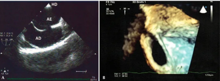
Importantly, these authors classiied the defects based on images of two-dimensional transthoracic echocardiograms, which introduce some margin of error, since they assessed defects that had mostly an oval or elliptical shape and, therefore, exhibited varying diameters, depending on the plan in which the septum was interrogated by the ultrasound beam (Figure 2).13-15
Static diameters Stretched diameters p < 0.01
4.74 ± 1.15
10.93 ± 3.09
In this study, only those defects considered as small-sized were included (≤ 6 mm diameter on two-dimensional TEE). This cut-off point was decided upon because it is a near-consensus in the literature, and because the searched studies reported absence of right cavity overload in these defects at a pre-procedural TEE. As demonstrated, there is wide variation between the static diameter and the SD of the defects. The use of the measuring balloon signiicantly increased the diameter of osASDs. There are three possible explanations: the low sensitivity of two-dimensional echo in determining the defect edge, particularly in non-spherical defects; excessive compliancy of the edges; and, while the balloon is theoretically a more compliant device, the possibility of edge rupture, resulting in increases in the diameter of the defect.
Most centers perform occlusive procedures in 4-or 5-year-old patients, based on the premise that there is a possibility of spontaneous closure in childhood. Reports of spontaneous closure up to adolescence, al-though much less frequent, have also been published, which reinforces the conservative approach of most authors.16,17 Hanslik et al.18 reported the occurrence of spontaneous closure in approximately 56% of defects measuring between 4 and 5 mm, 30% between 6 and 7 mm, and 12% between 8 and 10 mm. None of the defects measuring > 10 mm resulted in spontaneous closure. Other authors reported similar observations, with spontaneous closure rates ranging from 12 to 82% in small ASDs.4,9,10,12
With respect to small defects, it is common belief that these defects remain asymptomatic and hemody-namically insigniicant; nevertheless, the possibility of increases in defect sight throughout life cannot be excluded. Among the defects without spontaneous clo-sure, the defect size may increase with age, and may even increase to a point that will defeat any attempt
of percutaneous occlusion.19 In the series of Hanslik et al.,18 18% of defects with < 4 mm increased in size. McMahon et al.10 reported that 63% of small defects increased in size, with an increase of 1.6 mm per year in patients with a mean follow-up of 38 months (9-85 months). In other studies, the small defects increased suficiently so as to require surgical or percutaneous occlusion with the use of devices.4,11,12
In adults, small osASD may be more harmful than in children. Symptoms rarely reported in childhood are most commonly found in adults, appearing on average at the age of 33 years.3 Adults suffering from congenital heart defect are more likely to suffer major adverse cardiovascular events (heart failure, arrhythmias, and stroke) compared with controls. Patients with osASD were those individuals who beneited most from surgi-cal correction, in terms of reducing the risk of major adverse cardiovascular events at follow-up (adjusted hazard ratio 0.3; 95% conidence interval 0.3-0.5;
p < 0.0001).7
Atrial arrhythmias are part of late natural history of osASD, and are associated with signiicant mor-bidity and mortality, mainly in patients of older age groups. These conditions are associated with congestive heart failure and embolic events, among them stroke. Tachyarrhythmias (lutter and AF) are uncommon events in individuals younger than 40 years, with a reported incidence of 1%. Over 40 years of age, their preva-lence increases disproportionately, when compared to the normal population, ranging from 15% between 40 and 60 years to 61% above the sixth decade of life.20,21
osASD occlusion is associated with a progressive normalization of the diameters of the right cavities and, as might be anticipated, the earlier the correc-tion, the higher its frequency.22 Although there may be a regression of atrial lutter after correction of osASD,
A B
the same does not occur with AF, which usually require electrophysiological procedures besides the occlusion of the defects for heart rhythm normalization.20
The incidence of AF onset after percutaneous oc-clusion with devices is estimated to be 7% for PFO and 12% for osASD, regardless of device type and size. Again, patients diagnosed with post-occlusion AF are older than other patients.23 This incidence is higher than that expected in general population; but these results may be challenged, in that it is unclear whether the occurrence of AF was indeed caused by the device, or whether these patients would have de-veloped changes that could cause AF, even without the correction of defects.24
Interestingly, the incidence of AF after surgical correction is comparable to that for percutaneous closure: 14% among patients who underwent per-cutaneous occlusion and 16% among those who underwent surgery.25
Regardless of patient’s rhythm, the presence of paradoxical embolism is also a complication of osASDs, especially defects of small dimensions. Patients present-ing with embolic events, with the exception of patients with PFO, tend to be female, younger, and with smaller defects, when compared to osASD patients, who did not exhibit paradoxical embolism (Figure 3).26
Studies have shown that, among patients with epi-sodes of paradoxical embolism and who were referred for percutaneous closure of their atrial septal defects, 11% had osASD, 9% had osASD and PFO, and 80% had PFO. These data suggest that the risk of paradoxi-cal embolism is considerable in patients with osASD (Figure 4).27
The wide variety of models and brands of prosthe-ses used was not due to any particular criterion. PFO occlusion prostheses were employed in those cases in which the initial diagnosis was mistaken. In these cases, the patients had small central oriices with appropriate
A B
Figure 3 – Color low mapping of a small ostium secundum type atrial septal defect in two phases of the cardiac cycle. In A, the color Doppler reveals a normal low from left-to-right atrium. In B, the color Doppler detects the reverse transit low, from right to left.
A B
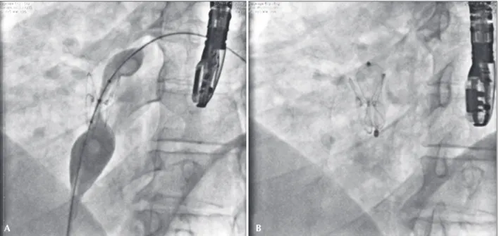
edges, and available prostheses were suited to the types of defects found. In a case of residual shunt with a long and tapered oriice, it was decided to use patent ductus arteriosus occlusion prosthesis, well suited to the anatomy of the defect (Figure 5).
CONCLUSIONS
The results of this study showed that, in line with the literature, the percutaneous occlusion of small
ostium secundum-type atrial septal defects was a safe and effective procedure, with very few complications in the short and medium term. It would be desirable a long-term longitudinal follow-up to establish a valid correlation between the defect size and its evolution in patients of older age groups. Randomized, double-blinded controlled studies are still needed to validate the use of percutaneously implanted devices targeting the treatment of atrial septal defects measuring ≤ 6 mm.
CONFLICTS OF INTEREST
The authors declare no conlicts of interest.
FUNDING SOURCES
None.
REFERENCES
1. Feldt RH, Avasthey P, Yoshimasu F, Kurland LT, Titus JL. Inci-dence of congenital heart disease in children born to residents of Olmsted County, Minnesota, 1950-1969. Mayo Clin Proc. 1971;46(12):794-9.
2. Keith J. Atrial septal defect: ostium secundum, ostium primum and atrioventricularis communis (common AV canal). In: Keith J, Rowe R, Vlad P, editors. Heart disease in infancy and child-hood. 3rd ed. New York: MacMillan; 1978. p. 380-404. 3. Forfang K, Simonsen S, Andersen A, Efskind L. Atrial septal
defect of secundum type in the middle-aged. Clinical results of surgery and correlations between symptoms and hemody-namics. Am Heart J. 1977;94(1):44-54.
4. Azhari N, Shihata MS, Al-Fatani A. Spontaneous closure of atrial septal defects within the oval fossa. Cardiol Young. 2004;14(2):148-55.
5. Cheng TO. All atrial septal defects should be closed. Int J Cardiol. 2014;175(2):224-5.
6. Meier B. Percutaneous closure of atrial septal defects—more roads lead to Rome than meets the eye. Catheter Cardiovasc Interv. 2011;78(5):675-6.
7. Lin YS, Liu PH, Wu LS, Chen YM, Chang CJ, Chu PH. Major cardiovascular events in adult congenital heart disease: a population-based follow-up study from Taiwan. BMC Cardiovasc Disord. 2014;14:38.
8. Chamie FCD, Ramos S, Tress JC, Victer R. Fechamento per-cutâneo das comunicações interatriais (CIA) complexas. Rev Bras Cardiol Invasiva. 2006;14(1):47-55.
9. Brassard M, Fouron JC, van Doesburg NH, Mercier LA, De Guise P. Outcome of children with atrial septal defect considered too small for surgical closure. Am J Cardiol. 1999;83(11):1552-5. 10. McMahon CJ, Feltes TF, Fraley JK, Bricker JT, Grifka RG,
Tortoriello TA, et al. Natural history of growth of secundum atrial septal defects and implications for transcatheter closure. Heart. 2002;87(3):256-9.
11. Geva T, Martins JD, Wald RM. Atrial septal defects. Lancet. 2014;383(9932):1921-32.
12. Saxena A, Divekar A, Soni NR. Natural history of secundum atrial septal defect revisited in the era of transcatheter closure. Indian Heart J. 2005;57(1):35-8.
A B
Figure 5 – Patient who had ostium secundum-type atrial septal defect occluded with a HELEX prosthesis. In A, the prosthesis in proper position and
coniguration can be observed. The residual oriice is outlined by a measuring balloon, between the two discs of the device. In B, the Cera
13. Ferreira SM, Ho SY, Anderson RH. Morphological study of defects of the atrial septum within the oval fossa: implica-tions for transcatheter closure of left-to-right shunt. Br Heart J. 1992;67(4):316-20.
14. Roberson DA, Cui W, Patel D, Tsang W, Sugeng L, Weinert L, et al. Three-dimensional transesophageal echocardiography of atrial septal defect: a qualitative and quantitative anatomic study. J Am Soc Echocardiogr. 2011;24(6):600-10.
15. Kim KH, Song J, Kang IS, Chang SA, Huh J, Park SW. Bal-loon occlusive diameter of non-circular atrial septal defects in transcatheter closure with amplatzer septal occluder. Korean Circ J. 2013;43(10):681-5.
16. Cockerham JT, Martin TC, Gutierrez FR, Hartmann AF Jr, Goldring D, Strauss AW. Spontaneous closure of secundum atrial septal defect in infants and young children. Am J Car-diol.1983;52(10):1267-71.
17. Hartmann AF Jr, Elliott LP. Spontaneous physiologic closure of an atrial septal defect after infancy. Am J Cardiol. 1967;19(2): 290-2.
18. Hanslik A, Pospisil U, Salzer-Muhar U, Greber-Platzer S, Male C. Predictors of spontaneous closure of isolated secundum atrial septal defect in children: a longitudinal study. Pediatrics. 2006;118(4):1560-5.
19. Tortoriello TA, McMahon C, Kovalchin JP, Bricker JT, Grifka RG. Growth of an atrial septal defect: missing the window for transcatheter closure. Pediatr Cardiol. 2002;23(5):542-4. 20. Berger F, Vogel M, Kramer A, Alexi-Meskishvili V, Weng Y,
Lange PE, et al. Incidence of atrial lutter/ibrillation in adults
with atrial septal defect before and after surgery. Ann Thorac Surg. 1999;68(1):75-8.
21. Oliver JM, Gallego P, Gonzalez AE, Benito F, Sanz E, Aroca A, et al. Surgical closure of atrial septal defect before or after the age of 25 years: comparison with the natural history of unoperated patients. Rev Esp Cardiol. 2002;55(9):953-61. 22. Veldtman GR, Razack V, Siu S, El-Hajj H, Walker F, Webb
GD, et al. Right ventricular form and function after percu-taneous atrial septal defect device closure. J Am Coll Car-diol.2001;37(8):2108-13.
23. Spies C, Khandelwal A, Timmermanns I, Schrader R. Incidence of atrial ibrillation following transcatheter closure of atrial septal defects in adults. Am J Cardiol. 2008;102(7):902-6. 24. Knirsch W, Dodge-Khatami A, Valsangiacomo-Buechel E, Weiss
M, Berger F. Challenges encountered during closure of atrial septal defects. Pediatr Cardiol. 2005;26(2):147-53.
25. Oliver JM, Gallego P, Gonzalez A, Benito F, Mesa JM, Sobrino JA. Predisposing conditions for atrial ibrillation in atrial sep-tal defect with and without operative closure. Am J Cardiol. 2002;89(1):39-43.
26. Bannan A, Shen R, Silvestry FE, Herrmann HC. Characteristics of adult patients with atrial septal defects presenting with paradoxi-cal embolism. Catheter Cardiovasc Interv. 2009;74(7):1066-9. 27. Khositseth A, Cabalka AK, Sweeney JP, Fortuin FD, Reeder