Arq Neuropsiquiatr 2008;66(2-B):428-430
428
InclusIon Body MyosItIs and HIV InfectIon
Marcos R. Gomes de Freitas
1, Marco A.O. Neves
1,3, Osvaldo J.M. Nascimento
1,
Mariana P. de Mello
3, John P. Botelho
3, Leila Chimelli
2MIosIte PoR coRPos de InclusÃo e InfecÇÃo PoR HIV
1Neurology Department, Fluminense Federal University, Niteroi RJ, Brazil; 2Neuropathology Department, Rio de Janeiro Federal University, Rio de Janeiro
RJ, Brazil; 3Serra dos Orgãos University. Rio de Janeiro RJ, Brazil.
Received 4 October 2007, received in inal form 25 March 2008. Accepted 1 April 2008.
Dr. Marcos R. Gomes de Freitas – Rua Gastão Ruch 16 / 1402 - 24220-100 Niterói RJ - Brasil. E-mail: mgdefreitas@hotmail.com
Neurological disorders are frequent complications of human immunodeiciency virus (HIV) type 1 infection, and include central nervoussystem (CNS) infections, neo-plasm, vascular complications, peripheralneuropathies, and myopathies1. Early series emphasized CNS diseases, with relative few reports of primary disorders of periph-eral nerve and muscle2.Myopathy may occur at any time during the course of HIV infection and is not associated with any particular stage of immunosuppression3. Before the introduction of zidovudine (azidothymidine, AZT) for the treatment of AIDS, muscle disease was considered a rare complication of HIV, found in less than 1% of cas-es of AIDS2.A variety of muscular disorders has been de-scribed in HIV infected patients3: polymyositis, myopa-thy induced by nucleoside reverse transcriptase inhibi-tors (NRTI), such as zidovudine, opportunistic infections including toxoplasmosis, iniltration by tumour, HIV asso-ciated vasculitis, and rhabdomyolysis caused by HIV itself or by drugs including didanosine5. A myopathy in every re-spect similar to inclusion body myositis (IBM) is observed in rare patients infected by HIV-1 or human T-cell leukae-mia virus type 1(HTLV-1)6,7. IBM is a chronic inlammatory muscle disease, and the typical clinical indings are mus-cle weakness and atrophy, most prominent in the quadri-ceps muscles and the wrist and inger lexors8.
We report a case of a male patient, who presented with signs and symptoms of IBM in association with HIV infection.
case
A 56 year-old French man was diagnosed as having HIV infec-tion in 2000. Initially the CD4 cell counts were 314 and the viral load was 626 copies. A treatment with HAART was started. Two months later the CD4 was normal and the viral load fell to 0. One year later he noticed dificulty in climbing stairs with slowly progression and in seven months he could walk only with aids of canes. He also noticed some dificulties with movements of the hands. A diagnostic of a muscle disease due to zidovudine was done and HAART was stopped. As there was no improvement in
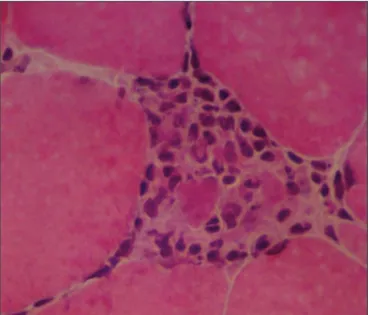
Fig 1. Parafin section stained with H&E showing endomysial lym-phocytic foci (x100).
Arq Neuropsiquiatr 2008;66(2-B)
429 Inclusion body myositis: HIV infection Freitas et al.
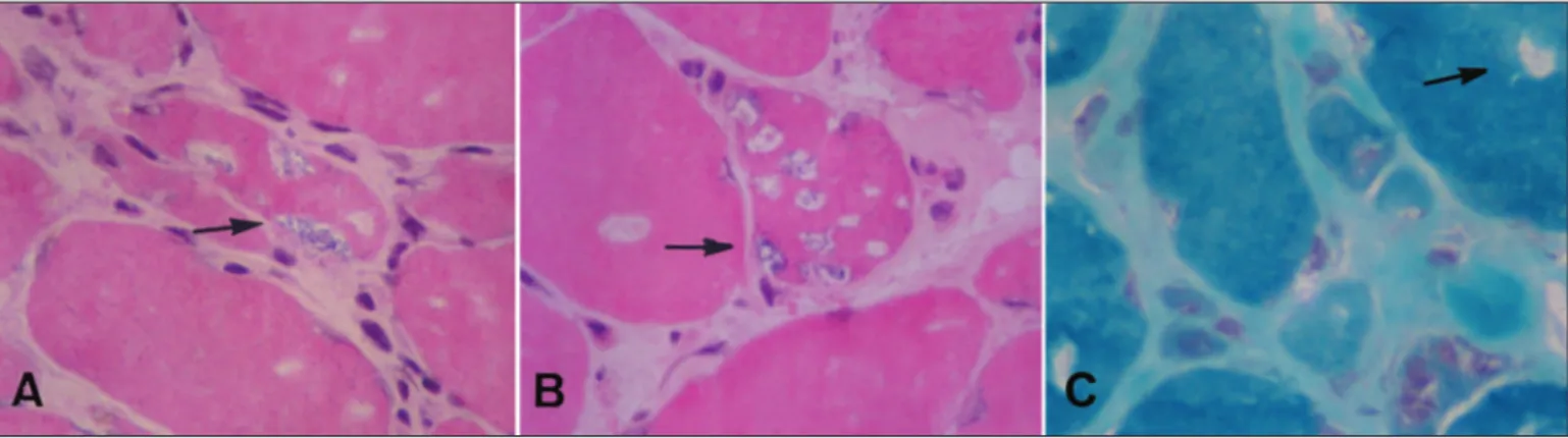
eight months the patient was referred to our service. The physi-cal examination was normal. There was proximal muscle atrophy in lower limbs mainly in the quadriceps. The strength was dimin-ished in proximal and distal muscles in lower limbs. In the upper limbs the weakness was localized in the hands, mainly in wrist and ingers lexor muscles (Table 1). The patellar relexes were abolished and the ankle relexes were diminished. In the upper limbs the tendon relexes were normal. The sensory and the cra-nial nerves examination were normal. The blood biochemical examination was normal except for a CK of 2600 U. The EMG examination revealed increased spontaneous activity, with ibril-lations, complex repetitive discharges and positive sharp waves. The motor units had low-amplitude polyphasic units, usually of short duration. A muscle biopsy was performed. Histochemical stains were done. Muscle ibres were irregular in size and shape; there were many atrophic ibres, some of them angulated, and mild increase in endomysial collagen. Endomysial lymphocytic foci (Fig 1) and necrotic ibres iniltrated by macrophages (Fig 2) were present, as well as some ibres containing rimmed vacuoles (Fig 3A,B), also shown with the Gomori’s trichrome (Fig 3C). The treatment consists of Immunoglobulin IV (IVIg) 400 mg/kg/day for ive days. The muscles weakness improved slowly and the CK decreased to 367 U (Table). The IVIg infusion was done once a month. After ive months of IVIG, the muscle weakness became stable till the last examination in 2007, August.
dIscussIon
IBM is one of the three main subsets of inflamma-tory myopathies, the other two being polymyositis and dermatomyositis8, and it’s considered the most common acquired, progressive and disabling myopathy in patients above the age of 50 years, and has a male predominance8.
IBM has a slow progression, affects both the proximal and the distal muscles. The amyotrophy can be asymmet-ric, and in typical cases muscle weakness and wasting are most profound in knee extensors, hip flexors and long inger lexors8. Most patients require an assistive device within several years of onset9. Neck lexors and extensors
and facial muscle are frequently affected8. The muscles of swallowing are affected in about 50% of the patients8. The tendon relexes can diminish in later stages when the atrophy of major muscle groups becomes evident8. Our case had the typical clinical indings of IBM.
Creatine kinase (CK) levels can initially be elevated up to 10-fold and remain slightly elevated as the disease pro-gresses. In our case the CK was very high what is described in IMB associated with retrovirus6. The EMG of our patient is typical of muscle affection.
The main histological features are red-rimmed vacu-oles, endomysial T cell iniltrates, cytoplasm inclusions,
atrophic ibres and amyloid deposits6. The
inlammato-ry iniltrates consist of CD8+ T cells and macrophages, suggesting involvement of a T cell mediated cytotoxic mechanism against muscle fibres10. Although we could not perform techniques for amyloid, as speciic antibod-ies against beta amyloid or immunocytochemical analysis
Fig 3. Sections stained with H&E (A, B) and Gomori’s trichrome (C) showing ibres with rimmed vacuoles (x400) (arrow).
Table. Strength examination (MRC) and CK.
Muscle First day of admission
Five months after IVIg Abductors of the shoulder R 5 L5 R 5 L5 Extensors of the forearm R 5 L5 R 5 L5 Flexors of the arms R 5 L5 R 5 L5 Flexors of the ingers R 3 L 3 R 4 L4 Abductors of the ingers R 5 L5 R 5 L5 Extensors of the thigh R 3 L3 R 4 L4 Flexors of the thigh R 3 L3 R 4 L4 Extensors of the legs R 4 L4 R 5 L4 Adductors of the thigh R 5 L5 R 5 L5 Extensors of the foot R 0 L0 R 1 L1 Flexors of the foot R 2 L2 R 4 L4 Extensors of the toes R 0 L0 R 1 L1 CK (U) 2600 367
Arq Neuropsiquiatr 2008;66(2-B)
430
Inclusion body myositis: HIV infection Freitas et al.
and ultrastructure techniques, the morphological changes seen in our case, particularly the lymphocytic iniltration and the rimmed vacuoles, although non-speciic, are high-ly suggestive of IBM.
The aetiology of IBM is unclear. The immunopatho-logical indings suggest an immune-mediated process but the lack of response to immunotherapy and the amyloid deposits have raised the possibility of a degenerative disorder. Viral aetiologies have been suggested11. A few reports of HIV or HTLV-1 positive patients with IBM indi-cates that the disease is more common in patients who live longer and harbour this virus for several years6,12. The IBM in HIV infected patients is like to the sporadic IBM, except for the earlier age of onset and the higher eleva-tion of muscle enzymes6.
The mechanism by which the retrovirus triggers the disease is unclear. Retroviral antigens have been detected in endomysial macrophages but not within the muscle ibers6,7.The activated CD8+ cells invade muscle ibres ex-pressing MHC class I, as seen in retrovirus-negative poly-myositis and IBM6,11.These cells are retrovirus-speciic, be-cause their CDR3 region contains amino acid residues that are speciic for viral peptide bound to HLA molecules12.
The myopathy due to AZT is different from IBM. It’s presumably due to an interference with mitochondrial function4. Typical features of this myopathy are ragged red ibres and paracrystalline inclusions in mitochondria that have been attributed to its DNA (mtDNA) depletion13. Ragged red ibres may be seen in rare cases of IBM suggest-ing that mitochondrial function is impaired in this disease10.
A direct link between NRTI, mitochondrial dysfunc-tion, and IBM is strongly suggested in a case of IBM in HIV infection7 NRTI prolonged use may contribute to the de-velopment of IBM in this type of patients. In these cases the discontinuation of NRTI may be a strategy for manage-ment, although whether the condition is reversible remain unknown7.
Because there is no effective medical treatment in IBM (steroid and other immunosuppressive treatments
have disappointing results), all other measures that could possibly be of beneit to patients should be considered8. Some authors think that IVIg may be useful for treatment
of IBM14. Our patient showed a modest but permanent
improvement with IVIg 400 mg/day for ive days and one month infusion for one day. Mild to moderate muscle training or aerobic endurance training, can be performed without adverse effects15.
RefeRences
1. Simpson DM, Tagliati M. Neurologic manifestations of HIV infection. Ann Intern Med 1994;121:769-785.
2. Levy RM, Bredesen DE, Rosenblum ML. Neurological manifestations of
the acquired immunodeiciency syndrome (AIDS): experience at UCSF
and review of the literature. J Neurosurg 1985;62:475-495.
3. Wulff EA, Simpson DM. Neuromuscular Complications of the human immunodeiciency virus type 1 infection. Semin Neurol 1999;19:157-164. 4. Dalakas MC, Illa I, Pezeschkpour GH, Laukaitis JP, Cohen B, Grifin JL.
Mitochondrial myopathy caused by long-term zidovudine therapy. N
Engl J Med 1990;322:1098-1105.
5. Roedling S, Pearl D, Manji H, Hanna MG, Holton JL, Miller RF. Unusual muscle disease in HIV infected patients. Sex Transm Infect 2004;80:315-317. 6. Cupler EJ, Leon-Monzon M, Miller J, Semino-Mora C, Anderson TL,
Dalakas MC. Inclusion body myositis in HIV-1 and HTLV-1 infected
patients. Brain 1996;119:1887-1893.
7. Authier FJ, Chariot P, Gherardi RK. Skeletal muscle involvement in hu
-man immunodeiciency virus (HIV)-infected patients in the era of high
-ly active antiretroviral therapy (HAART). Muscle Nerve 2005;32:247-260.
8. Dalakas MC. Sporadic inclusion body myositis: diagnosis, pathogene
-sis and therapeutic strategies. Nat Clin Pract Neurol 2006;2:437-447. 9. Peng A, Koffman BM, Malley JD, Dalakas MC. Disease progression in
sporadic inclusion body myositis: observations in 78 patients. Neurol-ogy 2000;55:296-298.
10. Scola RH, Werneck LC, Iwamoto FM, Messias IT, Tsuchiya LV. Análise imunocitoquímica do iniltrado inlamatório na miosite por corpo de
inclusão citoplasmática e outras doenças neuromusculares com vacúo
-los marginados. Arq Neuropsiquiatr 1998;56:388-397.
11. Dalakas MC. Inlammatory, immune and viral aspects of inclusion-body myositis. Neurology 2006;66(Suppl):S33-S38.
12. Ozden S, Cochet M, Mikol J, Teixeira A, Gessain A, Claudine Pique C. Direct evidence for a chronic CD8+-T-cell-mediated immune reaction to tax within the muscle of a human T-cell leukemia/lymphoma virus
type 1-infected patient with sporadic inclusion body myositis. J Virol 2004;78:10320-10327.
13. Arnaudo E; Dalakas MC; Shanske S; Moraes CT; Di Mauro S; Schon
EA. Depletion of muscle mitochondrial DNA in AIDS patients with zi
-dovudine-induced myopathy. Lancet 1991;337:508-510.
14. Dalakas MC, Sonies B, Dambrosia J, Sekul E, Cupler E, Sivakumar K.
Treatment of inclusion-body myositis with IVIg: a double-blind, pla-cebo-controlled study. Neurology 1977;48:712-716.
15. Alexanderson H, Lundberg IE. The role of exercise in the rehabilitation of idiopathic inlammatory myopathies. Curr Opin Rheumatol 2005;17: