Functional and morphological changes in
the quadriceps muscle induced by eccentric
training after ACL reconstruction
Alterações funcionais e morfológicas do quadríceps induzidas
pelo treinamento excêntrico após reconstrução do LCA
Jamilson S. Brasileiro1, Olga M. S. F. Pinto2, Mariana A. Ávila2, Tania F. Salvini2
Abstract
Objectives: The purpose of this study was to investigate the contributions of functional and morphological factors in the recovery of the quadriceps muscle after anterior cruciate ligament (ACL) reconstruction. Methods: Nine subjects (31.3±5.8 years) underwent eccentric exercise sessions twice a week for 12 weeks. Quadriceps muscle function was evaluated using an isokinetic dynamometer (isometric and eccentric peak torque) and electromyography (RMS). Morphological changes were measured using magnetic resonance imaging. Results: The initial evaluation showed a significant deficit in knee extensor torque in the involved limb and significant muscle atrophy along the length of the quadriceps. EMG activity was lower in all tested situations. Eccentric training significantly increased isokinetic torque (from 199±51 to 240±63, p<0.05, respectively) and quadriceps area, with the greatest hypertrophy in the proximal thigh region (from 169±27 to 189±25.8 cm2, p<0.01). The EMG activity of vastus medialis increased after the first six weeks of eccentric training. The
increased extensor torque was correlated with quadriceps cross-sectional area (r=0.81, p<0.01) and EMG activity (r=0.69, p<0.05). After twelve weeks of training, there was a correlation only between torque and cross-sectional area (r=0.78, p<0.01). Conclusions: 1) eccentric training proved to be a potent resource for the quadriceps recovery, both morphologically and functionally, 2) the contributions of functional and morphological factors varied according to the length of training.
Key words: anterior cruciate ligament; electromyography; magnetic resonance; quadriceps muscle.
Resumo
Objetivos: O propósito deste estudo foi avaliar as contribuições dos fatores funcionais e morfológicos na recuperação da força muscular do quadríceps femoral após reconstrução do Ligamento Cruzado Anterior (LCA). Métodos: Nove indivíduos (31,3±5,8 anos) foram treinados por meio de contrações excêntricas máximas, duas vezes por semana, durante 12 semanas. A função do quadríceps foi avaliada pela dinamometria isocinética (pico de torque isométrico e excêntrico) e pela eletromiografia (EMG). As alterações morfológicas foram mensuradas por meio de ressonância magnética (RNM). Na avaliação inicial, observou-se significativo déficit no torque extensor do joelho do membro acometido, com hipotrofia muscular de todo o quadríceps e redução na atividade
EMG, quando comparado ao membro não-acometido. Resultados: O treinamento excêntrico produziu aumento no torque excêntrico
a 30º/s (de 199±51 Nm para 240±63 Nm, p<0,05) e no volume muscular, sendo que maiores hipertrofias ocorreram na região proximal da coxa (de 169±27 para 189±25,8 cm2,p<0,01). A atividade EMG do Vasto Medial (VM) aumentou nas primeiras seis semanas de
treinamento. O aumento no torque extensor demonstrou correlação positiva com o aumento no volume (r=0,81, p<0,01) e na atividade eletromiográfica (EMG) (r=0,69, p<0,05) nas primeiras seis semanas. Após 12 semanas de treinamento, houve correlação apenas entre o aumento do torque e do volume (r=0,78, p<0,01). Conclusões: 1) O treinamento excêntrico mostrou-se como potente recurso tanto na recuperação dos fatores morfológicos como funcionais do músculo quadríceps; 2) A contribuição dos fatores neurais e morfológicos varia em função do período de treinamento.
Palavras-chave: ligamento cruzado anterior; eletromiografia; ressonância magnética; quadríceps femoral.
Received: 31/05/2010 – Revised: 26/11/2010 – Accepted: 30/03/2011
1 Physical Therapy Department, Universidade Federal do Rio Grande do Norte (UFRN), Natal, RN, Brazil 2 Physical Therapy Department, Universidade Federal de São Carlos (UFSCar), São Carlos, SP, Brazil
Correspondence to: Jamilson Simões Brasileiro, Universidade Federal do Rio Grande do Norte, Campus Universitário, Departamento de Fisioterapia, Av. Senador Salgado Filho, 3000, Lagoa Nova, CEP 59.078-970, Natal, RN, Brasil, e-mail: [email protected]
Introduction
he anterior cruciate ligament (ACL) is one of the most frequently injured ligaments1. Several studies have
demonstra-ted that even after ACL reconstruction patients are discharged from rehabilitation, signiicant deicits in quadriceps muscle strength and cross-sectional area still remain. hese deicits can persist for months or even years2-7. It has been shown that
the recovery of knee extensor torque is an essential element for functional rehabilitation of the lower limb after reconstruction since the return to functional activity is strongly correlated with the ability of the quadriceps femoris to generate force3,5,8.
Understanding the physiological basis of muscle strength recovery in patients submitted to ACL reconstruction is funda-mental for developing an appropriate rehabilitation program. Without correct identiication of the factors involved in the loss and recovery of strength, it is diicult to select the most adequate physical therapy resources to use.
Neural and morphological participation have been sug-gested as the two main factors in the recovery of quadriceps strength. Neural factors are largely related to better motor unit eicacy during muscle contraction. It is known that the greater the number of motor units activated, the greater the resulting strength. Moreover, better timing of muscle iber activation associated with an increase in triggering rate optimizes the excitation-contraction coupling which leads to further eleva-tion of muscle force produceleva-tion9-13. It has been observed that
in post-traumatic situations, inhibitory mechanisms act to re-duce neuromuscular excitement, which leads to a reduction in muscle contractile capacity9. his activity can be measured by
the RMS (Root Mean Square) of the electromyographic signal14.
On the other hand, morphological factors generally refer to the muscle cross-sectional area. he larger the iber diameter, the greater the number of cross-bridges built and, thus, the higher the capacity to generate force9,11. Reduced trophism is often associated
with traumatic injuries and can be measured by imaging techni-ques such as ultrasound, computerized tomography or, preferably, magnetic resonance imaging (MRI). Isokinetic dynamometry can reveal changes in neuromuscular function, such as those related to peak torque, average torque, power, total work, time and peak torque angle. In current practice, it is an important tool for func-tional assessment due to its high level of reliability and validity.
It has been suggested that initial strength gains would result from the contribution of neural factors and that later phase gains would be due to muscular hypertrophy9,11,13. However, studies
evaluating these factors have been restricted to healthy subjects. Studies involving subjects submitted to this kind of rehabilitation are scarce. Lieber13 has called attention to the lack of studies with
rehabilitation subjects, suggesting that if strength deicits occur principally due to neural factors, rehabilitation should focus,
above all, on resources that stimulate neural activation, such as neuromuscular electrical stimulation and the use of stretch rele-xes and balance reactions. However if deicits are due to morpho-logical factors such as hypertrophy, he states that the treatment focus should be directed to maximal resistance exercise.
he adequate identiication of the factors responsible for strength deicit in this population will allow the physical thera-pist to select the best resource for each phase of the rehabilita-tion process. For this reason, the present study investigated the contribution of neurological and morphological factors to the recovery of quadriceps femoris function in patients submitted to ACL reconstruction.
Methods
Subjects
Nine sedentary male subjects (31.3±5.8 years-old) who had been submitted to unilateral ACL reconstruction involving the middle third of the patellar tendon participated in this study. All the procedures were performed by the same surgeon. he sub-jects were between the 9 and 10 months post-surgery (mean of 9.4±0.7 months), which can be considered in the late rehabilita-tion phase. All the subjects were submitted to the same rehabi-litation protocol, which began immediately after surgery. hose with other associated injuries, a previous history of trauma in the contralateral knee or joint pain during training were exclu-ded from the study.
All the volunteers were informed about the objectives of this study and gave written informed consent before participating. his study was approved by the Research Ethics Committee of the Universidade Federal de São Carlos (UFSCar), São Carlos, SP, Brazil under protocol number 013/2004.
Procedures
Analysis of the cross-sectional area of the
quadriceps (CSA)
To obtain the images of the quadriceps femoris, a Torm 0.5 Tesla MRI scanner (São Carlos, SP, Brazil) was used with the subject in the supine position. he muscle image acquisition parameters were as follows: a) ield intensity = 0.5 tesla; b) time of repetition (TR) = 430 ms; time to echo (TE) = 26 ms; d) acqui-sition matrix = 256 x 192; e) slice thickness = 8 mm; f) slice gap = 0 mm; g) radio frequency bandwidth = 16 KHz.
he images were measured every 3.2 cm15 with Axionvision
v3.0 software (Carl Zeiss, Germany).
In each evaluated plane, the muscle area was measured consecutively three times by the same rater and the arithmetic mean of these three measures was considered as the CSA of the quadriceps (in cm2). Previous analysis revealed an intra-rater
reliability level above 0.95.
Isokinetic dynamometry
he subjects initiated the isokinetic tests after a 5 min warmup on a stationary cycle ergometer with 25 W resistance at 20 Km/h. hen they performed passive self-stretching of the quadriceps femoris in both limbs. he stretching maneuver consisted of maintaining the knee in complete lexion with the hip extended to the maximal tolerable amplitude while in the orthostatic position. hree 30 s repetitions were performed with a 30 s interval between each repetition.
An isokinetic dynamometer (Biodex Multi-Joint System 3, Biodex Biomedical System Inc., New York) was used for isometric and isokinetic torque assessment as well as for training. Sub-jects were seated in the chair, which was inclined 5º backwards from vertical, with their trunks stabilized by two belts while the resistance arm was positioned at the distal portion of the leg. he mechanical axis of the dynamometer was aligned with the lateral epicondyle of the femur. Adjustments to correct for the efects of gravity on torque were made with the knee at 60º of lexion and calculated with the dynamometer’s software, as recommended by Dvir16. he assessment was initially
perfor-med with the non-injured limb and then with the injured one. After a short period of familiarization with the equipment, the subject performed three 5 s maximal isometric voluntary contractions (MIVC) with the knee at 60º of lexion, resting for 2 min between each contraction. he extensor peak torque between the three trials was registered for further analysis.
he isokinetic evaluation was initiated after an interval of three minutes, with the dynamometer adjusted to eccentric mode and with a speed of 30 and 120º/s. he subject’s knee was passively extended to 20º (considering zero as a complete exten-sion) and then the subject was asked to perform a full extension until reaching a 90º angle, totaling an active range of 70º. Each subject performed ive repetitions at 30º/s and ive repetitions at 120º/s. During the execution of maximal voluntary contrac-tions, verbal encouragement was given by the same evaluators.
Electromyographic activity (EMG)
heEMG measurement procedure was as follows: the surface of the skin was shaved and cleaned with alcohol. he receiving elec-trodes (Lynx Tecnologia Eletrônica®
, Brazil) were then positioned over the rectus femoris (RF), vastus lateralis (VL) and vastus me-dialis obliquus (VMO) muscles according to recommendations of
SENIAM (SurfaceElectromyography for Non-Invasive Assessment of Muscles), with the reference electrode ixed at the tibial tuberosity. EMG activity was captured simultaneously with the torque records by means of a 12 bit analog-to-digital converter (CAD, 12/36-60K – Lynx Tecnologia Eletrônica) with a 16-channel signal conditioner module (MCS 1000). Signals were captured with Aqdados, v5.0(Lynx Tecnologia Eletrônica, Brazil), sampled at 1 KHz and iltered (20 to 500 Hz) according to DeLuca17. he
electrode had a 20x internal gain and a common mode rejection rate of over 80 dB. Since the gain programmed in the analog-to-digital converter was 50x, the signal was ampliied 1000 times.
he obtained data was stored and subsequently converted to ASCii format. After processing, the iles were analyzed in MatLabv5.0, by means of which the RMS value was identiied for EMG signal analysis.
he initial evaluation procedures were repeated after six weeks of training (evaluation 2) and again after 12 weeks of training (evaluation 3).
Training protocol
Each subject was submitted to a maximal eccentric exer-cise training session twice a week for 12 weeks. Each session began with a short warmup followed by stretching like that performed before the evaluations.
he subject was then positioned in the isokinetic dynamo-meter as described above. he same procedures used in the isokinetic evaluation were used for eccentric training, with three sets of ten repetitions performed. Only the injured limb was trained and only at the speed of 30º/s.
Statistical analysis
For the analysis and interpretation of the results, Statistical Package for Social Sciences (SPSS) v15.0 was used. he normality of all data was initially veriied with Kolmogorov-Smirnov. he peak torque analysis, EMG activity and CSA obtained in the three evaluations were submitted to a new repeated-measures ANOVA for comparison. For all signiicant causes of variation, post-hoc Tukey was used. Pearson’s Correlation was used to determine the relation between the dynamometric, EMG and CSA data. he signiicance level was set at 5% for all tests.
Results
Peak torque (PT)
he highest PT was found in the eccentric contraction at 30°/s, followed by the eccentric contraction at 120°/s. he
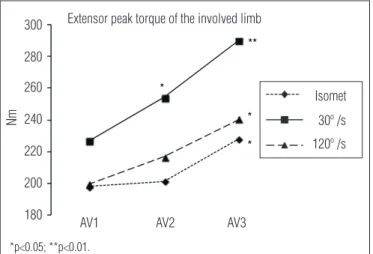
Figure 1. Isometric and eccentric (30 and 120º/s) knee extensor peak torque of the involved limb, pre-training (AV1), after six (AV2) and twelve weeks (AV3).
300
280
260
240
220
Nm
AV1 AV2 AV3
Extensor peak torque of the involved limb
200
180
30º /s Isomet
120º /s
*p<0.05; **p<0.01.
lowest PT was registered during the isometric contractions (Figure 1).
In the eccentric evaluation at 30°/s, the training had al-ready produced an increase in PT by the sixth week ( from 227±56 to 254±65 Nm, p=0.031) that continued until the end of the training (290±64 Nm, p=0.007). Signiicant increases in PT were also observed for isometric and eccentric contractions at 120°/s (velocity not used in training), which was veriied only after 12 weeks of training ( from 198±37 to 228±48 Nm, p=0.041 and from 200±51 to 240±63 Nm, p=0.039, respectively).
PT from the non-injured limb did not show signiicant va-riation over the course of the study.
Analysis of CSA
MRI images of the entire thigh showed that there was a sig-niicant increase in quadriceps CSA in the injured limb by the second evaluation that continued until the end of the study. In the non-injured limb, no alteration in the CSA of any of the measured regions was observed.
Individual analysis of each CSA level along the length of the thigh revealed that the trained limb presented non-homogeneous behavior with respect to recovery of quadriceps trophism (Figure 2).
No signiicant hypertrophy was observed in the distal por-tion of the thigh in the irst six weeks of training ( from 31.5±5.9 to 31.6±6.2 cm2, p=0.38). By the end of the training period, an
increase of 11.5% in measured area was observed (35.1±7 cm2,
p=0.043). he increase in this region was occurred mainly in the VM. In an intermediate region of the thigh, hypertrophy was observed by the end of the irst six weeks of training ( from 123±23 to 128±25 cm2, p=0.029) and increased until the third
evaluation (134±25 cm2, p=0.009).
he studied region includes the vastus intermedius (VI), vastus medialis (VM) and VL. In the proximal region of the thigh, hypertrophy was already observed after the six irst we-eks of training ( from 166±26 to 175±28 cm2, p=0.009), with a
continuous increase until the 12th week (182±29 cm2, p=0.003).
In this region, are evidences the VI, VL and RF.
EMG activity
here was a signiicant increase in the RMS value of the VMO and VL only during the irst six weeks of training ( from 213±107 to 289±81 µV, p=0.037 and from 207±65 to 229±69 µV, p=0.042, respectively). he RMS and RF values did not undergo signiicant modiications during the training (Figure 3).
In contrast, the statistical analysis did not reveal signiicant diferences between the EMG values registered during the iso-metric and eccentric contractions at 30º/s for the same muscle portion (p>0.05).
Correlations between peak torque, cross-sectional
area and EMG activity
In the irst six weeks of training, signiicant increases in isokinetic PT at 30º/s were observed, followed by an increase in the average EMG activity of the three analyzed vastus mus-cles and a signiicant increase in average CSA along the length
Figure 2. Representative axial T2-weighted MRI of the involved limb,
before (A and C) and after training (B and D) of the same subject. The images were obtained between 6 cm (A and B) and 20 cm (C and D) from the proximal border of the patella.
VM=vastus medialis; VL=vastus lateralis; VI=vastus intermedius; RF=rectus femoris.
A
VM
VL
VL VI
VI
RF RF
VM
VM
B
C D
of the thigh. here was a strong correlation between the varia-bles PT and CSA (r=0.81, p=0.020) and a moderate correlation between the variables PT and EMG (r=0.69, p=0.037).
In the second six weeks, PT also varied signiicantly from the values observed in the second evaluation. his gain was followed by an increase in muscle CSA, but no change in EMG activity was observed. In this phase, a strong correlation was also maintained between PT and CSA (r=0.78, p=0.01), although no correlation was observed between PT and EMG (r=0.28, p=0.13).
Discussion
he purpose of the present study was to analyze the contri-bution of neural and morphological factors to the recovery of quadriceps strength after a training period involving maximal eccentric contractions.
At the end of the training, although torque gains were sig-niicant, a residual deicit of 18 to 29% persisted between the injured and non-injured limbs. Hiemstra et al.5 found a global
deicit of 25.5% in extensor torque when evaluating 24 subjects submitted to ACL reconstruction after one year of recovery. he same authors emphasized the scarcity of studies involving eccentric evaluations. his is important since, theoretically, greater deicits could be revealed in this form of contraction, since greater muscle tension is generated.
he existence of such a torque deicit may reveal a change in recruitment pattern, mechanical joint changes or modii-cations in muscle properties due to physical deconditioning. Urbach et al.18 found torque deicits without the presence of
signiicant changes in neural recruitment in patients like those evaluated in the present study, which suggests that, in addition
to possible deicits of voluntary activation, disuse atrophy may still be present.
he proposed eccentric training program was eicacious in increasing muscle trophism, with gains that appeared after six weeks and gradually increased until the end of the 12th week.
Some studies have suggested that maximal eccentric trai-ning is more eicient for increasing muscle trophism than concentric treatment19-21. Since more strength can be
genera-ted eccentrically, this training modality would produce more overload in the muscle, which would induce higher protein synthesis22,23. It has also been suggested that this would
oc-cur due to a greater recruitment of type II ibers during the contractions10,24,25, since these ibers typically demonstrate
gre-ater potential for hypertrophy than type I ibers13,19,20.
Lieber13 andEnoka24 have suggested that few muscle
tro-phism changes can be observed during the initial weeks of trai-ning and that the recorded torque gains are almost exclusively attributed to neural changes. However, our study demonstrated a signiicant increase in CSA in the irst six weeks of training, which suggests that the process of hypertrophy in these sub-jects may be diferent from that observed in healthy subsub-jects.
Another result observed in our study was the diference in CSA increase between the proximal and distal extremes of the quadriceps. he proximal region presented signiicant hyper-trophy after the irst six weeks of training, whereas no gains were registered in the distal region.
Signiicant changes in hypertrophy between diferent parts of the quadriceps have been demonstrated in other studies10,26.
One possible justiication for this would be the diference in the proportion of type I and II ibers between the diferent beams of the quadriceps muscle group27, bearing in mind that type II
ibers have the greatest potential for hypertrophy13,19,20. Narici
et al.26 not only found signiicant diferences among portions,
but also within the same portion. In their study, the greatest gains were found in the RF (27.9%), followed by the VL (19.5%), VM (18.7%) and VI (17.4%). hese authors also observed that a parallel increase in the torque and CSA of the quadriceps only occurred after the second month of training.
he proposed training program was also efective in in-creasing the amplitude of the EMG signal in the trained limb. Signiicant increases in RMS values were recorded in VMO and VL during the irst six weeks of training. After this period, the values remained stable until the end of the study. here was no signiicant change in the RF RMS value throughout the training period.
Several studies have demonstrated an increase in EMG am-plitude after periods of training, suggesting that in response to exercise there is a correspondent increase in neural discharge in the muscle ibers24,28. Data from the present study show
incre-ased RMS values after training, both in isometric and eccentric
Figure 3. Electromyographic activity (RMS) of the VMO, VL and RF
during isometric contractions of the involved limbs, pre-training (AV1), after six (AV2) e twelve weeks (AV3).
300 350 400 450
250 200 150 100
Microvolts
VMO VL RF
2AV 1AV EMG / Involved limb - Isometric
3AV
50 0
*p<0.05.
evaluations. Any apparent quadriceps neural dysfunction se-ems to be restored in the initial phases of training.
It has been proposed that neural factors would have greater importance in strength development during the initial stages of training and that the gradual subsequent increase in hyper-trophy would gain inluence until becoming the main factor responsible for changes in muscle strength9,11-13,25. he present
study evaluated both factors, and our results diverge from those of some authors.
he majority of studies do not show signiicant gains in muscular trophism in the initial phases of training. Hortobágyi et al.25 observed that the initial adaptations to resistance
trai-ning are almost exclusively neural. MacDougall et al.29 found
increases in muscle strength before any measurable sign of hypertrophy could be observed. Enoka24 suggests that
signii-cant increases in cross-sectional area were not apparent before the eighth week of training.
However, one factor that could explain these divergent results is that all of the above-cited studies used healthy sub-jects. It is possible that the mechanisms involved in strength increases in non-injured limbs are diferent from those found after a period of disuse that includes neural inhibition and atrophy. Lieber13 has pointed out the “urgent necessity” of this
type of study in patients submitted to rehabilitation programs. If strength recovery is primarily due to neural factors, the tre-atment should emphasize mechanisms of neuromuscular ac-tivation. If, however, recovery is mainly due to morphological factors, the emphasis should be directed to muscle strengthe-ning exercises.
Our data show that in the initial training phase both neural and trophic factors contributed to torque increase. However, in the second half of the training period, only hypertrophy mecha-nisms inluenced recovery. hus, our data suggest that, in the
initial phases of muscle strength training programs, strength recovery would result from a combination of factors, involving increases in muscle cross-sectional area and contractile capa-city. During this period, resources emphasizing neuromuscular activation (such as the use of electrical stimulation, stretch relexes and balance reactions) should be associated with resources for increasing cross-sectional muscle area (such as maximal eccentric exercises). In the posterior phase, training should emphasize trophic factors, focusing on counter-resisted exercises. hese factors should be considered when prescribing rehabilitation programs.
Conclusion
he results of the present study suggest that the increased knee extensor torque of patients submitted to ACL reconstruc-tion is due to an initial associareconstruc-tion of neural and morphological factors, while trophic changes are predominant in later stages. he study also showed that maximal eccentric exercises are a powerful kinetic therapy resource, facilitating both muscle strength and trophic recovery. It should be pointed out that it is necessary to consider the level of transplant maturation when applying a rehabilitation program after an ACL reconstruc-tion. In other post-traumatic situations, factors such as pain, swelling and joint efusion must also be taken into account.
Acknowledgements
To the Fundação de Amparo a Pesquisa do Estado de São Paulo (FAPESP) and to the Conselho Nacional de Desenvolvi-mento Cientíico e Tecnológico (CNPq) for support.
References
1. Ernst GP, Saliba E, Diduch DR, Hurwitz SR, Ball DW. Lower-Extremity Compensations Following Anterior Cruciate Ligament Reconstruction. Phys Ther. 2000;80(3):251-60.
2. Lephart SM, Kocher MS, Harner CD, Fu FH. Quadriceps strength and functional capacity after anterior cruciate ligament reconstruction: ligamentum patellae autograft versus allograft. Am J Sports Med. 1993; 21:738-43.
3. Bach BR, Jones GT, Sweet FA, Hager CA. Arthroscopically-assisted anterior cruciate ligament reconstruction using ligament patellae substitution: two to four-year follow-up results. Am J Sports Med. 1994:22(6):758-67.
4. McHugh MP, Tyler TF, Nicholas SJ, Browne MG, Gleim GW. Electromyographic Analysis of Quadriceps Fatigue After Anterior Cruciate Ligament Reconstruction. J Orthop Sports Phys Ther. 2001;31(1):25-32.
5. Hiemstra LA, Webber S, MacDonald PB, Kriellaars DJ. Knee strength deficits after hamstring tendon and patellar tendon anterior cruciate ligament reconstruction. Med Sci Sports Exerc. 2000;32(8):1472-9.
6. Ejerhed L, Kartus J, Sernert N, Köhler K, Karlsson J. Patellar tendon or semitendinosus tendon autografts for anterior cruciate ligament reconstruction? A prospective randomized study with a
two-year follow-up. Am J Sports Med. 2003;31(1):19-25.
7. Konishi Y, Fukubavashi T, Takeshita D. Possible Mechanism of quadriceps femoris weakness in patients with ruptured anterior cruciate ligament. Med Sci Sports Exerc. 2002;34(9):1414-8.
8. Wilk KE, Romaniello WT, Soscia SM, Arrigo CA, Andrews JR. The relationship between subjective knee scores, isokinetic testing in the ACL-reconstructed knee. J Orthop Sports Phys Ther. 1994;20(2):60-73.
9. Enoka RM, Behm DG. Strength training: foundation and strategies. In: Bergfeld JA, Halpern B. Textbook of Sports Medicine. Cambridge: Blackwell Science Editors; 1996.
10. Higbie EJ, Cureton KJ, Warren GL 3rd, Prior BM. Effects of concentric and eccentric training on muscle strength, cross-sectional area and neural activation. J Appl Physiol. 1996;81(5):2173-81.
11. Akima H, Takahashi H, Kuno SY, Masuda K, Masuda T, Shimojo H, et al. Early phase adaptations of muscle use and strength to isokinetic training. Med Sci Sports Exerc. 1999;31(4):588-94.
12. Rich C, Cafarelli E. Submaximal motor unit firing rates after 8 wk of isometric resistance training. Med Sci Sports Exerc. 2000;32(1):190-6.
13. Lieber RL. Skeletal muscle structure, function and plasticity. Philadelphia: Lippincott Williams & Wilkins; 2002.
14. DeLuca CJ, Knaflitz M. Surface Electromiyography: What’s new? Torino: C.L.U.T; 1992.
15. Tracy BL, Ivey FM, Jeffrey Metter E, Fleg JL, Siegel EL, Hurley BF. A more efficient magnetic resonance imaging-based strategy for measuring quadriceps muscle volume. Med Sci Sports Exerc. 2003;35(3):425-33.
16. Dvir Z. Isokinetics – Muscle testing, interpretation, and clinical applications. Orlando: Harcourt Brace and Company; 1995.
17. DeLuca CJ. The use of surface electromyographic in biomechanics. Wartenweiler Conference. Boston: International Society Eletromyographic and Kinesiology; 1993.
18. Urbach D, Nebelung W, Weiler HT, Awiszus F. Bilateral deficit of voluntary quadriceps muscle activation after unilateral ACL tear. Med Sci Sports Exerc. 1999;31(12):1691-6.
19. Gerber JP, Marcus RL, Dibble LE, Greis LE, Burks RT, LaStayo PC. Effects of early progressive eccentric exercise on muscle structure after anterior cruciate ligament reconstruction. J Bone Joint Surg Am. 2007;89(3):559-70.
20. Gerber JP, Marcus RL, Dibble LE, Greis LE, Burks RT, LaStayo PC. Safety, feasibility, and efficacy of negative work exercise via eccentric muscle activity following anterior cruciate ligament reconstruction. J Orthop Sports Phys Ther. 2007;37(1):10-8.
21. Gerber JP, Marcus RL, Dibble LE, Greis PE, Burks RT, LaStayo PC. Effects of early progressive eccentric exercise on muscle size and function after anterior cruciate ligament reconstruction: A 1-year follow-up study of a randomized clinical trial. Phys Ther. 2009;89(1):51-9.
22. LaStayo PC, Woolf JM, Lewek MD, Snyder-Mackler L, Reich T, Lindstedt SL. Eccentric muscle contractions: their contribution to injury, prevention, rehabilitation, and sport. J Orthop Sports Phys Ther. 2003;33(10):557-71.
23. Kraemer WJ, Adams K, Cafarelli E, Dudley GA, Doody C, Feigenbaum MS, et al. American College of Sports Medicine position stand Progression models in resistance training for healthy adults. Med Sci Sports Exerc. 2002;34(2):364-80.
24. Enoka RM. Neural adaptations with chronic physical activity. J Biomech. 1997;30(5):447-55.
25. Hortobágyi T, Devita P, Money J, Barrier J. Effects of standard and eccentric overload strength training in young women. Med Sci Sports Exerc. 2001;33(7):1206-12.
26. Narici MV, Hoppeler H, Kayser B, Landoni L, Claassen H, Gavardi C. Human quadriceps cross-sectional area, torque and neural activation during 6 months strength training. Acta Physiol Scand. 1996;157(2):175-86.
27. Travnik L, Pernus F, Erzen I. Histochemical and morphometric characteristics of the normal human vastus medialis longus and vastus medialis obliquus muscles. J Anat. 1995;187(Pt 2): 403-11.
28. Aagaard P, Simonsen EB, Andersen JL, Magnusson SP, Halkjaer-Kristensen J, Dyhre-Poulsen P. Neural inhibition during maximal eccentric and concentric quadriceps contraction: effects of resistance training. J Appl Physiol. 2000;89(6):2249-57.
29. MacDougall JD, Gibala MJ, Tarnopolsky MA, MacDonald JR, Interisano SA, Yarashesky KE. The time course for elevated muscle protein synthesis following heavy resistance exercise. Can J Appl Physiol. 1995;20(4):480-6.