77
J
ournal of Epilepsy and ClinicalNeurophysiology
J Epilepsy Clin Neurophysiol 2010;16(2):77-79
Review Article
Non-traumatic Fracture of the Femur in a Infant
with Recurrent Epileptic Seizures – Case report
Áurea Nogueira de Melo*, Lívia Lucena de Medeiros**, Rogério Maciel Nobre***, Manuel Moreira Neto****, Antônio Sérgio Macedo Fonseca*****
Departamento de Pediatria, Universidade Federal do Rio Grande do Norte (UFRN)
ABSTRACT
Objective: To report a rare non traumatic fracture of femur after a partial secondarily generalized tonic seizure in a infant. Description – A 7-month-old female patient was referred to the Pediatric Hospitalwith two complaints: 1) frequent epileptic seizures since the age of 3 months and 2) intermittent crying when the left leg is moved or manipulated after last seizures. Computerized tomography of the hipbone revealed left spontaneous transepiphyseal fracture of the femur (Delbet type 1). Clinical, metabolic and radiological investigation did not reveal osteopenia or rickets. A family study ruled out battered-child syndrome. Conclusion: Given the non-traumatic nature of the fracture, a rare comorbidity in epileptic children mainly in infant, the authors describe the case calling attention to a complication that may be overlooked.
Keywords: Non-traumatic fracture; seizures; infant; childhood epilepsy; femur.
RESUMO
Fratura não traumática do fêmur em lactente com crises epilépticas frequentes
Objetivo: Descrever uma fratura não traumática do fêmur, num lactente, após uma crise focal com generalização secundaria. Descrição: Uma menina de 7 meses de idade foi encaminha ao hospital de pediatria com duas queixas: 1) crises epilépticas frequentes desde idade de 3 meses; 2) passou apresentar choro a movimentação do membro inferior esquerdo após última crise . A tomografia computadorizada do quadril revelou fratura espontânea transepifisária do fêmur esquerdo (Delbet Tipo I). A investigação clínica, metabólica e radiológica não revelou osteopenia ou raquitismo. O estudo da família afastou síndrome da criança espancada. Conclusão: Por se tratar de uma fratura não traumática, comorbidade rara em criança com epilepsia e na fase de lactente, os autores descrevem o caso chamando a atenção para uma complicação que pode estar sendo subdiagnosticada.
Unitermos: Fratura não traumática; crises epilépticas; lactente; epilepsia da criança; fêmur.
***** Professora de Neurologia Infantil do Departamento de Pediatria da Universidade Federal do Rio Grande do Norte (UFRN).
***** Neurologista Infantil – Campinas, SP, Brazil.
***** Ortopedista Infantil do Hospital de Pediatria Professor Heriberto Bezerra (HOSPED), UFRN.
***** Médico Assistente do Hospital Universitario Onofre Lopes – Diretor da Neurorradiologia. ***** Professor de Reumatologia Infantil do Departamento de Pediatria da UFRN.
78
INTRODUCTION
Traumatic fracture is a comorbidity that has been reported in patient with epilepsy mainly during the ictal event when falling or direct trauma occurs or as a consequence of the seizure itself.1
A detailed cohort study of fracture incidence in epileptic and non-epileptic patients showing that fractures are twice as frequent in epileptics, a clinically relevant finding, and three times as frequent in individuals of both sexes over the age of 50 years.2
A full systematic review of the literature was conducted in order to identify suitable paper using Medline, Web of Science and Scielo data banks showed no reports of non-traumatic fractures in infant related to epileptic seizures themselves. This verification motivated the case report of an infant with non-traumatic fracture of the femur, a rare comorbidity that may be overlooked in children with non-controlled epilepsy.
CASE REPORT
A 7-month-old female patient was referred to the Pediatric Hospital of Universidade Federal do Rio Grande do Norte, Brazil with two complaints: 1) frequent epileptic seizures since the age of 3 months and 2) intermittent crying when the left leg is moved or manipulated after the last seizures. Past neonatal history of the child was uneventful and her development milestone was normal. The type seizure was classified in partial seizure secondarily generalized. The seizures begin spontaneously with the
following characteristics: oculocephalic deviation to the right followed by global tonic hyperextension of the four limbs. Transient postictal sleepiness has been observed. Sometimes the onset of seizures is accompanied by momentary fixation of gaze without eye deviation and evolves into global tonic hyperextension seizure. On other occasions, she presented with momentary fixation of gaze without eye deviation followed by repetitive sucking and subsequent generalized tonic hyperextension. The duration of the seizure was from 1 to 3 min. Frequency since onset of 3 to 4 episodes per day. The mother also reports three episodes of status epilepticus with secondary generalized global tonic seizure. She was treated with phenobarbital, carbamazepine, clonazepam and sodium valproate without success. Neurologic examination was normal except for diminished movement of the lower left limb due to pain . General pediatric examination was normal. Computerized tomography of the hipbone revealed transepiphyseal fracture of the left femur Delbet type I3
(Figs. 1-2). This was immobilized and corrective surgery was scheduled. EEG showed right temporal focus with associated electroclinical discharge. Cerebral MRI showed no abnormalities. Investigation for osteopenia and rickets, with radiographs examinations, serum calcium (Ca), magnesium (Mg), phosphorus (Pi) and PTH (22 pg/ml) as well as screening test to Inborn Errors of Metabolism were normal. A family study ruled out battered-child syndrome. The child’s epileptic seizures are currently under control with Oxcarbazepine. The parents have formally given authorization for the publication of this case report.
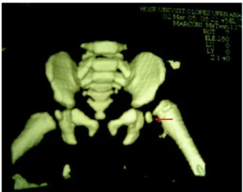
Figura 1. Asymmetry due to transepiphyseal fracture of the left femoral neck observed by computerized tomography. Age 7 months , with recurrent focal seizures evolving to secondarily generalized tonic seizures.
Figura 2. Type 1 fracture (transepiphyseal), according to Delbet’s classification, of the left femoral neck, verified by tridimensional tomographic reconstruction. Age 7 months , with recurrent focal seizures evolving to secondarily generalized type tonic.
79
DISCUSSION
The incidence of fractures is higher in the epileptic population.1,2,4 A series of circumstances act as risk factors
for fractures in this population and they occur: during the seizure itself, in the course of a fall or other accident.4 The
pathological fractures without direct trauma occur in 0.3% of cases of convulsive seizure.5 They may also be related or
not to the use of antiepileptic drugs that act as co-factors for bone demineralization.6,7
The infant in question did not present with a history of trauma, falls or battered-child syndrome, but she did use three antiepileptic drugs: phenobarbital, carbamazepine and clonazepam. Shet8 argues that phenobarbital and
carbamazepine are two drugs that may interfere in the bone health of epileptic children. However, the patient presented with no sign whatsoever of bone demineralization when submitted to metabolic and radiological investigation. Moreover, the child used these drugs for a short time 1-3 months, a fact that surely did not alter bone mineralization.
A review of the literature also showed the following as risk factors: seizure recurrence and the multiple deficiencies associated, type of pathology, treatment duration, type of epilepsy, family history of epilepsy and the use of phenytoin;9
type of drug used.6,7 However, seizure recurrence is the
most significant cause of accidental injury referred to in the majority of studies.
In our case, we believe that the main risk factor was the type of epileptic seizure: partial with secondary generalized tonic seizures associated to poor seizure control. The child had a history of three emergency hospital admissions with convulsive status epileptic. The child may have developed this comorbidity during the period in which she presented with convulsive status epileptic. Regarding the type of spontaneous fracture of the femur following the epileptic crisis itself, we found no reports in the literature on infants with non-controlled seizures. This fact reveals a comorbidity that pediatrician and or pediatric neurology may overlooked in neurologically normal or abnormal epileptic children. This type of fracture involving the proximal femur is one of the most frequent in a epileptic population of hospitalized patients in the 11-to-85 year age group with no history of falling during the seizures and the tonic seizures was the second most frequent type observed.10
Another important factor to be considered in our case is the future orthopedic consequences. While fracture of neck of the femur is rare in children, the high incidence of
complications that can lead to life-long disability makes it an important clinical entity.
Shet,8 sustains that preventing the devastating effect of
fractures in epileptic patients, whether children or adults, must be carried out by recognizing bone status and through investigating the therapeutic effects of antiepileptic drugs so as not to compromise bone health. This child presented with a type 1 transepiphyseal fracture of the left femoral neck, according to Delbet’s classification, an important fracture from the orthopedic point of view, which may result in deficits if not duly diagnosed and treated.4
In conclusion, the case reported here demonstrates the following: first, that there are few reports in the literature on epileptic infants with atraumatic fractures without previous risk factors for osteopenia or rickets, or metabolic alterations; second, that adequate control of seizures is the main fracture prevention measure in this population of infants; and third, the likely non-existence of reports in the literature on this comorbidity in the pediatric population with epilepsy supports the idea that it may overlooked.
REFERENCES
1. Vestergaard P, Tigaran S, Rejnmark L, et al. Fracture risk is increased in epilepsy. Acta Neurol Scand 1999;99:269-75.
2. Souverein PC, Webb DJ, Petri H, Weil J, Van Staa TP, Egberts T. Incidence of Fractures among Epilepsy Patients: A Population-based Retrospective Cohort Study in the General Practice Reseach.
Epilepsia 2005;46(2):304-10.
3. Mirdad T. Fractures of the neck of femur in children: an experience at the Aseer Central Hospital, Abha, Saudi Arabia. Injury 2002;33(9):823-7.
4. Finneli P and Cardi JK. Seizure as a causae of fracture. Neurology 1989;39:858-60.
5. Ribacoba-Montero R and Salas-Puig J. Silmultaneous bilateral fractures of the hip following a grand mal seizure. An unusual complication. Seizure 1997;6:403-4.
6. Mattson RH, Gidal BE. Fractures, epilepsy, and antiepileptic drugs. Epilepsy & Behavior. 2004; 5:Suppl 2, S36-40.
7. Drezner MK. Treatment of anticonvulsant drug-induced bone disease. Epilepsy & Behavior 2004;5:Suppl 2, S41-7.
8. Sheth RD. Metabolic concerns associated with antiepileptic medications. Neurology 2004;63(10)Suppl 4:S24-9.
9. Vestergaard P, Rejnmark L, Mosekilde L. Fracture Risk Associated with Use of Antiepileptic Drugs. Epilepsia 2004;45(11):1330-7. 10. Desai KB, Ribbans WJ and Taylor GJ. Incidence of five common
fractures types in an institutional epileptic population. Injury 1996;27(2):97-100.
Address for correspondence: Áurea Nogueira de Melo