127
J
ournal of Epilepsy and ClinicalNeurophysiology
J Epilepsy Clin Neurophysiol 2005; 11(3):127-130
Memory Tests Are Not Good Predictors of Surgical Outcome
in Patients with Mesial Temporal Lobe Epilepsy Associated
with Hippocampal Sclerosis
Sara Escorsi-Rosset*, Marino M. Bianchin*, Roger Walz****, Vera C. Terra-Bustamante*, Carlos G. Carlotti Jr.**, João A. Assirati Jr.**, Antonio C. Santos***, Américo C. Sakamoto*
Department of Neurology, Psychiatry and Psychology, Ribeirão Preto School of Medicine – USP
ABSTRACT
Introduction: One of the objectives of pre-surgical evaluation in mesial temporal epilepsy associated to hippoocampal sclerosis is the identification of patients with bad surgical prognosis for seizure control. At least theoretically, neuropsychological tests could be used in this venue. Objective: To evaluate whether verbal and visual memory tests can be used as isolate predictors of the post-surgical seizure outcome in patients with mesial temporal lobe epilepsy associated with hippocampal sclerosis refractory to pharmacological treatment. Methods: In a retrospective cohort study using the control of epileptic seizures as end-point, we evaluated 187 patients and calculated the correlation of clinical variables, cognitive evaluation, neuroimaging data, demographic data and electrophysiological findings with the result of seizure control after lobectomy in these patients. Results: An unfavorable prognosis during the postoperative period was observed only in association with low visual reproduction scores (visual memory). However, after Bonferroni correction, which was necessary to reduce the chance of type I error, this result was found to be spurious. Conclusion: We conclude that neuropsychological tests of verbal and visual memory such as those used in the routine presurgical evaluation of our patients with temporal lobe epilepsy are not good isolated predictors of surgical outcome.
Key words: temporal lobectomy, cognitive tests, epilepsy, evolution.
RESUMO
Testes de memória como preditores de resultado cirúrgico em pacientes com epilepsia do lobo temporal mesial associada à esclerose hipocampal
Introdução: Um dos objetivos da avaliação pré-cirúrgica de pacientes com epilepsia do lobo temporal é detectar pacientes com mau prognóstico cirúrgico para o controle das crises epilépticas. Teoricamente, tes-tes neuropsicológicos poderiam ser empregados com tal finalidade. Objetivo: Avaliar se tes-testes-tes de memória verbal e visual podem ser utilizados como preditores isolados de resultado cirúrgico do controle de crises em pacientes com epilepsia do lobo temporal mesial associada a esclerose hipocampal refratária ao tratamento farmacológico. Métodos: Em um estudo de coorte retrospectivo, usando como end-point o controle de cri-ses epilépticas, nós avaliamos 187 pacientes, correlacionando aspectos clínicos, avaliação cognitiva, dados de neuroimagem, dados demográficos e achados eletrofisiológicos com o resultado do controle de crises após a lobectomia nesses pacientes. Resultados: Um prognóstico desfavorável no período pós-operatório foi ob-servado apenas em associação com baixos escores de reprodução visual (memória visual). Contudo, após uma correção de Bonferroni, necessária para reduzir a chance de erro tipo I, esse resultado mostrou-se espú-rio. Conclusão: Nós concluímos que os testes neuropsicológicos de memória verbal e visual, tais como utili-zados na avaliação pré-cirúrgica de rotina dos nossos pacientes com epilepsia do lobo temporal, não são bons preditores isolados de resultado cirúrgico.
Unitermos: lobectomia temporal, testes cognitivos, epilepsia, evolução.
* Epilepsy Surgery Center (CIREP), Department of Neurology, Psychiatry and Psychology, Ribeirão Preto School of Medicine, University of São Paulo, Brazil.
** Division of Neurosurgery, Department of Surgery and Anatomy, Ribeirão Preto School of Medicine, University of São Paulo, Brazil. *** Division of Radiology, Department of Internal Medicine, Ribeirão Preto School of Medicine, University of São Paulo, Brazil. **** Division of Neurology, Department of Internal Medicine, Federal University of Santa Catarina, Brazil.
128 INTRODUCTION
Neuropsychological tests have long been used as part of the presurgical evaluation of patients with temporal lobe epilepsy associated with hippocampal sclerosis refractory to pharmacological treatment. They are valuable tools for the presurgical evaluation of these patients since they have important implications regarding the cognitive prognosis during the postoperative period.
Indeed, a poor performance in neuropsychological tests may reflect patients who present multiple brain lesions or dysfunctions that may not be detectable by other auxiliary exams commonly used for the preoperative evaluation of these patients. Along this same line of reasoning, it is possible that the magnitude of these lesions or dysfunctions may be correlated with the prognosis of surgical seizure control, as well as with the cognitive prognosis of these patients. On this basis, neuro-psychological tests could be used as measures of the underlying neurological damage or dysfunction and could theoretically be correlated with the surgical prognosis.
Varying results have been obtained in different studies regarding the possible use of cognitive measurements as separate predictors of surgical outcome. While some authors have found no prognostic value for these evaluations, others have demonstrated their potentials. For example, Loring et al.(1), Sperling et al.(2) and Salanova
et al.(3), using the sodium amytal test, observed that
asymmetrical memory performance was of no value as a presurgical predictor of the prognosis of seizure evolution in temporal lobe epilepsy. Jeong et al.(4) and Kim et al.(5)
observed a correlation with electrophysiological or imaging exams, but not with postoperative seizure evolution. In contrast, Blume et al.(6) found a correlation between
neuropsychological tests and side of surgery although they did not find a correlation with postoperative seizure prognosis. Holmes et al.(7) reported that
neuropsycho-logical tests can help to select the side of the lesion when the electroencephalogram is poorly lateralizing or when it presents bilateral discharges.
In the present study we analyzed the possible role of verbal memory and visual memory as separate predictors of seizure prognosis in patients with temporal lobe epilepsy associated with hippocampal sclerosis in a cohort of patients surgically treated at the Ribeirão Preto Epilepsy Surgery Center (CIREP).
PATIENTS AND METHODS
In a classic retrospective cohort study, we first analyzed 187 eligible patients submitted to anterior and mesial tem-poral lobectomy for the treatment of epilepsy refractory to medications during the period from 1995 to 2001. We classified the patients as having good surgical control or
poor surgical control according to the criteria described below. We then analyzed and correlated the results of the Logical Memory and Visual Reproduction tests (routinely used for the presurgical evaluation of these patients) with the post-surgical seizure outcome. All patients included in the study had been followed up for at least four years after surgery.
The inclusion criteria were as follows: 1) seizure semiology consistent with mesial temporal sclerosis, usually with epigastric malaise and psychic auras followed by behavioral arrest and oro-alimentary and gestual auto-matisms. 2) interictal temporal spikes; 3) no other lesion except atrophy and increased signal in the hippocampus revealed by nuclear magnetic resonance (NMR); 4) histo-pathological examination compatible with hippocampal sclerosis; 5) absence of double pathology that might be identified by clinical, electrophysiological or neuroimaging methods. The exclusion criteria were as follows: 1) abnor-mal physical and neurological examination; 2) extra-temporal or generalized spikes in the electroencepha-logram.
Clinical parameters and presurgical evaluation of the patients
Clinical characteristics such as sex, age at surgery, age at the onset of epilepsy (recurrent seizures), duration of epilepsy, a positive history of an initial precipitating event (IPE), frequency of seizures per month and side of surgery were analyzed. A positive family history of epilepsy was defined when at least one first-degree relative reported at least two spontaneous seizure during life. Presurgical evaluation was performed by a multidisciplinary team, consisting of a detailed clinical history and neurological examination, analysis of ictal and interictal video-electroencephalogram, structural and functional imaging, psychiatric evaluation, neuropsychological examination, social evaluation, and, when appropriate, the intracarotid test with sodium amytal (Wada test) for memory and language representation. All tests were carried out at the same center (CIREP).
129 seizures after surgery was defined on the basis of seizure status determined by clinical interviews with the patients. Patients were characterized as seizure free if they did not present partial complex or generalized seizures after surgery. Auras (Engel 1b) were not considered in our analysis. Patients who presented a partial complex seizure or a generalized tonic-clonic seizure clearly related to non-compliance with the medication (Engel 1d) or during the first month after surgery (perioperative period), but who later maintained a good seizure control with adequate use of the medication were also considered to be seizure free.
Statistical analysis
The differences between the seizure-free group (Engel 1) and the group that continued to have seizures (Engel 2, 3, 4) were analyzed by the Mann-Whitney test for continuous variables and by the Chi-square test for categorical variables. Due to the multiple correlations, Bonferroni correction was used to reduce the chance of type I error. The level of significance was set at p < 0.05. The SPSS program (Chicago, IL, USA), version 10, was used for all statistical analyses.
RESULTS
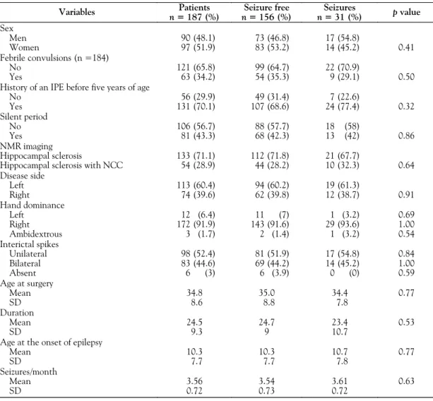
The results are presented in Tables 1 and 2. Sex dis-tribution was similar in both groups. Male patients (n = 90) represented 48% of a total of 187 patients, and 73 (81.1%) of them became seizure free after surgical treatment. Female patients (n = 97) represented 52% of the total number of patients and 83 (85.6%) became seizure free after surgical treatment. None of the variables analyzed (sex, febrile convulsions, history of an IPE before five years of age, silent period, neuroimaging findings, side of disease, hand dominance, interictal spikes, age at surgery, duration of disease, or age) proved to be predictive of the surgical prognosis of seizure control in our patients.
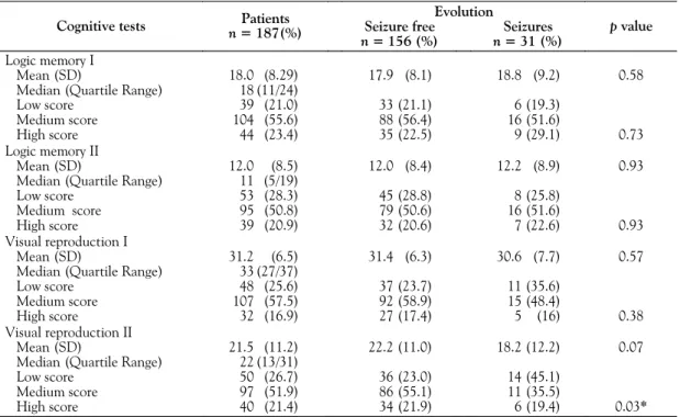
The results of cognitive performance for patients with temporal lobe epilepsy are presented in Table 2. The results of the Visual Reproduction II test showed that the patients with the highest performance tended to have a better surgical prognosis regarding seizure control (p = 0.03) com-pared to patients with a lower performance. However, this result was probably spurious (alpha error) since this signi-ficance disappeared after Bonferroni correction (p = 0.48).
Table 1. Demographic data correlated with seizure evolution during the postoperative period.
Variables Patients
n = 187 (%)
Seizure free
n = 156 (%)
Seizures
n = 31 (%) p value Sex
Men 90 (48.1) 73 (46.8) 17 (54.8)
Women 97 (51.9) 83 (53.2) 14 (45.2) 0.41
Febrile convulsions (n =184)
No 121 (65.8) 99 (64.7) 22 (70.9)
Yes 63 (34.2) 54 (35.3) 9 (29.1) 0.50
History of an IPE before five years of age
No 56 (29.9) 49 (31.4) 7 (22.6)
Yes 131 (70.1) 107 (68.6) 24 (77.4) 0.32
Silent period
No 106 (56.7) 88 (57.7) 18 (58)
Yes 81 (43.3) 68 (42.3) 13 (42) 0.86
NMR imaging
Hippocampal sclerosis 133 (71.1) 112 (71.8) 21 (67.7)
Hippocampal sclerosis with NCC 54 (28.9) 44 (28.2) 10 (32.3) 0.64 Disease side
Left 113 (60.4) 94 (60.2) 19 (61.3)
Right 74 (39.6) 62 (39.8) 12 (38.7) 0.91
Hand dominance
Left 12 (6.4) 11 (7) 1 (3.2) 0.69
Right 172 (91.9) 143 (91.6) 29 (93.6) 1.00
Ambidextrous 3 (1.7) 2 (1.4) 1 (3.2) 0.54
Interictal spikes
Unilateral 98 (52.4) 81 (51.9) 17 (54.8) 0.84
Bilateral 83 (44.6) 69 (44.2) 14 (45.2) 1.00
Absent 6 (3) 6 (3.9) 0 (0) 0.59
Age at surgery
Mean 34.8 35.0 34.4 0.77
SD 8.6 8.8 7.8
Duration
Mean 24.5 24.7 23.4 0.53
SD 9.3 9 10.7
Age at the onset of epilepsy
Mean 10.3 10.3 10.7 0.77
SD 7.7 7.7 7.8
Seizures/month
Mean 3.56 3.54 3.61 0.63
SD 0.72 0.73 0.72
130 DISCUSSION
In the present study we report the value of visual and verbal memory tests when used as prognostic factors of seizure control after anterior and mesial temporal lobectomy in patients with mesial temporal lobe epilepsy associated with hippocampal sclerosis. These tests are commonly used for the evaluation of such patients. We conclude that neither the Logic Memory nor the Visual Reproduction tests are useful to predict the surgical prognosis of these patients regarding the control of epileptic seizures.
Performance in cognitive tests may be directly associated with different factors, among them an early onset of epilepsy, the initial precipitating event, the duration of epilepsy, the use of antiepileptic drugs in monotherapy or polytherapy, febrile convulsion, and number of epileptic seizures per month. These tests are particularly relevant for the postsurgical prognosis regarding memory (verbal and non-verbal) functions, language, visuospatial organization, attention, executive functions, as well as intellectual efficiency. On this basis, we can inform the patients about possible cognitive changes after mesial and anterior temporal lobectomy. In addition, these tests can be used to assess in an objective manner the changes in these functions due to surgery.
In the present study, we conclude that, even though neuropsychological tests are important in different aspects of the evolution of patients with temporal lobe epilepsy associated with mesial sclerosis, they are not efficient predictors of seizure control after temporal lobectomy.
ACKNOWLEDGMENTS
MMB was supported by FAPESP (02/03743-0). The authors are indebted to Elettra Greene for English review.
REFERENCES
1. Loring DW, Meador KJ, Lee GP, Nichols ME, King DW, Gallagher BB, Murro AM, Smith JR. Wada memory performance predicts seizure outcome following anterior temporal lobectomy. Neurology 1994 Dec; 44(12):2322-4.
2. Sperling MR, Saykin AJ, Glosser G, Moran M, French JA, Brooks M, O’Connor MJ. Predictors of outcome after anterior temporal lobectomy: The intracarotid amobarbital test. Neurology 1994 Dec; 44(12):2325-30.
3. Salanova V. Markand ON, Worth R. Clinical characteristics and predictive factors in 98 patients with complex partial seizures treated with temporal resection. Arch Neurol 1994 Oct; 51(10):1008-13.
4. Jeong SW, Lee SK, Kim KK, Kim H, Kim JY, Ching CK. Prognostic factors in anterior temporal lobe resections for mesial temporal epilepsy: multivariate analysis. Epilepsia 1999 Dec; 40(12):1735-9. 5. Kim H, Yi S, Son EI, Kim J. Lateralization of epileptic foci by neuropsychological testing in mesial temporal lobe epilepsy. Neuropsychologia 2004 Jan; 18(1):141-51.
6. Blume WT, Grabow JD, Darley FL, Aronson AE. Intracarotid amobarbital test of language and memory before temporal lobectomy for seizure control. Neurology 1973 Aug; 23(8):812-9. 7. Holmes MD, Miles AN, Dodrill CB, Ojemann GA, Willensky AJ.
Identifying potential surgical candidates in patients with evidence of bitemporal epilepsy. Epilepsia 2003 Aug; 44(8):1075-9.
Corresponding Author:
Américo C. Sakamoto
Department of Neurology, Psychiatry and Psychology Campus Universitário
14048-900, Ribeirão Preto, SP, Brazil Fax: (55) 16-633-0760
E-mail: [email protected]
Table 2. Cognitive performance and seizure evolution during the postoperative period.
Evolution Cognitive tests n = 187(%) Patients Seizure free
n = 156 (%)
Seizures
n = 31 (%)
p value
Logic memory I
Mean (SD) 18.0 (8.29) 17.9 (8.1) 18.8 (9.2) 0.58 Median (Quartile Range) 18 (11/24)
Low score 39 (21.0) 33 (21.1) 6 (19.3)
Medium score 104 (55.6) 88 (56.4) 16 (51.6)
High score 44 (23.4) 35 (22.5) 9 (29.1) 0.73
Logic memory II
Mean (SD) 12.0 (8.5) 12.0 (8.4) 12.2 (8.9) 0.93
Median (Quartile Range) 11 (5/19)
Low score 53 (28.3) 45 (28.8) 8 (25.8)
Medium score 95 (50.8) 79 (50.6) 16 (51.6)
High score 39 (20.9) 32 (20.6) 7 (22.6) 0.93
Visual reproduction I
Mean (SD) 31.2 (6.5) 31.4 (6.3) 30.6 (7.7) 0.57
Median (Quartile Range) 33 (27/37)
Low score 48 (25.6) 37 (23.7) 11 (35.6)
Medium score 107 (57.5) 92 (58.9) 15 (48.4)
High score 32 (16.9) 27 (17.4) 5 (16) 0.38
Visual reproduction II
Mean (SD) 21.5 (11.2) 22.2 (11.0) 18.2 (12.2) 0.07 Median (Quartile Range) 22 (13/31)
Low score 50 (26.7) 36 (23.0) 14 (45.1)
Medium score 97 (51.9) 86 (55.1) 11 (35.5)
High score 40 (21.4) 34 (21.9) 6 (19.4) 0.03*
*p = 0.48 after Bonferroni correction.