ORIGINAL ARTICLE
DOI: 10.1590/1516-3180.2014.1322544Incidence and risk factors for retinopathy
of prematurity in a Brazilian reference service
Incidência e fatores de risco para retinopatia da prematuridade
em um serviço de referência brasileiro
Eduardo Gonçalves
I, Luciano Sólia Násser
II, Daniella Reis Martelli
III, Isadora Ramos Alkmim
IV, Thalita Veloso Mourão
V,
Antônio Prates Caldeira
VI, Hercílio Martelli-Júnior
VIUniversidade Estadual de Montes Claros (Unimontes)
, Montes Claros, Minas Gerais, Brazil
ABSTRACT
CONTEXT AND OBJECTIVE: Retinopathy of prematurity (ROP) is a known cause of blindness in which diagnosis and timely treatment can prevent serious harm to the child. This study aimed to evaluate the incidence of ROP and its association with known risk factors.
DESIGN AND SETTING: Longitudinal incidence study in the neonatal intensive care unit (NICU) of Univer-sidade Estadual de Montes Claros.
METHODS: Newborns admitted to the NICU with gestational age less than 32 weeks and/or birth weight less than 1,500 grams, were followed up over a two-year period. The assessment and diagnosis of ROP were deined in accordance with a national protocol. The chi-square test or Fisher’s exact test were used to determine associations between independent variables and ROP. Analysis on the independent efect of the variables on the results was performed using multiple logistic regression.
RESULTS: The incidence of ROP was 44.5% (95% conidence interval, CI = 35.6-46.1) in the study popu-lation. The risk factors associated with the risk of developing the disease were: birth weight less than 1,000 grams (odds ratio, OR = 4.14; 95% CI = 1.34-12.77); gestational age less than 30 weeks (OR = 6.69; 95% CI = 2.10-21.31); use of blood derivatives (OR = 4.14; 95% CI = 2.99-8.99); and presence of sepsis (OR = 1.99; 95% CI = 1.45-2.40).
CONCLUSIONS: The incidence of ROP was higher than that found in the literature. The main risk factors were related to extreme prematurity.
RESUMO
CONTEXTO E OBJETIVO: A retinopatia da prematuridade (ROP) é causa conhecida de cegueira e diag-nóstico e tratamento oportunos podem evitar graves danos à criança. Este estudo objetivou avaliar a incidência da ROP e sua associação com fatores de risco conhecidos.
TIPO E LOCAL DE ESTUDO: Estudo longitudinal de incidência na Unidade de Terapia Intensiva Neonatal (UTIN) da Universidade Estadual de Montes Claros.
MÉTODOS: Foram acompanhados neonatos admitidos na UTIN, com idade gestacional menor que 32 semanas e/ou peso ao nascimento inferior a 1.500 gramas, por um período de dois anos. Avaliação e diag-nóstico de ROP foram deinidos conforme protocolo nacional. Utilizou-se o teste qui-quadrado ou teste exato de Fisher para determinar a associação entre as variáveis independentes e ROP. A análise do efeito independente das variáveis sobre o resultado (ROP) foi realizada por meio de regressão logística múltipla.
RESULTADOS: A incidência de ROP foi de 44,5% (intervalo de coniança, IC 95% = 35,6-46,1) entre a população estudada. Os fatores de risco pesquisados que apresentaram relação de risco para o de-senvolvimento da doença foram: peso de nascimento menor que 1000 gramas (odds ratio, OR = 4,14; IC 95% = 1,34;12,77), idade gestacional menor que 30 semanas (OR = 6,69; IC 95% = 2,10;21,31), uso de hemoderivados (OR = 4,14; IC 95% = 2,99;8,99) e presença de sepse (OR = 1,99; IC 95% = 1,45;2,40).
CONCLUSÃO: A incidência de ROP foi maior que a encontrada na literatura. Os principais fatores de risco estão relacionados à prematuridade extrema.
IMD. Doctoral Student and Professor,
Postgraduate Health Science Program, Universidade Estadual de Montes Claros (Unimontes), and Faculdades Integradas Pitágoras (FIPMoc), Montes Claros, Minas Gerais, Brazil.
IIMD. Master’s Student, Postgraduate Health
Science Program, Universidade Estadual de Montes Claros (Unimontes), Montes Claros, Minas Gerais, Brazil.
IIIMD. Doctoral Student and Professor,
Postgraduate Health Science Program, Universidade Estadual de Montes Claros (Unimontes), Montes Claros, Minas Gerais, Brazil.
IVMedical Student, Universidade Estadual de
Montes Claros (Unimontes), Montes Claros, Minas Gerais, Brazil.
VMedical Student, Faculdades Integradas Pitágoras
(FIPMoc), Montes Claros, Minas Gerais, Brazil.
VIMD, PhD. Professor, Postgraduate Health
Science Program, Universidade Estadual de Montes Claros (Unimontes), Montes Claros, Minas Gerais, Brazil.
KEY WORDS:
Retinopathy of prematurity. Incidence.
Infant, premature. Risk factors. Gestational age.
PALAVRAS-CHAVE: Retinopatia da prematuridade. Incidência.
INTRODUCTION
Retinopathy of prematurity (ROP) is a vasoproliferative eye dis-ease of multifactorial etiology that afects the retinal vasculariza-tion of premature infants.1,2 he importance of ROP lies in its frequency and in prevention of blindness due to this condition, given that, once diagnosed and treated, it is unlikely to develop into complete loss of vision.3-6 he incidence of blindness varies between countries, and it is inluenced by the level of perinatal care and the existence of screening programs for early diagnosis.7
In the United States, around 0.12% of all live births develop ROP, or one case for every 820 newborns, and there are an esti-mated 300 new cases of blindness annually due to ROP in that country.3 In Brazil, studies have shown increased numbers of ROP cases, especially in large centers.7 It is known that ROP is multifactorial, due to the immaturity of the preterm retina,3,8 and that the risk factors are: prematurity, low birth weight, oxygen therapy, intracranial hemorrhage and persistent ductus arterio-sus, among others.8-10
OBJECTIVE
he aim of this study was to evaluate the incidence of ROP at a Brazilian reference service, along with the main known risk factors for this important condition and its association with morbidity.
METHODS
This was a prospective longitudinal incidence study and it was approved by the Ethics Committee for Institutional Research of Montes Claros State University (Universidade Estadual de Montes Claros, Unimontes), in Minas Gerais, Brazil (protocol 2013/10).
In this study, a convenience sample limited to a period of time was used. he study included preterm infants who had been admitted to the neonatal intensive care unit (NICU) of the Unimontes university hospital, between May 2009 and April 2011. he inclusion criteria were that the birth weight should be
≤ 1,500 g and/or the gestational age should be ≤ 32 weeks, with survival at least until the sixth week of life;6 and that the infants were treated as outpatients for follow-up care in the eye clinic. he study excluded infants born with ocular malformation or ocular genetic alterations and those who did not survive past the sixth week of life.
here was no sample calculation. Over the period from May 2009 to April 2011, 124 patients who met the inclusion criteria were admitted. However, 12 patients died and two did not return to the clinic for monitoring. he eye examination took place in the sixth week of life, or between 32 and 36 weeks of corrected gestational age. It consisted of using a binocular indirect ophthal-moscope (BIO) under pupil dilation produced by the eye drops
0.5% tropicamide (Midriacyl, Alcon) and 2.5% phenylephrine (Fenilefrina, Allergan), which were applied twice with an inter-val of ten minutes. he test was completed one hour aterwards and, when necessary, anesthetic eye drops (Anestalcon, Alcon) were also used. A 28-diopter lens (Nikon, Melville, NY, USA) and lid speculum (Alfonso Eye Speculum, Storz, Bausch & Lomb Inc., San Dimas, CA, USA) were used. Scleral indentation was used when necessary.
Children who did not present any degree of retinopathy of pre-maturity were not considered to be ROP patients. Children who had some degree of ROP were considered to have ROP. he degree of retinopathy assigned to each patient was that of the severest degree seen in the eyes of the infant under examination.6
The evaluations were repeated periodically, in accordance with the procedures laid out in the Brazilian Examination and Treatment Guidelines for individuals with ROP.6 The ophthal-mological examinations were always performed by the same ophthalmologist, who had had specialized retinal training, in the neonatology center. The ophthalmologist did not have any prior knowledge of the medical histories of any of the patients. The examinations were conducted at the neonatal unit and the patients were monitored until reaching 42 weeks of cor-rected gestational age, or until complete retinal stabilization had been achieved.
he ophthalmological monitoring of the patients was per-formed based on the stage of the disease. Patients with incom-plete vascularization of the retina were monitored at intervals of one to three weeks until vascularization had been completed. Infants with ROP of degrees I, II or III4 (excluding threshold dis-ease) underwent weekly monitoring until complete vascular-ization of the retina had been achieved. hose with threshold disease underwent treatment for retinal photo-coagulation or cryotherapy. Infants with ROP of degree IV underwent evalua-tion of scleral explant with or without associated photo-coagu-lation or cryotherapy or posterior vitrectomy. For infants with ROP of degree V, possible surgical treatment needed to be dis-cussed. Because of technical limitations, patients requiring treat-ment by means of photo-coagulation, cryotherapy or another surgical procedure were referred to another unit of the hospital.
Incidence and risk factors for retinopathy of prematurity in Brazilian reference service | ORIGINAL ARTICLE
In relation to the therapies used in this study, the follow-ing factors were taken into consideration: maximum fraction of inspired oxygen (FiO2) level; use of oxygen therapy by means of continuous positive airway pressure (CPAP) or mechanical ven-tilation; use of indomethacin; use of a surfactant, aminophylline or cafeine; phototherapy use; blood transfusions; use of diuret-ics; and corticosteroid use.
For diseases detected during hospitalization, presence of the following was recorded: initial respiratory distress, broncho-pulmonary dysplasia (use of oxygen for 28 days or more), sep-sis (clinical or laboratorial diagnosep-sis), patent ductus arterio-sus (diagnosis by Doppler echocardiography) and intracranial hemorrhage (transfontanellar ultrasound between ive and ten days of life).
For statistical analysis, the study population was classiied as individuals with ROP and those without ROP, i.e. patients who did not show the illness. he descriptive analysis was performed using absolute numbers and percentages of the qualitative vari-ables and of the central trend averages: means and their respec-tive standard deviations (SD) of the quantitarespec-tive variables. In fur-ther analysis, all the variables were dichotomized.
In the univariate analysis, the chi-square test and the Fisher exact test were used to determine any associations between the independent variables and the outcome (ROP). he magnitude of the efect of the risk factors on the outcome was expressed as the odds ratio (OR), with its respective 95% conidence interval (CI). he analysis on the independent efects of the intervening vari-ables on the outcome (ROP) was performed by means of mul-tiple logistic regression, using forward modeling. All variables that showed P < 0.25 in the association test were included in the modeling process. To build the database and to do the statisti-cal analysis, the Statististatisti-cal Package for the Social Sciences (SPSS) sotware (SPSS 18.0 for Windows, SPSS Inc., Chicago, IL, USA) and the Epi Info sotware (CDC Epi Info 3.5.4, Atlanta, Georgia, USA) were used.
RESULTS
Between May 2009 and April 2011, 124 children weighing less than 1,500 grams were born, of which 12 died before the irst evaluation and 2 did not return for examination ater being dis-charged. hus, 110 infants were evaluated. he population stud-ied was classistud-ied as individuals with ROP, i.e. those who showed
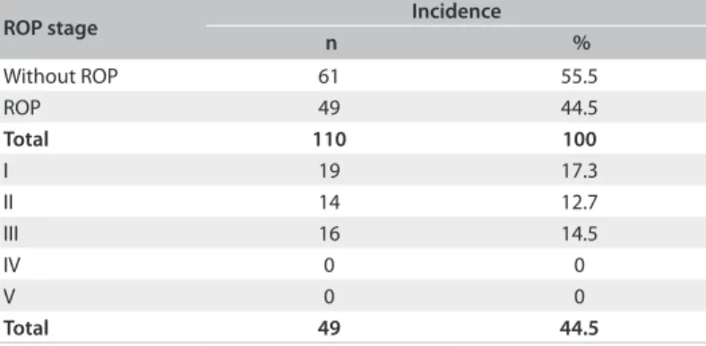
some degree of retinopathy of prematurity, and individuals with-out ROP, i.e. those who did not show retinopathy of prematu-rity. Among these 110 infants, the prevalence of ROP was 44.5% (95% CI = 35.6-46.1). Considering all the births that took place in the department during the study period, the incidence of ROP was 1.1%. Stage I ROP was the form with the highest incidence among these infants (Table 1), afecting 19 children (17.3% of
those with ROP), followed by stage III with 16 children (14.5%) and stage II with 14 children (12.7%). here were no patients with stages IV or V. Two premature infants (1.8%) needed laser treatment because they showed threshold disease. he aver-age weights and gestational aver-ages of all the patients are shown in Table 2.
In the study population, 49.1% of the patients were male. Seventy-four percent of the mothers reported that they had attended prenatal care consultations, 72% presented some com-plications during pregnancy and 43% had some comcom-plications during childbirth. he most common form of delivery was cesar-ean (65.5%). he SNAPPE II score was obtained for all the pre-term infants ater they had reached an average of 12 hours of life, ranging from 0 to 88. About half of the population studied was small for the gestational age (49.8%).
Among the risk factors studied, the following correlated sig-niicantly (P < 0.05) with the development of ROP, in univariate analysis: gestational age less than 30 weeks, Apgar scores at the irst and ith minute less than 7, SNAPPE II score less than 12, use of blood transfusions, use of diuretics, use of aminophylline or cafeine, use of surfactants, presence of sepsis and presence of bronchopulmonary dysplasia (Table 3).
Table 1. Incidence of retinopathy of prematurity (ROP) and disease stage
ROP stage Incidence
n %
Without ROP 61 55.5
ROP 49 44.5
Total 110 100
I 19 17.3
II 14 12.7
III 16 14.5
IV 0 0
V 0 0
Total 49 44.5
Table 2. Means and standard deviations of the birth weight and gestational age risk factors of the diferent stages of retinopathy of prematurity (ROP)
Risk factors Without ROP
(n = 61)
ROP I (n = 19)
ROP II (n = 14)
ROP III
(n = 16) P-value
Weight (grams) 1153.43 ± 241.48 1012.13 ± 183.70 1097.30 ± 213.43 814.35 ± 159.39 0.004
Table 3. Risk factors for development of retinopathy of prematurity (ROP) in infants with weight ≤ 1,500 grams and/or gestational age
≤ 32 weeks; univariate analysis
Risk factors
With ROP (n = 49)
Without ROP
(n = 61) P-value OR (CI)
n (%) n (%)
Male gender 22 (44.9) 32 (52.5) 0.430 0.74 (0.35-1.57)
Gestational age less than 30 weeks 36 (73.5) 16 (26.2) < 0.001 7.79 (3.32-18.28)
Birth weight less than 1,000 grams 14 (28.6) 10 (16.4) 0.124 2.04 (0.81-5.11)
Presence of multiple pregnancies 5 (10.2) 4 (6.6) 0.508 0.62 (0.16-2.44)
Use of prenatal corticosteroids 13 (26.5) 10 (16.4) 0.194 1.84 (0.73-4.66)
Apgar score at one minute less than 7 38 (77.6) 35 (57.4) 0.026 2.57 (1.11-5.95)
Apgar score at ive minutes less than 7 17 (34.7) 9 (14.8) 0.014 3.07 (1.22-7.70)
Initial respiratory discomfort 46 (93.9) 58 (95.1) 1.000 0.79 (0.15-4.12)
SNAPPE II score less than 12 34 (69.4) 21 (34.4) < 0.001 4.32 (1.93-9.66)
Oxygen therapy using headpiece (HOOD) for more than 5 days 12 (24.5) 10 (16.4) 0.290 1.65 (0.65-4.23)
Oxygen therapy using CPAP for more than 5 days 13 (26.5) 8 (13.1) 0.075 2.39 (0.90-6.36)
Oxygen therapy using mechanical ventilation for more than 5 days 29 (59.2) 21 (34.4) 0.001 2.76 (1.27-6.01)
Use of blood transfusions 27 (55.1) 13 (21.3) < 0.001 4.53 (1.97-10.41)
Use of diuretics 30 (61.2) 20 (32.8) 0.003 3.24 (1.48-7.09)
Use of indomethacin 10 (20.4) 8 (13.1) 0.304 1.69 (0.61-4.69)
Use of aminophylline/cafeine 36 (73.5) 33 (54.1) 0.037 2.35 (1.05-5.28)
Use of surfactant 29 (59.2) 20 (32.8) 0.006 2.97 (1.36-6.49)
Presence of sepsis 48 (98.0) 50 (82.0) 0.001 1.98 (1.63-2.41)
Presence of bronchopulmonary dysplasia 26 (53.1) 15 (24.6) 0.002 3.47 (1.54-7.78)
Presence of intracranial hemorrhage 10 (20.4) 10 (16.4) 0.589 1.31 (0.49-3.45)
Presence of patent ductus arteriosus (diagnosed using Doppler
echocardiography) 14 (28.6) 13 (21.3) 0.379 1.48 (0.62-3.53)
Use of phototherapy 45 (91.8) 60 (98.4) 0.170 1.10 (0.02-1.73)
OR = odds ratio; CI = conidence interval; CPAP = continuous positive airway pressure; SNAPPE = Score for Neonatal Acute Physiology and SNAP Perinatal Extension.
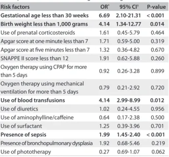
Table 4. Risk factors for retinopathy of prematurity (ROP) development in infants with weight < 1,500 grams
Risk factors OR* 95% CI† P-value
Gestational age less than 30 weeks 6.69 2.10-21.31 < 0.001 Birth weight less than 1,000 grams 4.14 1.34-12.77 0.014 Use of prenatal corticosteroids 1.61 0.45-5.79 0.464 Apgar score at one minute less than 7 1.71 0.59-5.00 0.319 Apgar score at ive minutes less than 7 1.32 0.36-4.82 0.670
SNAPPE II score less than 12 1.91 0.62-5.88 0.260
Oxygen therapy using CPAP for more
than 5 days 0.92 0.26-3.28 0.899
Oxygen therapy using mechanical
ventilation for more than 5 days 0.79 0.21-2.92 0.720 Use of blood transfusions 4.14 2.99-8.99 0.012
Use of diuretics 1.02 0.24-4.55 0.956
Use of aminophylline/cafeine 0.64 0.17-2.38 0.500
Use of surfactant 1.25 0.39-3.96 0.701
Presence of sepsis 1.99 1.45-2.40 < 0.001
Presence of bronchopulmonary dysplasia 1.92 0.68-5.46 0.219
Use of phototherapy 0.27 0.69-1.07 0.062
*OR = odds ratio; †CI = conidence interval; SNAPPE = Score for Neonatal
Acute Physiology and SNAP Perinatal Extension; CPAP = continuous positive airway pressure.
he multivariate analysis using hierarchical logistic regres-sion (the forward method) is represented in Table 4. All of
the variables that showed P < 0.25 in the association test were included in the modeling process. Among the risk factors inves-tigated, we found that the following showed a risk of develop-ing ROP: birth weight less than 1,000 grams (OR = 4.14; 95% CI = 1.34-12.77); gestational age less than 30 weeks (OR = 6.69; 95% CI = 2.10-21.31); use of blood transfusions (OR = 4.14; 95% CI = 2.99-8.99); and presence of sepsis (OR = 1.99; 95% CI = 1.45-2.40). here was no veriied association with any risk factors, except for the signiicant trend of the gestational age fac-tor (P = 0.06).
DISCUSSION
Incidence and risk factors for retinopathy of prematurity in Brazilian reference service | ORIGINAL ARTICLE
his includes all preterm infants assessed. In the Brazilian litera-ture, ROP incidence rates of 29.1%,13 28.5%9 and 27.2%14 have been described. hese studies fell within a similar range of inci-dence rates and showed the highest rate for stage I ROP, which was about the same as what was found in the present study. his could explain the higher ROP incidence rate found.15
he Cryotherapy for Retinopathy of Prematurity (CRYO-ROP) study showed an incidence of 65.8%.16 However, the weight criterion required for inclusion in the study was lower than that of the present study (weight < 1,250 grams). Another impor-tant American study, the Early Treatment for Retinopathy of Prematurity Study (ETROP), also included infants with weights of less than 1,250 grams and showed an incidence of 68%.17
Although gestational age can sometimes be hard to pin-point as it is oten imprecise and sometimes unknown, gesta-tional age and birth weight were still used as the criteria for this study. hese criteria were set because the Brazilian guidelines for screening ROP, published in 2007,14 deine the inclusion crite-ria as birth weight < 1,500 g and/or a gestational age < 32 weeks. Additionally, low gestational age and low birth weight have been associated with and have the same consequence as immature ret-inal tissue.9,18,19 he results from these studies showed that both gestational age and birth weight were associated with ROP.
Several studies in the literature have shown that the lower the birth weight and the lower the gestational age are, the higher the chance of developing ROP also is.20 Moreover, lower birth weight and lower gestational age are associated with the devel-opment of more serious forms of ROP. In the current study, stage I of the disease was the most frequently observed form, account-ing for 17.3% of the cases when all infants with birth weight < 1,500 grams are included. his proportion rises to 25.8% if only the infants with birth weight < 1,250 grams are included. hese ind-ings were not reported in the CRYO-ROP or ETROP studies, which showed higher rates of the severer forms of ROP in infants with lower birth weights and in those with lower gestational age. By ana-lyzing the average birth weight and gestational age among individu-als with varying stages of ROP in the present study, it was noted that there was a statistically signiicant diference between the preterm infants with stage III ROP and those without ROP, thus conirm-ing the association between the immature retina and the ROP stage.
Just 1.8% of the premature infants in the present study were treated. his small number can be explained by the decrease in stage III cases. It has been questioned whether there is any asso-ciation between the severity of the SNAPPE II score and retinop-athy of prematurity.14 he present study showed that although a signiicant association was observed in univariate analysis, it did not remain in the inal model of the multivariate analysis.
The use of oxygen therapy in this study was evaluated in terms of number of days and form of administration. After
multivariate analysis, none of the forms of oxygen admin-istration (mechanical ventilation, CPAP or HOOD) showed any statistical association with ROP. Other studies have con-firmed the association between the risk of ROP and the use of mechanical ventilation21 and CPAP. Oxygenation, regard-less of the form of use, has been implicated in causing ROP.22 The less mature the preterm infant is, the greater the need for mechanical ventilation and CPAP is, which explains this finding.
he treatment for bronchopulmonary dysplasia includes use of diuretics and corticosteroids. he present study showed that there was an association between the risk of ROP and the use of diuretics, but only in univariate analysis. In the multicenter STOP-ROP study,23 it was found that supplemental use of oxygen therapy for preterm infants with pre-threshold disease increased the risk of chronic pulmonary diseases and also increased the use of diuretics and length of hospital stay. Use of prenatal corticoste-roids did not show any statistical impact on development of ROP, which has not been shown in other studies.24
he presence of sepsis in our study was considered to be a risk factor for ROP and showed signiicance. In the literature, this association has already been described.25 A study by Chen et al. showed that sepsis can be considered to be an important risk fac-tor for ROP, in screening for preterm infants weighing between 1,501 and 2,000 grams.26
he presence of blood transfusions was another risk factor that showed a signiicant relationship between the groups. his inding has also been reported by several authors in the litera-ture.5,9,19 It is believed that fetal hemoglobin has a greater ain-ity to oxygen than does adult hemoglobin. hus, a transfusion of adult hemoglobin could generate possible hyperoxia due to increased oxygen delivery to tissues.27 Another theory is that there could be an increase in free radicals ater the transfusions due to an increase in plasma free iron.28-30
CONCLUSIONS
he observed incidence was higher than that found in the litera-ture, thus showing that occurrences of retinopathy of prematu-rity remain high among infants with very low birth weight. he development of ROP was inversely proportional to the weight and gestational age at birth. Prevention of prematurity and cau-tion in using oxygen in neonatal intensive care units may help reduce the future incidence of retinopathy of prematurity.
REFERENCES
1. Fortes Filho JB, Valiatti FB, Eckert GU, et al. Ser pequeno para a idade gestacional é um fator de risco para a retinopatia da prematuridade? Estudo com 345 pré-termos de muito baixo peso [Is being small for gestational age a risk factor for retinopathy of prematurity? A study with 345 very low birth weight preterm infants]. J Pediatr (Rio J). 2009;85(1):48-54.
2. Fortes Filho JB, Barros CK, Costa MC, Procianoy RS. Resultados de um programa de prevenção da cegueira pela retinopatia da prematuridade na Região Sul do Brasil [Results of a program for the prevention of blindness caused by retinopathy of prematurity in southern Brazil]. J Pediatr (Rio J). 2007;83(3):209-16.
3. Lad EM, Nguyen TC, Morton JM, Moshfeghi DM. Retinopathy of prematurity in the United States. Br J Ophthalmol. 2008;92(3):320-5. 4. Shinsato RN, Paccola L, Gonçalves WA, et al. Frequência de retinopatia
da prematuridade em recém-nascidos no Hospital das Clínicas da Faculdade de Medicina de Ribeirão Preto da Universidade de São Paulo [Frequency of retinopathy of prematurity at newborns at the Clinical Hospital, Ribeirão Preto Medical School, University of São Paulo]. Arq Bras Oftalmol. 2010;73(1):60-5.
5. Fortes Filho JB, Eckert GU, Valiatti FB, et al. The inluence of gestational age on the dynamic behavior of other risk factors associated with retinopathy of prematurity (ROP). Graefes Arch Clin Exp Ophthalmol. 2010;248(6):893-900.
6. Zin A, Florêncio T, Fortes Filho JB, et al. Proposta de diretrizes brasileiras do exame e tratamento de retinopatia da prematuridade (ROP) [Brazilian guidelines proposal for screening and treatment of retinopathy of prematurity (ROP)]. Arq Bras Oftalmol. 2007; 70(5):875-83.
7. Fortes Filho JB, Eckert GU, Procianoy L, Barros CK, Procianoy RS. Incidence and risk factors for retinopathy of prematurity in very low and in extremely low birth weight infants in a unit-based approach in southern Brazil. Eye (Lond). 2009;23(1):25-30.
8. Jalali S, Matalia J, Hussain A, Anand R. Modiication of screening criteria for retinopathy of prematurity in India and other middle-income countries. Am J Ophthalmol. 2006;141(5):966-8.
9. Lermann VL, Fortes Filho JB, Procianoy RS. The prevalence of retinopathy of prematurity in very low birth weight newborn infants. J Pediatr (Rio J). 2006;82(1):27-32.
10. Sears NC, Sears JE. Oxygen and retinopathy of prematurity. Int Ophthalmol Clin. 2011;51(1):17-31.
11. Ballard JL, Khoury JC, Wedig K, et al. New Ballard Score, expanded to include extremely premature infants. J Pediatr. 1991;119(3):417-23. 12. Vanpée M, Walfridsson-Schultz U, Katz-Salamon M, et al. Resuscitation
and ventilation strategies for extremely preterm infants: a comparison study between two neonatal centers in Boston and Stockholm. Acta Paediat. 2007;96(1):10-6; discussion 8-9.
13. Branco de Almeida MF, Guinsburg R, Martinez FE, et al. Fatores perinatais associados ao óbito precoce em prematuros nascidos nos centros da rede brasileira de pesquisas neonatais [Perinatal factors associated with early death in preterm infants born in the centers of the Brazilian Neonatal Research Network]. Rev Soc Boliv Pediatr. 2010;49(1):48-57.
14. Fortes Filho JB, Dill JC, Ishizaki A, et al. Score for Neonatal Acute Physiology and Perinatal Extension II as a predictor of retinopathy of prematurity: study in 304 very-low-birth-weight preterm infants. Ophthalmologica. 2009;223(3):177-82.
15. Hård AL, Löfqvist C, Fortes Filho JB, et al. Predicting proliferative retinopathy in a Brazilian population of preterm infants with the screening algorithm WINROP. Arch Ophthalmol. 2010;128(11):1432-6. 16. Palmer EA, Flynn JT, Hardy RJ, et al. Incidence and early course of
retinopathy of prematurity. The Cryotherapy for Retinopathy of Prematurity Cooperative Group. Ophthalmology. 1991;98(11):1628-40. 17. Good WV, Hardy RJ, Dobson V, et al. The incidence and course of
retinopathy of prematurity: indings from the early treatment for retinopathy of prematurity study. Pediatrics. 2005;116(1):15-23. 18. Bonotto LB. Workshop de Retinopatia da Prematuridade em
Fortaleza. Oftalmopediatria. Available from: http://www. oftalmopediatria.com/texto.php?cs=17&n=1&ct=64&ano=2010. Accessed in 2013 (May 14).
19. Hellström A, Ley D, Hansen-Pupp I, et al. New insights into the development of retinopathy of prematurity--importance of early weight gain. Acta Paediatr. 2010;99(4):502-8.
20. Liu L, Tian T, Zheng CX, et al. Risk factors and laser therapy for retinopathy of prematurity in neonatal intensive care unit. World J Pediat. 2009;5(4):304-7.
21. Al-Amro SA, Al-Khari TM, Thabit AA, Al-Mofada SM. Risk factors for acute retinopathy of prematurity. Compr Ther. 2007;33(2):73-7. 22. Karkhaneh R, Mousavi SZ, Riazi-Esfahani M, et al. Incidence and risk
factors of retinopathy of prematurity in a tertiary eye hospital in Tehran. Br J Ophthalmol. 2008;92(11):1446-9.
23. Supplemental Therapeutic Oxygen for Prethreshold Retinopathy of Prematurity (STOP-ROP), a randomized, controlled trial. I: primary outcomes. Pediatrics. 2000;105(2):295-310.
Incidence and risk factors for retinopathy of prematurity in Brazilian reference service | ORIGINAL ARTICLE
25. Chen ML, Guo L, Smith LE, Dammann CE, Dammann O. High or low oxygen saturation and severe retinopathy of prematurity: a meta-analysis. Pediatrics. 2010;125(6):e1483-92.
26. Yanovitch TL, Siatkowski RM, McCafree M, Corf KE. Retinopathy of prematurity in infants with birth weight > or = 1250 grams-incidence, severity, and screening guideline cost-analysis. J AAPOS. 2006;10(2):128-34.
27. Vinekar A, Dogra MR, Sangtam T, Narang A, Gupta A. Retinopathy of prematurity in Asian Indian babies weighing greater than 1250 grams at birth: ten year data from a tertiary care center in a developing country. Indian J Ophthalmol. 2007;55(5):331-6.
28. Tlucek PS, Corf KE, Bright BC, et al. Efect of decreasing target oxygen saturation on retinopathy of prematurity. J AAPOS. 2010;14(5):406-11. 29. York JR, Landers S, Kirby RS, Arbogast PG, Penn JS. Arterial oxygen
luctuation and retinopathy of prematurity in very-low-birth-weight infants. J Perinatol. 2004;24(2):82-7.
30. Saugstad OD. Oxygen and retinopathy of prematurity. J Perinatol. 2006;26 Suppl 1:S46-50; discussion S63-4.
Sources of funding: None
Conlict of interest: None
Date of irst submission: June 25, 2012
Last received: June 6, 2013
Accepted: June 14, 2013
Address for correspondence: Eduardo Gonçalves
Rua Gabriel Passos, 116 — apto 201 Centro — Montes Claros (MG) — Brasil CEP 39400-112
Tel. (+55 38) 8822-2575 Fax. (+55 38) 3224-8372