Endovascular therapeutic options for the treatment of aortoiliac
aneurysms
Opções terapêuticas endovasculares para o tratamento dos aneurismas aortoilíacos
Bernardo Massière1,2; arnovon-ristow1; alBerto vescovi1; daniel leal1; lea Mirian BarBosa Fonseca2.
INTRODUCTION
T
he involvement of common iliac arteries occurs in 20% of patients with abdominal aortic aneurysms (AAA) and is a challenge to endovascular treatment because it compromises sealing and distal fixation of endoprostheses1. Several techniques have beende-veloped to achieve the goal of sealing the aneurysmal sac. However, long-term treatment efficacy is depen-dent on careful selection2. The purpose of this review is
to discuss the endovascular techniques for preserving internal iliac artery patency in the treatment of aortoil-iac aneurysms (AIA).
Embolization of the internal iliac artery
Internal iliac artery (IIA) embolization prevents type-2 endoleak, resulting from the IIA retrograde flow into the aneurysm sac. Occlusion coils are implanted in the IIA prior to the implantation of an endoprosthesis to cover its origin and to extend to the external iliac artery (EIA)3,4. An occluder (nitinol plug) may also be
used instead of the coils, with a better cost-effective-ness and a lower incidence of complications due to greater position control during release5. Complications
of this procedure are due to the ischemic effects of IIA embolization. Gluteal claudication is the dominant symptom, and may manifest with different intensities and eventually regress over time. Its incidence varies from 13% to 50% and the risk is lower when the coils are positioned proximally to the IIA bifurcation3,4,6.
In the literature, there are other complication reports of IIA embolization, such as sexual dysfunction, neurological deficit, urinary retention, gluteal necrosis and colonic ischemia6,7.
Cerclage of the common iliac artery
Puech-Leo8, in 2000, reported treatment of
AIAs by adapting the previously described9 technique
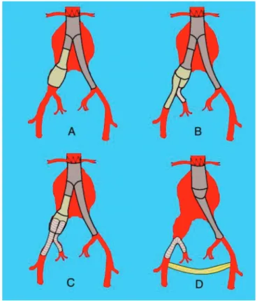
of common iliac artery (CIA) cerclage for the treatment of endoleak in patients undergoing AAA endovascular treatment (Figure 1). Initially, extraperitoneal surgical access to CIA is performed. The artery is dissected cra-nially at its bifurcation, extending 2 to 3cm and two vascular tapes are passed around the vessel with a distance of 1cm between them. After completion of the retroperitoneal approach, inguinal incisions are made to expose the femoral arteries. The endopros-thesis is introduced and a forceps is placed at the level
1 - Pontifical University of Rio de Janeiro, CENTERVASC-RIO, Department of Vascular Surgery, Rio de Janeiro State, Brazil. 2 - Federal University of Rio de Janeiro, Department of Radiology, Rio de Janeiro State, Brazil.
A B S T R A C T
About 20% of patients with abdominal aortic aneurysms have associated iliac aneurysms. Distal sealing during the endovascular treatment of aortic-iliac aneurysms is a challenge that has led to the emergence of several technical options to achieve this goal over the years. Inter-nal iliac artery embolization is associated with the risk of ischemic complications, such as gluteal necrosis, lower limb neurological deficit, colonic ischemia, impotence and gluteal claudication. This article summarizes the technical options for endovascular treatment of aortoiliac aneurysms with different approaches to preserving the patency of internal iliac arteries.
of the most caudal vascular tape, to be used as a ra-diopaque reference to fluoroscopy. After positioning and expanding the device, a balloon catheter of the same diameter as the endoprosthesis is introduced over fluoroscopy and inflated to the caudal end of the endoprosthesis. The two vascular tapes are tied until the resistance promoted by the balloon is perceived. The balloon is then deflated and removed for control angiography8.
Iliac branches in bell-bottom
The bell-bottom technique was originally described with the use of a proximal aortic extension (cuff) selected based on the diameter of the iliac artery and positioned with at least 1cm overlap within the distal iliac branch and by at least 1cm in an area of the ectasized CIA (Figure 2A)10. The presence of a
thrombus in the ectasia segment is a contraindication to this technique. The cuffs described in this configuration are expanded to adapt to the iliac branch, promoting adequate distal sealing in the ectasiated or aneurysmatic CIA. Thereafter, broad-diameter iliac branches specifically developed for this purpose have become available for use in the bell-bottom technique11. There is no consensus on the CIA limiting
diameter recommended for the use of this technique. A study analyzing common iliac arteries up to 30mm in diameter submitted to the implantation of bell-bottom endoprostheses demonstrated a satisfactory
long-term outcome (type-1B endoleak in 4% of cases)11. A
comparative analysis showed no evolutionary difference between a group of patients with AICs submitted to the implantation of bell-bottom endoprostheses with a diameter between 20mm and 22mm and a group with a diameter be tween 24mm and 28mm12. The incidence
of type-1B endoleak observed in the literature ranged from 0 to 11%10-15. However, the Mayo Clinic group
reports late dilation of CIA affecting up to 86% of patients10-12,14. Nonetheless, some authors relate this
late expansion to over-dimensioning greater than 15% of the endoprostheses implanted in the CIA16, without
correlation with adverse effects14.
Sandwich
The sandwich technique for the treatment of aortoiliac aneurysms was initially described by Lobato17. This technique consists of the following
steps: 1) femoral implantation of a bifurcated stent graft, with position of the contralateral iliac branch 1cm cranial to the CIA ostium; 2) IIA catheterization Figure 1. Iliac cerclage technique. Representative scheme of
the cerclage technique in the treatment of aortoiliac aneurysms.
through left brachial access; 3) implantation of a coated, self-expanding stent in the IIA, with adequate overlap in the iliac branch, followed by implantation of the iliac branch of the endoprosthesis; 4) Modeling of the iliac branch and expanding the stent using balloon catheters; 5) implantation of the contralateral iliac branch (Figure 2C)18. Lobato17 indicates overlapping of
more than 6cm between endoprostheses to minimize the formation of gutters and the risk of leakage. Limitations of this technique include the need to use long iliac endoprostheses, the potential risk of compression of one of the parallel endoprostheses, and the absence of controlled data with a long follow-up period2. Despite these considerations, the technique is
used in several centers with reports of low prostheses occlusion rates or leaks19-22. Lobato describes a series
with 40 patients, with an average follow-up time of 12 months, observing 100% technical success rates, primary patency of 93.8%, and type-3 endoleak (associated with the technique) of 2.5%23.
Branched internal iliac artery endoprosthesis The IIA branching technique consists of the implantation of a bifurcated iliac stent combined with a coated stent connecting the latter with IIA. The ZBIS® endoprosthesis is available with external branches in the helical and straight configurations, the latter being the most used. This device is generally combined with an endoprosthesis implanted in the abdominal aorta (Figure 2B)18,24. In our country, Massière and
von Ristow24 developed a bifurcated branched device
based on the Apollo platform, and used the Viabahn® connecting stent.
The following morphological criteria are described for the use of the ZBIS device technique: the presence of a non-aneurysmal segment of the EIA of at least 20mm in length and a diameter between 8mm and 11mm, with the length of the CIA greater than 50mm, luminal diameter of the CIA greater than 16mm and the presence of a non-aneurysmal segment of the IIA at least 10mm in length and with a diameter between 6mm and 9mm2,18,25. Using these criteria, a study of
51 patients undergoing AIAs treatment determined
that only 38% of the patients analyzed fully met the requirements determined by the manufacturer26.
Ferreira et al27 published a study investigating
47 bifurcated devices implanted in 37 patients with an average follow-up time of 11.6 months. Technical success was obtained in 97.3% of the cases and a secondary patency rate in 22 months of 85.4%. They observed stent occlusion in five patients (10.6%), gluteal claudication in one case (2.7%) and no endoleaks. The incidence of complications associated with the device, evaluated by the combined incidence of type-1endoleak, type-3 endoleak and branch occlusion, varies from 7% to 13.8% in the literature. The incidence of gluteal claudication varies from 2.7 to 5.6%26-31.
The main limitation to this technique is the anatomical prerequisites necessary for the device implanting32.
A meta-analysis analyzing the performance of 236 connection, coated stents in five studies on branched endoprosthesis reported an incidence of occlusion in 6% of the cases and gluteal claudication in 3.4%, with the main causes being the IIA small diameter, IIA perioperative dissection due to excessive dilation, use of long stents, concomitance of IIA atherosclerotic disease, and compression associated with the use of the helical model branched device. A lower occlusion rate was observed in cases in which the expandable balloon stent was used as a connection stent. However, statistical methods could not be used due to the heterogeneity of the studies31.
Until the moment of submission of this article, only initial results of the use of the Excluder® iliac branched endoprosthesis were published32.
Retrograde endovascular revascularization of the internal iliac artery
The technique of retrograde endovascular revascularization of the internal iliac artery was initially described by Hoffer et al33. It consists of the implantation
Massière et al34 described a series of 21
patients submitted to this technique for the treatment of complex aortoiliac aneurysms, unable of being submitted to another endovascular technique and with a high surgical risk. They used the Viabahn® connecting stent. The mean follow-up time was 52 months, with technical success in all cases, endoleak associated with the connection stent in one patient (4.7%), type-IB endoleak in one patient (4.7%) and occlusion of the coated stent in one case (4.7%) in 30 days31.
The need for the extra-anatomical bridge constitutes a limitation to this technique due to the risk of infection and thrombosis, being reserved for
selected cases. However, no complications associated with cross-bridge were observed and the technique allows the treatment of cases with complex anatomy, offering few anatomical restrictions to its use.
CONCLUSION
Each one of the various endovascular techniques available for the treatment of aortoiliac aneurysms presents its anatomical limitations. The complexity of these cases requires adequate selection of the technique that will allow the exclusion of the aneurysm, offering lower risk and better outcome in the long term.
REFERENCES
1. Brunkwall J, Hauksson H, Bengtsson H, Berqqvist D, Takolander E, Bergentz SE. Solitary aneurysms of the iliac arterial system: an estimate of their frequency of occurrence. J Vasc Surg. 1989;10(4):381-4.
2. Fatima J, Correa MP, Mendes BC, Oderich GS. Pel-vic revascularization during endovascular aortic an-eurysm repair. Perspect Vasc Surg Endovasc Ther. 2012;24(2):55-62.
3. Criado FJ, Wilson EP, Velazquez OC, Carpenter JP, Barker C, Wellons E, et al. Safety of coil emboliza-tion of the internal iliac artery in endovascular graft-ing of abdominal aortic aneurysms. J Vasc Surg. 2000;32(4):684-8.
4. Cynamon J, Lerer D, Veith FJ, Taragin BH, Wahl SI, Lautin JL, et al. Hypogastric artery coil embolization prior to endoluminal repair of aneurysms and fistu-las: buttock claudication, a recognized but possi-bly preventable complication. J Vasc Interv Radiol. 2000;11(5):573-7.
5. Vandy F, Criado E, Upchurch GR Jr, Williams DM, Rectenwald J, Eliason J. Transluminal hypogastric artery occlusion with an Amplatzer vascular plug during endovascular aortic aneurysm repair. J Vasc Surg. 2008;48(5):1121-4.
6. Farahmand P, Becquemin JP, Desgranges P, Allaire E, Marzelle J, Roudot-Thoraval F. Is hypogastric ar-tery embolization during endovascular aortoiliac aneurysm repair (EVAR) innocuous and useful? Eur J Vasc Endovasc Surg. 2008;35(4):429-35.
7. Engelke C, Elford J, Morgan RA, Belli AM. In-ternal iliac artery embolization with bilateral occlusion before endovascular aortoiliac aneu-rysm repair-clinical outcome of simultaneous and sequential intervention J Vasc Interv Radiol. 2002;13(7):667-76.
8. Puech-Leao P. Banding of the common iliac artery: an expedient in endoluminal correction of aortoiliac aneurysms. J Vasc Surg. 2000;32(6):1232-4. 9. Chuter TA, Risberg B, Hopkinson BR, Wendt G,
Scott RA, Walker PJ, et al. Clinical experience with a
Cerca de 20% dos pacientes com aneurismas de aorta abdominal apresentam aneurismas ilíacos associados. A obtenção do selamento distal, durante o tratamento endovascular dos aneurismas aortoilíacos, constitui-se em um desafio que suscitou, ao longo dos anos, o surgimento de diversas opções técnicas para alcançar esse objetivo. A embolização da artéria ilíaca interna é associada ao risco de desenvolvimento de complicações isquêmicas, tais como: necrose glútea, déficit neurológico dos membros inferiores, isquemia colônica, impotência e claudicação glútea. Esse artigo resume as opções técnicas de tratamento endovascular dos aneurismas aortoilíacos com diferentes formas de abordagem de preservação da perviedade das artérias ilíacas internas.
Descritores: Aneurisma Aórtico. Artéria Ilíaca. Procedimentos Endovasculares.
bifurcated endovascular graft for abdominal aortic aneurysm repair. J Vasc Surg. 1996;24(4):655-66. 10. Karch LA, Hodgson KJ, Mattos MA, Bohannon
WT, Ramsey DE, McLafferty RB. Management of ectatic, nonaneurysmal iliac arteries during en-doluminal aortic aneurysm repair. J Vasc Surg. 2001;33(2 Suppl):S33-8.
11. Torsello G, Schönefeld E, Osada N, Austermann M, Pennekamp C, Donas KP. Endovascular treat-ment of common iliac artery aneurysms using the bell-bottom technique: long-term results. J Endo-vasc Ther. 2010;17(4):504-9.
12. Naughton PA, Park MS, Kheirelseid EA, O’Neill SM, Rodriguez HE, Morasch MD, et al. A com-parative study of the bell-bottom technique vs hy-pogastric exclusion for the treatment of aneurys-mal extension to the iliac bifurcation. J Vasc Surg. 2012;55(4):956-62.
13. Kritpracha B, Pigott JP, Russell TE, Corbey MJ, Whalen RC, DiSalle RS, et al. Bell-bottom aortoili-ac endografts: an alternative that preserves pelvic blood flow. J Vasc Surg. 2002;35(5):874-81. 14. England A, Butterfield JS, McCollum CN,
Ash-leigh RJ. Endovascular aortic aneurysm repair with the talent stent-graft: outcomes in patients with large iliac arteries. Cardiovasc Intervent Radiol. 2008;31(4):723-7.
15. Alvarez Marcos F, Garcia de la Torre A, Alonso Perez M, Llaneza Coto JM, Camblor Santervas LA, Zanabili Al Sibbai AA, et al. Use of aortic exten-sion cuffs for preserving hypogastric blood flow in endovascular aneurysm repair with aneurysmal involvement of common iliac arteries. Ann Vasc Surg. 2013;27(2):139-45.
16. Kirkwood ML, Saunders A, Jackson BM, Wang GJ, Fairman RM, Woo EY. Aneurysmal iliac arteries do not portend future iliac aneurysmal enlargement after endovascular aneurysm repair for abdominal aortic aneurysm. J Vasc Surg. 2011;53(2):269-73. 17. Lobato AC. Sandwich technique for aortoiliac an-eurysms extending to the internal iliac artery or isolated common/internal iliac artery aneurysms: a new endovascular approach to preserve pelvic
circulation. J Endovasc Ther. 2011;18(1):106-11. 18. Geisbüsch P, Attigah N, Hyhlik-Dürr A, Hakimi
M, Müller-Eschner M, Böckler D. Decision-mak-ing and techniques in hypogastric artery revascu-larization. J Cardiovasc Surg (Torino). 2013;54(1 Suppl 1):71-9.
19. Friedman SG, Wun H. Hypogastric preservation with Viabahn stent graft during endovascular an-eurysm repair. J Vasc Surg. 2011;54(2):504-6. 20. Mosquera Arochena N, Rodríguez Feijoo G,
Car-ballo Fernandez C, Molina Herrero F, Fernandez Lebrato R, Barrios Castro A, et al. Use of modified Sandwich-graft technique to preserve hypogas-tric artery in EVAR treatment of complex aortic aneurysm anatomy. J Cardiovasc Surg (Torino). 2011;52(5):643-9.
21. Heckenkamp J, Brunkwall J, Luebke T, Aleksic M, Schöndube F, Stojanovic T. Novel chimney-graft technique for preserving hypogastric flow in com-plex aortoiliac aneurysms. J Cardiovasc Surg (Tori-no). 2012;53(6):773-6.
22. Yoshida Rde A, Yoshida WB, Kolvenbach R, Vieira PR. Modified “stent-graft sandwich” technique for treatment of isolated common iliac artery an-eurysm in patient with Marfan syndrome. Ann Vasc Surg. 2012;26(3):419.e7-9.
23. Lobato AC, Camacho-Lobato L. The sandwich technique to treat complex aortoiliac or isolated iliac aneurysms: results of midterm follow-up. J Vasc Surg. 2013;57(2 Suppl):26S-34S.
24. Massière B, von Ristow A, Cury J, Gress M, Vescovi A, Marques M. Internal iliac artery branch stent grafting for aortoiliac aneurysms using the Apollo branched device. Ann Vasc Surg. 2010;24(3):417. e15-8. 25. Physician’s pocket reference guide. Zenith®
En-dovascular Grafts. Queensland, Australia: Cook Medical; 2012. p. 45-50.
27. Ferreira M, Monteiro M, Lanziotti L. Technical as-pects and midterm patency of iliac branched de-vices. J Vasc Surg. 2010;51(3):545-50.
28. Karthikesalingam A, Hinchliffe RJ, Holt PJ, Boyle JR, Loftus IM, Thompson MM. Endovascular an-eurysm repair with preservation of the internal il-iac artery using the ilil-iac branch graft device. Eur J Vasc Endovasc Surg. 2010;39(3):285-94.
29. Donas KP, Torsello G, Pitoulias GA, Austermann M, Papadimitriou DK. Surgical versus endovascular re-pair by iliac branch device of aneurysms involving the iliac bifurcation. J Vasc Surg. 2011;53(5):1223-9. 30. Parlani G, Verzini F, De Rango P, Brambilla D, Co-scarella C, Ferrer C, et al. Long-term results of iliac aneurysm repair with iliac branched endograft: a 5-year experience on 100 consecutive cases. Eur J Vasc Endovasc Surg. 2012;43(3):287-92.
31. Donas KP, Bisdas T, Torsello G, Austermann M. Technical considerations and performance of bridging stent-grafts for iliac side branched de-vices based on a pooled analysis of single-center experiences. J Endovasc Ther. 2012;19(5):667-71.
32. Ferrer C, De Crescenzo F, Coscarella C, Cao P. Early experience with the Excluder® iliac branch
endoprosthesis. J Cardiovasc Surg (Torino). 2014;55(5):679-83.
33. Hoffer EK, Nicholls SC, Fontaine AB, Glickerman DJ, Borsa JJ, Bloch RD. Internal to external iliac artery stent-graft: a new technique for vessel ex-clusion. J Vasc Interv Radiol. 1999;10(8):1067-73. 34. Massière B, von Ristow A, Vescovi A, Pedron C, Fonseca LM. Management of aortoiliac aneu-rysms by retrograde endovascular hypogastric artery preservation. Vascular. 2014;22(2):116-20.
Received in: 12/07/2016
Accepted for publication: 26/09/2016 Conflict of interest: none.
Source of funding: none.
Mailing address: Bernardo Massière
E-mail: [email protected]