Spectroscopic Properties of pigment Li
2-xZn
1-xPr
xTi
3O
8Maria Suely Costa da Câmaraa*, Adaiane Spinellib, Elson Longoc, Luciano Nobrega Azevedod,
Asenete Frutuoso Costae, Patricia Mendonça Pimentelf, Dulce Maria de Araújo Melod
aUniversidade Federal Rural de Pernambuco – UFRPE, Serra Talhada, PE, Brazil
bFundação Universidade Federal de Rondônia – UNIR, Porto Velho, RO, Brazil
cInstituto de Química, Universidade Estadual Paulista – UNESP, Araraquara, SP, Brazil
dInstituto de Química, Universidade Federal do Rio Grande do Norte – UFRN, Natal, RN, Brazil
ePrograma de Pós-graduação em Ciência e Engenharia de Materiais – PPGCEM, Universidade
Federal do Rio Grande do Norte – UFRN, Natal, RN, Brazil
fUniversidade Federal Rural do Semi-Árido – UFERSA, Angicos, RN, Brazil
Received: October 20, 2015; Revised: April 3, 2016; Accepted: May 23, 2016
Inorganic compounds doped with rare earths (Ce3+, Pr3+, Nd3+, Sm3+, Eu 3+, Gd 3+, Tb 3+, Tm3+)
have been used in various applications including light-emitting devices such as luorescent lamps,
cathode ray tubes, lasers and inorganic pigments. In this study, Pr3+ is doped in spinel Li 2ZnTi3O8
system and synthesized by the polymeric precursor method, which is based on the process developed
by the Pechini, and characterized by X-ray difraction, UV-visible and CIE-L colorimetric measures * a * b *., in order to study the efect of doping and thermal treatment on its colorimetric properties. With three diferent samples of Pr3+ doping (0.01; 0.05 and 0.1 mol%) were prepared and calcined
at 500°C, 600°C, 700°C, 800°C and 900°C for 4 h. The analysis of X-ray difraction conirmed the formation of pure phases with spinel structure and average crystallite size of less than 46 nm. It was found that the colorimetric properties ranging from green to red, in accordance with the increase in the concentration of Pr3+ and thermal processing temperature.
Keywords: Ceramics, inorganic compounds, nanostructures, chemical synthesis, X-ray difraction
1. Introduction
Inorganic compounds doped with trivalent rare earth (RE) ions (Ce3+, Pr3+, Nd3+, Sm3+, Eu3+, Gd3+, Tb3+, Tm3+)
have been used in many different applications, including luminescent devices such as fluorescent lamps, cathode
ray tubes and lasers.In these applications are particularly
preferred ions displaying 4fn transitions1,2. Among the rare
earth ions, trivalent praseodymium shows a particular
feature due to close energy separation between the low edge of the 4f5d configuration and the 1S
0 level of the 4f 2
configuration. The radiative de-excitation between these two configurations gives rise a broad band emissions between 220 and 450 nm approximately, depending on the
host lattice3. Moreover, emitting species can be excited,
either directly or by two-step excitation processes related
to the 3P 0 or
1D
2 metastable intermediate levels, located
in the blue or orange spectral domains, respectively.
The two-steps excitation of the 4f5d states of Pr3+ ions
has been demonstrated in YAlO3:Pr3+ crystals, as well
as in fluoride crystals such as KY3F10:Pr 3+, BaY
2F8:Pr 3+,
LiYF4:Pr3+ and LiLuF
4:Pr3+.
The Pr3+ emission spectra strongly depend on the
crystalline field which the ion is allocated. For instance,
while Gd2O2S:Pr3+ emits green color due to the 3P 0 →
3H
4
transition, LiYF4:Pr3+ displays a red color associated to
the 3P 0 →
3H 6,
3F
2 transitions
3. The chromatic properties
of rare earth sesquisulfides Ln2S3 and Aln2S4 (A = Ca,
Sr) have been recently investigated. These compounds
exhibit pronounced color, ranging from yellow to red depending on the rare earth. The color of these compounds
has been related to various energetic parameters, such as
ionization energies, crystal field splitting or nephelauxetic effect, the positions of the energy levels at the valence and the conduction bands, which are a function of the
crystal structure5. Metal oxides have been extensively
used as host matrices due to their much better chemical
stability than conventional sulfide compounds. The spinel structure is an interesting class of oxides with general formula AB2O4 which can act as a host matrix for rare
earth ions. A refers to cations in tetrahedral sites and B represents the cations in the octahedral positions. It is a cubic structure with space group symmetry Fd3m6.
This elaborate crystallographic structure, which can
accommodate significant cation disorder, has a unique perspective for studies of substitutions and their relations
with the chemical and physical properties7,8. Spinel
crystal structures can be divided into two types: normal (or direct) and inverse spinel. In the normal spinels, the A2+ ions are contained in the tetrahedral holes, and
the B3+ in the octahedral holes. In the inverse spinels,
on the other hand, the A2+ ions and half the B3+ ions
in tetrahedral holes9-12 In fact, very few spinels have
exactly the normal or inverse structure, and these are sometimes called mixed spinels. Li2ZnTi3O8 presents a derived spinel structure with P4332 space group, in
which the octahedral 12d and 4b sites are occupied by
Ti and Li respectively, and the tetrahedral 8c sites are shared by Zn and Li, suggesting an intermediary spinel structure13-15.
The present paper presents an optical properties study
of the Li2-xZn1-xPrxTi3O8 spinels, where x = 0.01; 0.05 and 0.1 mol%, synthesized by the Pechini method14. This study
was based on analyses of difuse relectance and colorimetric coordinates and focused in the energy levels of the Pr3+ ions.
It should be emphasized that no publication was found in the literature about the energy levels of the Pr3+ ions in the
Li2ZnTi3O8 host lattice.
2. Experimental Procedure
The polymeric precursor solution was prepared by the Pechini method16, which has been used to synthesize
polycationic powders. The process is based on the metallic citrate polymerization using ethylene glycol in order to
promote polymerization of the metallic citrate. Due to the formation of high viscosity polyester, the segregation of
the cations during thermal decomposition is minimal17.
A hydrocarboxylic acid, such as citric acid, is used to chelate cations in an aqueous solution. The addition of a glycol such as ethylene glycol leads to the formation of
an organic ester. Polymerization, promoted by heating
the mixture, results in a homogeneous resin in which
metal ions are uniformly distributed throughout the organic matrix.
The polymeric precursor aqueous solution was prepared
using the 3:1 molar ratio between citric acid and metallic cations. First, a citric acid solution was kept under stirring
for 5 min at 70°C, then lithium carbonate (Li2CO3, Merck),
zinc acetate dihydrate (Zn(CH3CO2)2.2H2O, Aldrich) and praseodymium carbonate (Pr2(CO3)3, Nuclemon) were added to the solution and kept under stirring until their
complete dissolution. Subsequently, ethylene glycol
(HOCH2CH2OH, Merck) was added in a ratio of 2:3 in
relation to the citric acid and stirred vigorously for 10 min at 100°C to promote polyesterification reactions and water evaporation, which lead to formation of a
polymeric resin. The resin was heat treated at 300°C
for 3 h, and then the obtained powder was ground in
a mortar and calcined in air at 500, 600, 700, 800 and
900°C for 4 h.
The produced powders were characterized by XRD
using a Siemens D5000 equipment operating with Cu Kα
radiation. The data were collected in the range 5-100o (2θ),
step size of 0.02o and count time of 10 s per step.
UV-vis-NIR spectroscopy (difuse relectance) of the samples was performed with a Varian 5G spectrophotometer. In addition, the L*, a* and b* color parameters and difuse relectance were measured by a Gretac Macbeth Color-eye spectrophotometer 2180/2180 UV, in the 300-800 nm range,
using the D65 illumination. The CIE-L*a*b* colorimetric
method, recommended by the CIE was followed. In this method, L* is the lightness axis [black (0)→ white (100)], b* is the blue (-) → yellow (+) axis, and a* is the green (-)
→ red (+) axis, and ∆E is the hue variation.
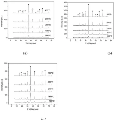
Figure 1: XRD patterns of the Li2ZnTi3O8 spinel doped with: (a) 0.01% mol % of Pr
Figure 2: Efect of the calcinations temperature on the crystallite size of the spinel phase.
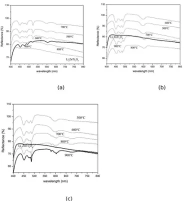
Figure 3: Difuse relectance of the Li2ZnTi3O8 spinel, calcined at the 500-900°C temperature range, and doped with Pr
3+ (a) 0.01mol%, (b)0.05mol% and (c) 0.1mol%.
3. Results and discussion
The X-ray difraction patterns conirmed the presence of
a single pure phase with spinel-type structure, Li2ZnTi3O8,
for all samples treated at the 500-900°C temperature range, independently of the Pr3+ ions concentration (Figure 1).
These results suggest that Pechini method is able to produce
oxide powders with a high crystallinity and nanometric size
crystallites. It was also observed crystallite size increases when the heat-treatment temperature increases (Figure 2).
Figure 3 presents the difuse relectance of Li2ZnTi3O8
doped with 0.01, 0.05 and 0.1 mol% of Pr3+ ions and
calcined at 500, 600, 700, 800 and 900°C. It can be seen by the spectra that the spinel phase presents absorption bands
at the spectral domains ranging from blue to yellow. The
CIE- L*a*b* chromatic coordinates are depicted in Figure
4. It is noticed that the color of the spinel phase is more intense, toward the direction of the green, when the Pr3+
ions concentration increases. On the other hand, the color becomes darker when the calcination temperature raises
and the compounds become more crystalline (Figure 4a). Figure 5 illustrates the difuse relectance of the spinel calcined at 800°C for diferentconcentrations of dopant. The spectra obtained at room temperature showed the 4f2
Stark levels characteristic of Pr3+ ions which are presented in
Table 1. It is noticed for all samples the characteristic spectral lines of Pr3+ ions in the range from 1.75 to 3 eV, attributed
to intercon igurational transitions of RE 4fn-15d→4fn ions,
due to the strong coupling between the 5d electrons of the
active ion and the host lattice9. It is well known that Pr3+
ion energy levels are shifted toward lower energies when in the presence of a crystalline ield. Usually, the diference between the energy levels of the rare earth ion when isolated and into a crystalline ield (∆E) is higher than 500 cm-1. It
was found (Table 1) that Pr3+ ions presents greater ∆E when
Figure 4: Chromatic coordinates of the of the Li2ZnTi3O8 spinel doped with Pr
3+ (a)“L” and (b) a*.
Figure 5: Difuse relectance of the Li2ZnTi3O8 spinel doped with Pr
3+ (a) 0.01, 0.05 and 0.1 mol% (b) ampliication of the 0.1% curve, calcined at 800°C.
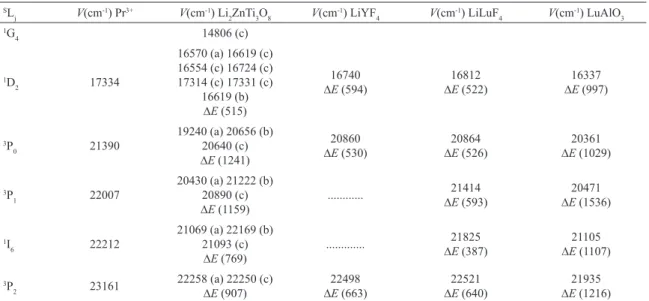
Table 1: Crystal ield splitting of the 2s+1L
j manifolds of Pr
3+ ion in Li
2ZnTi3O8, LiYF4, LiLuF4 and LuAlO3. SL
j V(cm
-1) Pr3+ V(cm-1) Li
2ZnTi3O8 V(cm -1) LiYF
4 V(cm-1) LiLuF4 V(cm-1) LuAlO3 1G
4 14806 (c)
1D
2 17334
16570 (a) 16619 (c)
16554 (c) 16724 (c) 17314 (c) 17331 (c)
16619 (b)
∆E (515)
16740 ∆E (594)
16812
∆E (522)
16337
∆E (997)
3P
0 21390
19240 (a) 20656 (b) 20640 (c) ∆E (1241)
20860
∆E (530)
20864 ∆E (526)
20361
∆E (1029)
3P
1 22007
20430 (a) 21222 (b)
20890 (c)
∆E (1159)
... ∆21414
E (593)
20471 ∆E (1536)
1I
6 22212
21069 (a) 22169 (b) 21093 (c)
∆E (769)
... ∆21825
E (387)
21105
∆E (1107)
3P
2 23161
22258 (a) 22250 (c)
∆E (907)
22498 ∆E (663)
22521
∆E (640)
21935
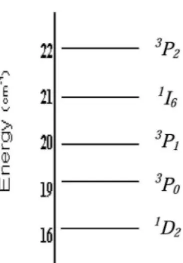
Figure 6: Energy level diagram of Pr 3+ in Li
2ZnTi3O8, obtained
from absorption spectra.
4. Conclusions
We have demonstrated that Li2-xZn1-xPrxTi3O8
nanocrystalline powders can be successfully synthesized
by a polymer precursor method. These compounds showed a green color directly related to the Pr3+ ion concentration.
By using a simple technique of difuse relectance it was possible to determinate the positions of the Stark 4f2 levels of the Pr3+ ion in the Li
2-xZn1-xPrxTi3O8 host
lattice. It was found that the spinel Li2-xZn1-xPrxTi3O8 is
a promising material to be used as luorescent material
and/or a pigment.
5. Acknowledgements
The authors gratefully acknowledge the inancial support of the Brazilian research agencies FAPESP, PRONEX, FINEP, CNPq E FACEPE.
6. References
1. Dorenbos P. The 4fn↔4fn-15d Transitions of the Trivalent Lanthanides in Halogenides and Chalcogenides. Journal of
Luminescence. 2000;91(1-2):91-106.
2. Barros BS, Melo SP, Gama L, Alves-Jr S, Fagury-Neto E, Kiminami RHGA, et al. Caracterização morfológica e luminescente de
nanopartículas de aluminato de zinco dopadas com Eu3+.
Cerâmica. 2005;51(317):63-69.
3. Nicolas S, Descroix E, Guyoto Y, Ejoubert M, Pédrini C, Ovanesyan ZKL, et al. Spectroscopic and colour centre studies of Pr-doped
LuAlO3. Optical Materials. 2002;19(1):129-137.
4. Sinha SP, ed. Systematics and the Properties of the Lanthanides. Dordrecht: Springer; 1983.
5. Demourgues A, Tressaud A, Laronze H, Macaudiére P. Rare earth luorosulides LnSF and Ln2AF4S2 as new colour pigments.
Journal of Alloys and Compounds. 2001;323-324:223-230.
6. Poleti D, Vasović D, Karanović LJ, Branković Z. Synthesis and Characterization of Ternary Zinc-Antimony-Transition Metal
Spinels. Journal of Solid State Chemistry. 1994;112(1):39-44. 7. Barth TFW, Posnjak E. The spinel structure: example of variate
atoms equipoints. Journal of the Washington Academy of
Sciences. 1931;21:255-258.
8. Sickafus KE, Wills JM, Grimes NW. Structure of Spinel. Journal
of the American Ceramic Society. 1999;82(12):3279-3292.
9. Shriver DF, Atkins PW, Langford CH. Inorganic Chemistry. 2nd
ed. Oxford: Oxford University Press; 1994. 819p.
10. Lisboa-Filho PN, Gama L, Paiva-Santos CO, Varela JA, Ortiz WA, Longo E. Crystallographic and magnetic structure of polycrystalline Zn 7− x Ni x Sb 2 O 12 spinels. Materials
Chemistry and Physics. 2000;65(2):208-211.
11. Braun PB. A Superstructure in Spinels. Nature. 1952;170:1123. 12. Diallo PT, Boutinaud P, Mahiou R, Cousseins JC. Red
Luminescence in Pr3+-Doped Calcium Titanates. Physica Status
Solidi. 1997;160(1):255-263.
13. Câmara MSC, Lisboa-Filho PN, Cabrelon MD, Gama L, Ortiz WA, Paiva-Santos CO, et al. Synthesis and characterization
of Li2ZnTi3O8 spinel using the modiied polymeric precursor method. Materials Chemistry and Physics. 2003;82(1):68-72.
14. Câmara MSC, Gurgel MFC, Lazaro SR, Bochi TM, Pizani OS, Beltran A, et al. Room temperature photoluminescence of the
Li2ZnTi3O8 spinel: Experimental and theoretical study. International
Journal of Quantum Chemistry. 2005;103(5):580-587.
15. Lucena PR, Pontes FM, Pinheiro CD, Longo E, Pizani OS, Lázaro S, et al. Fotoluminescência em materiais com desordem estrutural. Cerâmica. 2004;50(314):138-144.
16. Pechini MP, inventor. Sprague Electric Co. Method of preparing
lead and alkaline earth titanates and niobates and coating method
using the same to form a capacitor. US Patent. 3.330.697. 1967. 17. Paris EC, Leite ER, Longo E, Varela JA. Synthesis of PbTiO3 by