1 – PhD and Lecturer in the Physiotherapy Course, University of Uberaba (UNIUBE), Uberaba, MG, Brazil. 2 – MSc and Lecturer in the Physiotherapy Course, University of Uberaba (UNIUBE), Uberaba, MG, Brazil.
3 – PhD and Lecturer in the Physiotherapy and Medicine Courses, University of Uberaba (UNIUBE), Uberaba, MG, Brazil. 4 – Undergraduate Student in the Physiotherapy Course, University of Uberaba (UNIUBE), Uberaba, MG, Brazil. Work performed at the University of Uberaba (UNIUBE), Uberaba, MG.
Correspondence: Rua Luís Abrão 508, Bairro Olinda, 38055-720 Uberaba, MG. E-mail: [email protected]; [email protected] Work received for publication: May 20, 2011; accepted for publication: July 20, 2011
EFFECT OF LOW-LEVEL LASER (GAAS, 904
η
M) FOR
BONE REPAIR ON FRACTURES IN RATS
Jorge Alfredo Léo1, Alessanda da Cunha2, Elias Félix de Oliveira3, Reuder Pereira Prado4
INTRODUCTION
Fractures may be caused by diseases, trauma or surgical procedures, through direct or indirect mechanisms. In direct mechanisms, the bone is hit by, or it hits a hard object or surface, thus producing a fracture at the same site as the trauma occurrence. In indirect mechanisms, there is a torsional or angular force that results in a fracture at the site where the tensions accumulate or where the bone is most fragile(1). Given the high incidence of fractures consequent to car accidents, falls and sports trauma, the bone repair
process has been a topic studied worldwide(2).
Bone consolidation does not always take place in the expected manner: it can become irregular, with delayed consolidation, and this may lead to
the appearance of pseudarthrosis(3). In this regard,
different therapeutic resources have been used with the aim of accelerating the repair process and
im-proving the quality of bone consolidation(4). Thus,
studies have been conducted to evaluate the influ-ence of application of electric currents, ultrasound
and laser on the bone repair process(5).
In view of the points raised above, the present study had the objective of analyzing the effects of gallium
arsenide low-level laser radiation (GaAs; 904 ηm) for
repairing tibial bone fractures in Wistar rats, by means of mechanical tests and radiological examinations.
MATERIALS AND METHODS
The project for this study was approved by the Ethics Committee for Animal Experimentation of the University of Uberaba (UNIUBE), at a meeting on September 17, 2009 (CEEA Official Letter 141/2009).
For this study, 40 Wistar rats were used. Their mean body weight was 247.1 ± 32.87 grams and their mean age was 45 days.
The authors declare that there was no conflict of interest in conducting this work
This article is available online in Portuguese and English at the websites: www.rbo.org.br and www.scielo.br/rbort
ABSTRACT
Objective: To analyze the effects of low-level laser therapy (GaAs, 904 nm) for bone repair on tibial fractures in rats. Methods: Forty rats were divided into four groups of 10 ani-mals: control group without fracture (CG); fracture group without treatment (EG II); fracture group treated with laser at 10 J/cm² (EG III); and fracture group treated with laser at 15 J/cm² (EG IV). The fracture was produced surgically and the treatment lasted 45 days, done on alternate days. After treatment completion, the rats were sacrificed. The tibias were radiographed and subjected to mechanical three-point flexion tests in order to evaluate the maximum force (N) required to break them. Results: The observed maximum force values (N)
were: control group (CG) of 51.5 N ± 7.9 N; EG II 17.2 N ± 7.8 N; EG III 16.6 N ± 12.1 N; and EG IV 30.3 N ± 7.8 N. There were statistically significant differences between the control group and the experimental groups and also between experimental group IV and the other experimental groups (II and III). Radiographs showed callus formation in all the fractured groups, thus indicating that they had undergone the normal tissue repair process. Conclusion: EG IV, which underwent laser therapy with a dosage of 15 J/cm², showed the highest maximum force value (N) among the experimen-tal groups, thus demonstrating the influence of higher laser dosage on bone repair.
The animals were divided into four experimental groups containing 10 animals each: control group (CG), without any fracture; experimental group II (EG II): the animals were subjected to fracturing of the right tibia by surgical means and did not receive any treatment up to the day of sacrificing; experimen-tal group III (EG III): the animals were subjected to fracturing of the right tibia by surgical means and were
treated with GaAs laser (gallium arsenide; 904 ηm) at
a dose of 10 J/cm²; experimental group IV (EG IV): the animals were subjected to fracturing of the right tibia by surgical means and were treated with GaAs
laser (gallium arsenide; 904 ηm) at a dose of 15 J/cm².
Before the fracturing procedure was performed, the animals were anesthetized with thiopental sodium at a dose of 0.1 mg for each 100 grams of the ani-mal’s body mass and the anterior region of the lower leg was shaved. Asepsis was performed at the site using 1% polyvinylpyrrolidone-iodine. The animals received antibiotic therapy consisting of applications of ampicillin (0.02 mg/100 g of the animal), intramus-cularly, every 12 hours, starting one day before the fracturing procedure was performed, and continuing for seven days after the procedure.
To reach the tibia, a surgical incision of approxi-mate length 1 cm was made in the anterolateral re-gion. The fracture was started in the region of the proximal to middle third of the tibia by drilling, and it was completed by using a scalpel blade to “saw” the tibia to divide it into two pieces.
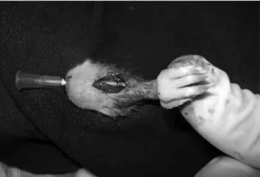
After the fracture had been produced, the tibia was stabilized using a 30 mm x 0.8 mm needle (Figure 1), which was used as an intramedullary nail. Starting on the day on which the fractures were produced,
the animals received applications of Ketofen® (0.02
mg/100 g of the animal), intramuscularly, every 24 hours for three days, which was used as an anti-in-flammatory agent and analgesic.
The animals in the two experimental groups (III and IV) were subjected to 22 sessions of applications of gallium
arsenide laser (GaAs; 904 ηm), using the Laserpulse
Special Diamond apparatus (IBRA-LC904), at a power of 30 mV, on alternate days. Point applications of laser irradiation were made at three sites on the anterior face of the lower leg over the focus of the fracture: one point at the fracture site, another 1 cm above it and the third 1 cm below it (Figure 2).
For radiological control and evaluation of the evo-lution of the bone repair, radiographs were produced
just after the fracturing and on the 15th, 30th and 45th days after this, using the Dabi Atlante Spectro 70X dentistry equipment in lateral view, with film placed below the lateral region of the tibia, and with a col-limator-leg distance of 8 cm. To produce the radio-graphs, the animals were anesthetized with an associa-tion of 0.2 mg/100 g of xylazine hydrochloride and 0.2 mg/100 g of ketamine hydrochloride, intramuscularly.
On the 46th day, the animals were sacrificed using
thiopental sodium at a dose of 2 mg for each 100 g of the animal’s body weight. The tibias were then dissected, the needles that had simulated intramedullary nails were removed and the bones were frozen at –27ºC for the subsequent procedure of mechanical testing. For this, the bones were defrosted over a 24-hour period, while kept in an ordinary refrigerator, and were maintained in a saline solution until the time of the test, as described by Pessan et al(6).
Figure 1 – Tibia stabilized with needle used as an intramedullary nail. Source: University of Uberaba.
The bones were positioned in a holder for the destruc-tive mechanical flexion test at three points to be carried out, such that the force imposed on the bone was applied perpendicularly to the focus of the fracture.
For this trial, a universal testing machine (EMIC®
model DL-3000) belonging to the Dental Materials Research Laboratory of the University of Uberaba
was used. It was equipped with a Kratos® load cell
of capacity 50 kgf. The machine was connected to a microcomputer equipped with software capable of registering the load and deformation values during the tests. Among the test parameters, a drop velocity of 0.2 mm/min was standardized. The force applied and the displacement were monitored and recorded by means os specific software for the equipment, which also supplied the force-deformation curve for each test (Figure 3). The resultant maximum force value (N) was recorded, and this was the parameter used for the present study.
RESULTS
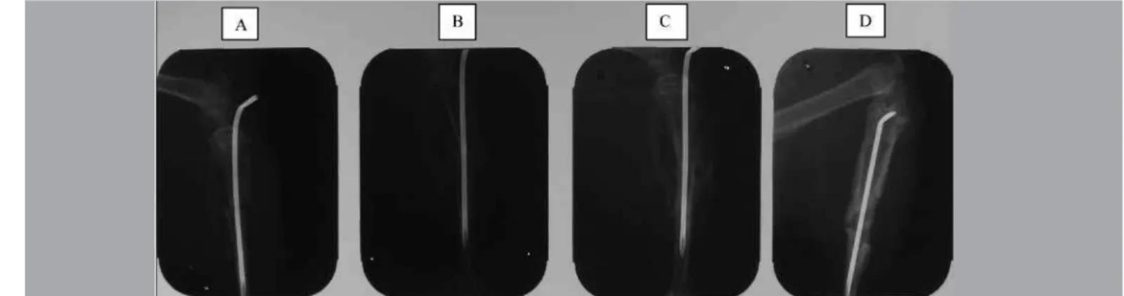
Figures 4, 5 and 6 show radiographic images of the right tibias, produced shortly after the surgery to
create the fracture (A), and on the 15th (B), 30th (C)
and 45th (D) days.
The radiographs produced on the 15th, 30th and 45th days demonstrated that bone callus was forming around the region of the fracture, in all the groups studied, with a gradual increase. Thus, it can be suggested that the bone consolidation process, which is preceded by for-mation of a fibrous callus, had begun. However, from the present analysis, it was not possible to qualify and quantify the bone repair process by comparing the di-fferent groups. Figure 7 presents the resultant means and respective standard deviations for the maximum force values (N) obtained in the mechanical flexion tests at three points for each group studied.
It could be seen from the analysis that there were statistically significant differences in comparing the mean maximum force values (N) between the experi-mental groups and the control group, which showed that effective fractures had been produced. Even with the laser stimulation, the values obtained in experimental groups III and IV, in comparison with the CG, indicated that the bones had not returned to the resistance level found in the bones that had not undergone fracturing.
There were statistically significant differences be-tween the experimental groups (EG II, EG III and EG IV), and the mean maximum force values (N) of experimental group IV were highest.
DISCUSSION
To carry out this study, Wistar rats were chosen as the experimental animals because they are easy to handle, have a low cost, need less space and can Figure 3 – Fractured tibia undergoing three-point mechanical flexion test.
Source: University of Uberaba.
Figure 4 – Lateral-view radiographs on experimental group II (EG II): (A) just after fracturing; (B) on 15th day; (C) on 30th day; and (d) on 45th day.
easily be obtained from the vivarium of the University of Uberaba. Male rates were chosen in order to avoid the bone alteration caused by female sexual hormones, the-reby maintaining a control pattern for increases in body weight, and for linear growth and growth in thickness(7).
Because bones, together with muscles, are respon-sible for support, movement and protection of vital organs, they are constantly subjected to traction, com-pression, torsion and flexion forces. They generally bear a combination of these forces, which makes it useful and necessary to know about their mechanical properties in order to assess their integrity(8).
Since the time of Galileo Galilei’s observations, it has been assumed that bone architecture is influenced by the mechanical tension associated with its
func-tion. From the 19th century onwards, this relationship
between structure and function became known as Wolff’s law. The principle of this law is based on the concept that there is a correlation between the trabecular alignment patterns and the directions of the
tensions that occur through functioning(6).
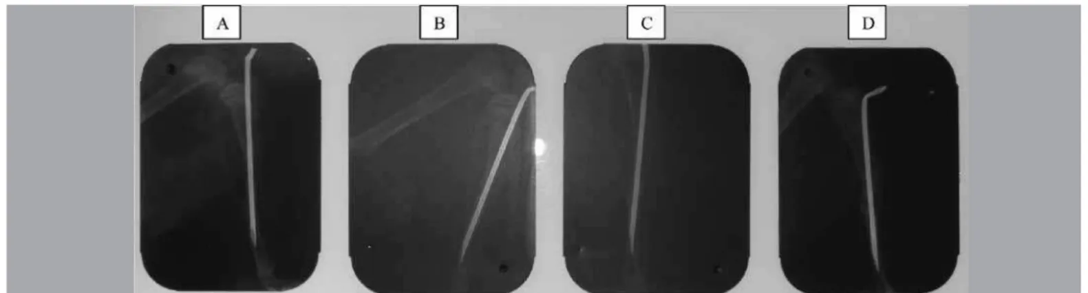
It is known that density and mechanical strength measurements are important characteristics in evaluating Figure 5 – Lateral-view radiographs on experimental group III (EG III): (A) just after fracturing; (B) on 15th day; (C) on 30th day; and (d) on 45th
day. Source: University of Uberaba.
Figure 6 – Lateral-view radiographs on experimental group IV (EG IV): (A) just after fracturing; (B) on 15th day; (C) on 30th day; and (d) on 45th
day. Source: University of Uberaba.
Figure 7 – Comparison of mean maximum force (N) between the groups. The symbol ♦ represents statistically significant differences shown by the
experimental groups II, III and IV (EG II, EG III and EG IV) versus the control group (CG); the symbol • represents statistically significant diffe -rences shown by experimental group II (EG II) and experimental group
III (EG III) versus experimental group IV (EG IV).
BIOMECHANICAL PArAMETEr OF MAXIMUM FOrCE (N)
Groups CG EG II EG III EG IV
Mean 51.6 17.28 16.7 30.38
Standard deviation 7.95 7.8 12.2 7.81
M
A
x
iM
u
M
f
o
r
C
e
(N
)
MAxiMuM forCe (N)
GroupS
CG
eG ii
eG iii
bone tissue. The guidelines proposed by the FDA (Food and Drug Administration) suggest that measurements should be made not only on bone density and biochemical markers of bone turnover, but also on bone strength through biomechanical tests. Such tests have been reported by various authors to be one of the ways of assessing bone consolidation(9).
During the three-point mechanical flexion test, the deformation over which force and displacement are proportion produces a linear graphical relationship known as the elastic phase of deformation. This de-formation is not permanent, which means that when the applied load is removed, the material will return to its original shape and remain undamaged. If the force continues to be applied (maximum force), per-manent deformation occurs (plastic phase). From an atomic perspective, this corresponds to breakage of bonds between neighboring trabeculae. Even if the force is then removed, the material will not return to its original position, thus leading to structural lesions
and consequently, bone fractures(10).
In the present study, the three-point mechanical flexion test was sufficient for the proposed analysis, since it is an easily applied and widely used test wi-thin scientific settings.
Several studies have reinforced the success of
low-level laser therapy for treating fractures(11-17).
However, despite the positive results found, these studies have used laser radiation emission devices with different wavelengths and very different doses and treatment durations, thus making it difficult to compare them and standardize a treatment protocol.
The doses of 10 J/cm2 and 15 J/cm2 used here were
chosen based on studies found in the literature, and these are the values most encountered, or close to
those most encountered(13,15,17).
The total treatment duration was set as 45 days, on alternate days, because this is the mean time needed for consolidation of the type of tibial fracture
pro-duced in this study(18) and because a pilot study
con-ducted previously using different lengths of treatment showed that the best quality of bone consolidation was on the 45th day.
The biomechanical evaluation was conducted by comparing the parameter of maximum force (N), and this showed that laser radiation treatment at a dose of 15 J/cm² for the length of time studied had positive effects regarding fracture repair. It facilitated formation
of a fibrous callus and consequently, bone tissue repair.
These results confirm those of Liriani(16), who found
a greater maximum force value (N) in the group of animals that received fractures and were treated with laser radiation than in the non-irradiated group.
On the other hand, laser radiation with a dose of 10 J/cm² was not shown to be effective in accelerating the bone repair process in tibial fractures in rats, since the maximum force values (N) obtained in the three-point mechanical flexion test were not statistically different from the values obtained in the EG II group. Pinheiro et al(12) found positive effects regarding bone realignment after infrared laser irradiation (AlGaAs;
830 ηm) and suggested, on the basis of previous
studies(19), that undifferentiated mesenchymal cells
were positively biostimulated to become osteoblasts, which more rapidly became osteocytes. The biological mechanisms involved in the improvement of bone tissue growth through low-level laser irradiation are still not clearly understood. The hypothesis advocated by Freitas(20), and corroborated by other studies, is that the laser energy may excite porphyrins and cytochromes (which are intracellular chromophores) and thus induce an increase in cell activity and consequently an increase in ATP concentration, which releases calcium. These morphological changes that occur in the bone structure after exposure to laser radiation may explain the increase in maximum force (N) in
the group treated with 15 J/cm2, given that through
facilitation of bone realignment, greater numbers of biostimulated cells in osteocytes and increased ATP and calcium concentrations, the bones probably were able to bear a greater load imposed on their structure during the three-point mechanical flexion test.
Through the 15-day intervals between radiographs used in this study, the evolution of the bone consoli-dation process of the fractures could be analyzed, thus simulating clinical practice for assessing bone repair in fracture cases.
The radiographic analysis demonstrated that bone callus formed in all the fractured bones, independent of whether the group was irradiated or not. However, this analysis was insufficient to quantify the bone re-pair process by comparing the different groups, given that all the groups presented bone callus formation in a similar manner.
deflection (mm) and stiffness (N/m) of the bone, along with analysis on bone density, which may complement the results encountered in the present study.
Lastly, we believe that there is a need for further studies on the effects of low-level laser therapy on the whole process of bone repair, especially analyzing the effect of the laser irradiation at cell and biomolecular levels, with the aim of establishing an effective proto-col for treating bone fractures, based on scientifically proven parameters.
CONCLUSION
It can be concluded from this study that low-level laser therapy showed positive results in the bone
re-pair process on tibial fractures in rats, thereby in-creasing the maximum force withstood by the group
treated with a dose of 15 J/cm2.
It was also observed that the dose of 10 J/cm2 was
not effective in accelerating the bone repair process in fractures, thus showing that bone tissue repair is dose--dependent, and that higher doses seem to be more effective for stimulating the bone repair process.
New methodology was established for carrying out the surgery, with the aim of obtaining fractures for conducting studies on bone lesions.
The three-point mechanical flexion test was shown to be efficient for analyzing the mechanical property studied here (maximum force for breakage), in the control and experimental groups.
REFERENCES
1. Gabriel MRS. Fisioterapia em traumatologia, ortopedia e reumatologia. Rio de Janeiro: Revinter; 2001.
2. Pinheiro AL, Limeira Júnior Fde A, Gerbi ME, Ramalho LM, Marzola C, Ponzi EA. Effect of low level laser therapy on the repair of bone defects grafted with inorganic bovine bone. Braz Dent J. 2003;14(3):177-81.
3. Guarniero R, Vaz CES, Santana PJ, Molin ED, Braum J, Harada MS. Avaliação do efeito do ibandronato na consolidação de fratura: estudo experimental em coelhos. Rev Bras Ortop. 2007;42(8):254-60.
4. Carvalho DCL, Rosin GC, Gama LOR, Tavares MR, Tribioli RA, Santos IR, Cliquet Júnior A. Non-pharmacological treatments in the stimulation of osteo-genesis. Rev Saude Publica. 2002; 36(5):647-54.
5. Liriani APR.; Lazaretti-Castro M. Evidences of physical agents action on bone metabolism and their potential clinical use. Arq Bras Endocrinol Metab. 2005;49(6):891-896.
6. Pessan VJO, Volpon JP, Shimano AC. Ensaio mecânico de flexão nas faces côn-cava e convexa da diáfise do fêmur de ratas. Rev Bras Ortop. 1996;31(7):600-4. 7. Carvalho DCL. Ação do ultra-som de baixa intensidade em ossos de ratas
osteopênicas [dissertação]. São Carlos: Escola de Engenharia de São Carlos, Faculdade de Medicina de Ribeirão Preto, Instituto de Química de São Carlos, Universidade de São Paulo; 2001.
8. Guagnelli RS. Propriedades mecânicas do osso esponjoso e cortical do rato, após período de imobilização por aparelho gessado ou pela cauda [disser-tação]. Ribeirão Preto: Escola de Engenharia de São Carlos, Faculdade de Medicina de Ribeirão Preto, Instituto de Química de São Carlos, Universidade de São Paulo; 2006.
9. Delgado-Martínez AD, Martínez ME, Carrascal MT, Rodríguez-Avial M, Munuera L. Effect of 25-OH-vitamin D on fracture healing in elderly rats. J Orthop Res. 1998;16(6):650-3.
10. Phillips RW. Skiner materiais dentários. Rio de Janeiro: Guanabara Koogan; 1993.
11. Silva Júnior AN, Pinheiro AL, Oliveira MG, Weismann R, Ramalho LM, Nicolau RA. Computerized morphometric assessment of the effect of low-level laser therapy on bone repair: an experimental animal study. J Clin Laser Med Surg. 2002;20(2):83-7.
12. Pinheiro ALB, Oliveira, MG, Martins, PPM, Ramalho LMP, Oliveira MAM, Novaes Júnior A, et al. Biomodulatory effects of LLLT on bone regeneration. Photomed Laser Surg. 2001;13(1):73-9.
13. Nicola RA, Jorgetti V, Rigau J, Pacheco MT, dos Reis LM, Zângaro RA. Effect of low-power GaAlAs laser (660 nm) on bone structure and cell activity: an experimental animal study. Lasers Med Sci. 2003;18(2):89-94.
14. Marino JAM, Taciro C, Zuanon JAS, Benatti Neto C, Parizotto NA. Efeito do LASER terapêutico de baixa potência sobre o processo de reparação óssea em tíbia de ratos. Rev Bras Fisioter. 2003;7(2):167-73.
15. Cerqueira A, Silveira RL, Oliveira MG, Sant’ana Filho M, Heitz C. Bone tissue microscopic findings related to the use of diode laser (830 nm) in ovine mandible submitted to distraction osteogenesis. Acta Cir Bras. 2007;22(2):92-7. 16. Liriani APR. Estudo comparativo dos efeitos do ultra-som e do LASER de
baixa intensidade no reparo ósseo de tíbia de rato [dissertação]. São Carlos: Escola de Engenharia de São Carlos, Faculdade de Medicina de Ribeirão Preto, Instituto de Química de São Carlos, Universidade de São Paulo; 2004. 17. Pes JV, Mazzanti A, Silveira AF, Agne JE, Ros DL, Trichez, KV. LASER arseneto
de gálio na estimulação da osteogênese. Fisioter Bras. 2007;8(5):313-316. 18. Hebert S. Ortopedia e traumatologia: princípios e prática. Porto Alegre: Artes
Médicas; 2009.
19. Pinheiro ALP, Frame JF. Laser em odontologia: seu uso atual e perspectivas futuras. Rev Gaucha Odontol. 1992;40(5):327-32.