Low-level laser therapy and in repairing diabetic foot ulcers
* Extracted from the theses “Efeito da Terapia a Laser de Baixa Intensidade com Calendula oicinalis no reparo de úlcera em pé diabético”, Universidade do Vale do Paraíba, 2015. 1 Universidade Estadual do Piauí, Faculdade de Ciências Médicas, Teresina, PI, Brazil. 2 DeVry|Facid,Teresina PI, Brazil.
3 Universidade do Vale do Paraíba, Instituto de Pesquisa e Desenvolvimento, São José dos Campos, SP, Brazil.
ABSTRACT
Objective: To evaluate the efects of low-level laser therapy isolated and associated with Calendula oicinalis oil in treating diabetic foot ulcers. Method: Anexperimental, randomized, controlled, prospective, interventional clinical case study using a quantitative approach. he sample consisted of 32 diabetic patients of both genders. Participants were randomly divided into four groups. Doppler Ultrasound evaluation of the Ankle-Brachial Index, brief pain inventory and analog pain scale were performed at baseline and after 30 days. Results: Reduced pain was observed in the level laser therapy and Low-level laser therapy associated with Essential Fatty Acids groups (p<0.01). Regarding the Ankle-Brachial Index and Doppler Ultrasound, all groups remained stable. By analyzing lesion area reduction, Low-level laser therapy associated with Essential fatty acids group showed a signiicance of p=0.0032, and the Low-level laser therapy group showed p=0.0428. Conclusion: Low-level laser therapy, performed alone or associated with the Calendula oicinalis oil was efective in relieving pain and accelerating the tissue repair process of diabetic foot.
DESCRIPTORS
Calendula Oicinalis; Diabetes Mellitus; Diabetic Foot; Laser herapy.
Low-level laser therapy and
Calendula officinalis
in repairing diabetic foot ulcers*
Terapia a laser de baixa intensidade e Calendula officinalis
no reparo de úlcera em pé diabético
Terapia con láser de baja intensidad y Calendula officinalis
en la reparación de úlcera en pie diabético
Ana Flávia Machado de Carvalho1, Maura Cristina Porto Feitosa1, Nayana Pinheiro Machado de Freitas Coelho1, Veruska Cronemberger Nogueira Rebêlo1, Juçara Gonçalves de Castro2, Patrícia Regina Gomes de Sousa2, Valrian Campos Feitosa2, Emilia Angela Lo Schiavo Arisawa3
How to cite this article:
Carvalho AFM, Feitosa MCP, Coelho NPMF, Rebêlo VCN, Castro JG, Sousa PRG, et al. Low-level laser therapy and Calendula officinalis in repairing diabetic foot ulcers. Rev Esc Enferm USP. 2016;50(4):626-632. DOI: http://dx.doi.org/10.1590/S0080-623420160000500013
Received: 11/29/2015 Approved: 06/15/2016
ORIGINAL ARTICLE DOI: http://dx.doi.org/10.1590/S0080-623420160000500013
Corresponding author:
Ana Flávia Machado de Carvalho DeVry|Facid
Rua Veterinário Bugyja Brito, 1354 – Horto Florestal
INTRODUCTION
Diabetes Mellitus (DM) is a metabolic syndrome char-acterized by the prevalence of hyperglycemia, resulting from a deiciency of insulin secretion and/or its inability to prop-erly perform its functions. Among the many consequences of this disease, we can highlight slow repair/healing of anatomical and functional tissue integrity, having negative repercussions on various biochemical and cellular events which are involved in tissue response to injury, which are also dependent on the repair quality(1-2).
DM complications have a degenerative character and usually occur in a time interval of 5 to 10 years after disease onset. In the eyes, the occurrence of retinopathy can be ob-served, being responsible for blindness; also, renal failure of the kidneys; acceleration of atherosclerosis (macrovas-cular disease) with higher risks for myocardial infarction or stroke; and peripheral neuropathy associated with athero-sclerosis of small vessels, making the individual susceptible to developing ischemic and infectious problems in the ex-tremities, which may develop into a condition of ulceration,
gangrene and even limb amputation(3).
In a study of amputations performed in England from surveying the National Health Service Hospitals data (UK), the importance of a multidisciplinary approach
has been provenin treating diabetic foot ulcers and the
infection itself by surgical procedures, revascularization and physical therapy rehabilitation with the application of electrical resources and phototherapy have been efec-tive in controlling edema, pain, metabolic disorders, tissue malnutrition, co-morbidities, meticulous wound care and
biomechanical decompression(4).
Low-level laser therapy (LLLT) is presented as a low-cost and eicient therapeutic resource proven in treating ulcers, able to accelerate the repair process in diferent tis-sues by employing low-power light sources such as Light Emitting Diode – LED, which may also be used in associa-tion with alternative therapies such as the use of Essential
Fatty Acids (EFAs)(5).
A type of EFA is Calendula oicinalis, being very
common in the Mediterranean and popularly known as marigold. Topical use of oil from this plant has been sug-gested as a therapeutic resource by the National Health Surveillance Agency (Agência Nacional de Vigilância Sanitária – ANVISA) due to its anti-inlammatory
thera-peutic and healing efects(6). It is recommended for treating
supericial lesions such as burns and bedsores, wounds and skin ulcers(7).
hus, this study aimed to assess the efects of isolated
LLLT and LLLT associated with Calendula oicinalis oil in
the healing process of diabetic foot ulcers.
METHOD
his is an experimental, randomized, controlled, pro-spective, interventional clinical case study using a quanti-tative approach and developed in the Diabetes Treatment Reference Center in Teresina-PI, Brazil, from March 2015 to October 2015. he sample was randomized and
simple, consisting of 32 patients. Eligibility criteria were decompensated type II diabetic patients of both gen-ders, aged 40-70 years with fasting blood glucose values between 150 and 350 mg/dL, presenting an ulcer in the lower limb and were being followed at the Diabetic foot outpatient clinic.
Participants were randomly distributed in four groups: 1. Control (C) 2. Low-Level Laser herapy (L) 3. Essential fatty acids (EFA) and 4. LLLT associated with EFA (LEFA).
At irst, patients from this diabetic patient care reference center were evaluated by an angiologist who characterized the ulcer through clinical evaluation, Doppler Ultrasound (US) (SAMSUNG) and Ankle-Brachial Index (ABI). his procedure was performed on the irst day and after 30 days of follow-up, corresponding to the expected time for com-pleting the treatment protocol.
Doppler US was used to evaluate peripheral circulation parameters of the lower limbs by a comparative analysis of the size (diameter in cm) of arteries: femoral, popliteal, anterior tibial, posterior tibial and ibular, throughout their
lengths(8). he posterior tibial artery was analyzed in this
study due to its importance in the distal vascularization of lower limbs.
ABI was performed since it is a simple, non-invasive, inexpensive and highly reliable method that provides a sys-tolic blood pressure ratio of the ankle and arm. he ABI cal-culation is obtained from the highest systolic blood pressure ratio of the posterior tibial artery and the dorsal artery of the foot, for both limbs or just one depending on which are afected, with the highest systolic pressure of the brachial arteries. ABI values <0.90 or >1.30 are considered abnormal
and represent cardiovascular risk markers(9).
After medical evaluation, careful kinetic and functional evaluation of the patients was performed by the physiother-apist consisting of personal data and history regarding their Diabetes and lower limb ulcers through a speciic evaluation form. Volunteers who agreed to participate were enrolled in the study only after the evaluations.
Before the start of the intervention protocol, patients were subjected to a procedure for ulcer characterization which consisted of measuring the wound using a measur-ing tape with cm division, conducted durmeasur-ing the irst and last appointment, after completing the treatment protocol.
Data were analyzed by Image J® software,which uses the
circumscription of the wound edges as reference for mea-surement in square centimeters (cm²), thereby calculating the total area of the injury. he wounds had to be located in the foot, or in the medial or distal third of the leg, and measuring between 1 and 5 cm length.
he Brief Pain Inventory Questionnaire and the Visual Analogue Scale (VAS) were applied in order to identify pain quantiication and its interference on quality of life and carrying out functions, the latter focused on patients with diiculties in measuring pain numerically.
Low-level laser therapy and in repairing diabetic foot ulcers
they returned to the physiotherapy service for the re-valuation process with Doppler US, ABI, pain scales and macroscopic imaging.
Group L participants were subjected to the following protocol: 658 nm, 30 mW power, 80s application time
(4 J/cm2), continuous wave, visible beam, on an area
equiv-alent to 12.566 mm² (Laser – HTM manufacturer). For the purposes of LLLT, the pen was held perpendicular to the wound with punctual contact, and at equidistant points around and on the wound bed. Wound protection was made
with transparent ilm.Initially, the wound was cleaned with
a sodium chloride solution (0.9% saline), removing the ex-cess with sterile gas. Both the physiotherapist and the pa-tient used goggles during each phototherapy intervention. Twelve meetings were held in total, corresponding to three weekly sessions, every other day.
For the EFA group, the therapeutic protocol imple-mented was: wound washed daily with sodium chloride solution (0.9% saline), removing the excess with the aid of sterile gas. Subsequently, 5 mL calendula oil (Embrafarma) was applied once a day for 30 days.
For the LEFA group, LLLT was initially applied accord-ing to the protocol mentioned in group L, and calendula oil (Embrafarma) was subsequently applied as described for the EFA group, followed by a dressing. he oil was applied during the 30 days of therapeutic protocol for this group; oil was applied alone in the days that LLLT was not applied.
he therapist applied the dressing at the end of each visit, being careful to cover the wound area with sterile gases, bandage and micropore (Nexcare®), independent of
protocol. he non-disposable materials used in the proce-dure were sanitized by immersion for 12 hours in 30% nitric
acid, rinsed in milli-QR water and dried in an oven.
Once collected, the data were organized in spreadsheets in the Microsoft Oice Excel 2010 program for distribution among speciic groups. Subsequently, the normality test was performed (Passed Normality Test), also being organized into charts and a table. For intragroup comparison, the vari-able averages were calculated by the “Student’s t” program, and the “One-way ANOVA post hoc Tukey test” was ap-plied for intergroup comparison, both with 95% conidence intervals and signiicance established at p<0.05. Finally, data analysis was performed using the Graph Pad Prism 5.0 sta-tistical program.
his study was developed according to the speciications of 466/12 resolution of the National Health Council and submitted for approval to the Research Ethics Committee, and started only after approval by the Research Ethics Committee of the Faculdade Integral Diferencial, proto-col number 40818114.4.0000.5211. Data proto-collection for the study was carried out with permission from the institution, and the signing of the Informed Consent Form (ICF) by the participants.
RESULTS
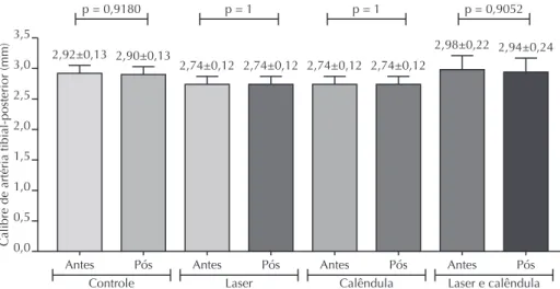
Posterior tibial artery caliber was recorded by Doppler US. Figure 1 shows that there was no statistically signiicant diference within groups, meaning the data remained constant until the end of the therapeutic protocols were performed.
Calibre de artéria tibial-posterior (mm)
3,5
2,5
1,5
0,5 3,0
2,0
1,0
0,0
Antes
Controle Laser Calêndula Laser e calêndula Pós Antes Pós Antes Pós Antes Pós p = 0,9180 p = 1 p = 1 p = 0,9052
2,92±0,13 2,90±0,13
2,74±0,12 2,74±0,12 2,74±0,12 2,74±0,12
2,98±0,22 2,94±0,24
Figure 1 – Comparative statistical analysis of the evaluation values of the caliber of the posterior tibial artery in each group – Teresina, Piauí, Brazil, 2015.
Figure 2 indicates that the pressure measurement by ABI showed no statistical diference for intragroup signiicance.
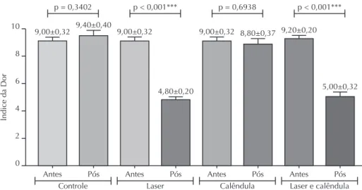
Data related to quantitative pain perception by the partici-pants from applying the Brief Pain Inventory and VAS analogue scale (represented in Figure 3) show that there was a statistically signiicant reduction for pain in L (p<0.001) and LEFA groups (p<0.001), evidencing that LLLT has an analgesic efect being isolated or associated with Calendula oicinalis oil.
here was a signiicant reduction of wound area in LEFA group (p=0.0032) and in L group (p = 0.0428). Group C showed signiicance contrary to the objective (p=0.3402) due to the absence of intervention. hese data are shown in Figure 4.
Índice tornozelo-br
aquial
0,6
0,2 0,8
0,4
0,0
Antes
Controle Laser Calêndula Laser e calêndula Pós Antes Pós Antes Pós Antes Pós p = 0,3402 p = 1 p = 0,7200 p = 0,6869 0,74±0,01 0,77±0,02 0,73±0,01 0,74±0,02
0,73±0,01 0,74±0,02 0,71±0,02 0,70±0,02
Figure 2 – Comparative statistical analysis of ABI values in each group – Teresina, Piauí, Brazil, 2015.
Índice da Dor
8
2 10
4
0
Antes
Controle Laser Calêndula Laser e calêndula Pós Antes Pós Antes Pós Antes Pós p = 0,3402 p < 0,001*** p = 0,6938 p < 0,001***
9,00±0,32 9,40±0,40 9,00±0,32
4,80±0,20
9,00±0,32 8,80±0,37 9,20±0,20
5,00±0,32 6
Figure 3 – Comparative statistical analysis of quantitative pain assessment values in the groups – Teresina, Piauí, Brazil, 2015.
Diâmetro (cm²)
8
2 10
4
0
Antes
Controle Laser Calêndula Laser e calêndula Pós Antes Pós Antes Pós Antes Pós p = 0,0145* p = 0,0428* p = 0,4658 p < 0,0032**
2,55±0,77
8,43±1,84 7,98±2,06
2,39±1,26
4,95±1,74
3,30±1,31
9,27±0,87
2,57±1,51 6
Low-level laser therapy and in repairing diabetic foot ulcers
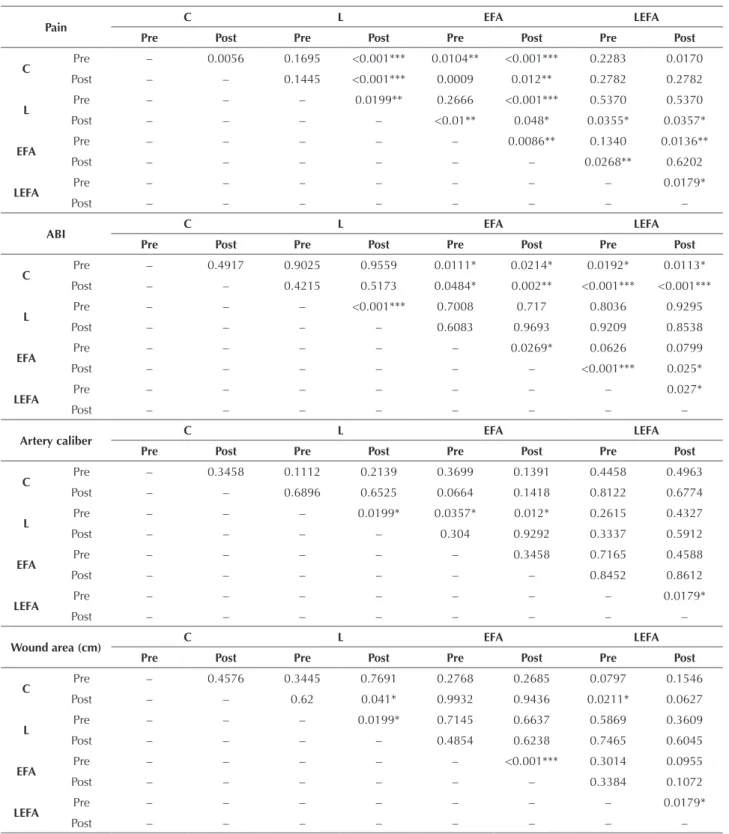
Table 1 – Intergroup assessment of pain, ABI, US, and wound area by Tukey test after ANOVA evaluation, with 95% confidence interval and significance at p<0.05 – Teresina, Piauí, Brazil, 2015.
Pain C L EFA LEFA
Pre Post Pre Post Pre Post Pre Post
C Pre – 0.0056 0.1695 <0.001*** 0.0104** <0.001*** 0.2283 0.0170
Post – – 0.1445 <0.001*** 0.0009 0.012** 0.2782 0.2782
L Pre – – – 0.0199** 0.2666 <0.001*** 0.5370 0.5370
Post – – – – <0.01** 0.048* 0.0355* 0.0357*
EFA Pre – – – – – 0.0086** 0.1340 0.0136**
Post – – – – – – 0.0268** 0.6202
LEFA Pre – – – – – – – 0.0179*
Post – – – – – – – –
ABI C L EFA LEFA
Pre Post Pre Post Pre Post Pre Post
C Pre – 0.4917 0.9025 0.9559 0.0111* 0.0214* 0.0192* 0.0113*
Post – – 0.4215 0.5173 0.0484* 0.002** <0.001*** <0.001***
L Pre – – – <0.001*** 0.7008 0.717 0.8036 0.9295
Post – – – – 0.6083 0.9693 0.9209 0.8538
EFA Pre – – – – – 0.0269* 0.0626 0.0799
Post – – – – – – <0.001*** 0.025*
LEFA Pre – – – – – – – 0.027*
Post – – – – – – – –
Artery caliber C L EFA LEFA
Pre Post Pre Post Pre Post Pre Post
C Pre – 0.3458 0.1112 0.2139 0.3699 0.1391 0.4458 0.4963
Post – – 0.6896 0.6525 0.0664 0.1418 0.8122 0.6774
L Pre – – – 0.0199* 0.0357* 0.012* 0.2615 0.4327
Post – – – – 0.304 0.9292 0.3337 0.5912
EFA Pre – – – – – 0.3458 0.7165 0.4588
Post – – – – – – 0.8452 0.8612
LEFA Pre – – – – – – – 0.0179*
Post – – – – – – – –
Wound area (cm) C L EFA LEFA
Pre Post Pre Post Pre Post Pre Post
C Pre – 0.4576 0.3445 0.7691 0.2768 0.2685 0.0797 0.1546
Post – – 0.62 0.041* 0.9932 0.9436 0.0211* 0.0627
L Pre – – – 0.0199* 0.7145 0.6637 0.5869 0.3609
Post – – – – 0.4854 0.6238 0.7465 0.6045
EFA Pre – – – – – <0.001*** 0.3014 0.0955
Post – – – – – – 0.3384 0.1072
LEFA Pre – – – – – – – 0.0179*
Post – – – – – – – –
Legend: C. control; L. Laser; EFA. Calendula; LEFA. Laser associated with Calendula. Source: Original data.
DISCUSSION
A study of 189 patients with diabetic foot used lower limb Doppler US to check the degree of stenosis of the arteries, classifying them in two ways: reduction in diam-eter indicated by values < or = to 50% and complete occlu-sion for values in between 51 and 99%. he results showed that the evaluated arteries were compromised, and that the
ultrasound was crucial in detecting vascular disorders caused
by DM, especially with risk of occlusion(10), thus agreeing
with the indings of this study in which a decrease in the diameter of the posterior tibial artery was detected, without a diference in post-intervention values.
cardiovascular and peripheral vascular disorders, since the observation of altered values in diabetic patients contrib-utes to determining or evidencing higher probability of the onset of Peripheral Arterial Occlusive Disease (POAD). Patients who have this index out of the normal range should be accompanied by a specialized medical team because they present a risk of recurrent ulcers and consequently serious
risk of lower limb amputation(11).
A study performed on 32 volunteers with the objec-tive to compare ABI values among older adult diabetic and non-diabetic patients showed that diabetics are more likely
to develop POAD(12). herefore, it can be said that this
re-search supports the cited authors, since this study was ac-curate in detecting pressure changes in the posterior tibial artery, despite the applied protocols in the three interven-tion groups not providing evidence of statistical signiicance regarding the improvement of the alterations, as ABI re-mained without signiicant intragroup variations. here was post-intervention signiicance in L and LEFA groups in the intergroup analysis.
he deinition of the therapeutic protocol and the choice for the wavelength of 658 nm, peak power of 30 mW, and
application time of 80s (4 J/cm2) in this study had published
articles as reference which also used LLLT as a proposal for pain treatment and tissue repair, suggesting that wavelengths between 600 and 1000 nm and power of 1 mW to 5 W/
cm2 have satisfactory efects. LLLT application has also been
mentioned in order to promote better healing of inlamma-tion, reduced pain, preventing occurrence of edema, as well as for preserving tissues and nerves adjacent to the injury site(13).
New drugs have additionally been used in the treatment of cutaneous lesions in patients with DM, with the purpose of speeding up the tissue repair process. Recently, studies of medicinal plants have been highlighted, as these favor the neovascularization process and collagen iber formation in the tissue repair process, with analgesic, anti-inlammatory
and antisepticaction(14-15). hese studies corroborate the
indings of the present study, since ulcer hydration in
dia-betic patients with vascular lesions using Calendula
oicina-lis oil was crucial for accelerating the tissue repair process,
for both intragroup and intergroup.
hus, the results of this study are in agreement with the abovementioned authors, since the efect of applying
Calendula oicinalis oil alone or associated with LLLT as
a therapeutic protocol presented positive results on the patients’ conditions. Figure 3 shows an increase in pain symptoms for group C. By analyzing the intergroup values, Groups L, EFA and LEFA showed signiicance. herefore, the need to implement care protocols for these patients in order to avoid the consequences of DM is evident.
CONCLUSION
We conclude that LLLT isolated or in combination
with Calendula oicinalis oil is efective in relieving pain
due to its anti-inlammatory action, and in reducing the total area of ulcers by stimulating neovascularization and accelerating cell proliferation, thereby contributing to im-proving possible morbidities that may occur as consequence of Diabetes Mellitus.
RESUMO
Objetivo: Avaliar os efeitos da Terapia a Laser de Baixa Intensidade isolada e associada ao óleo de Calendula oicinalis no reparo de úlceras em pé diabético. Método: Estudo de caso clínico, experimental, controlado, randomizado, prospectivo, intervencional, de caráter quantitativo. A amostra foi composta de 32 pacientes diabéticos, de ambos os gêneros. Os participantes foram distribuídos aleatoriamente em quatro grupos. Ultrassom Doppler, avaliação do Índice Tornozelo-Braquial, Inventário breve de dor e escala de dor analógica foram realizados no início e após 30 dias. Resultados: Houve redução da dor nos grupos Terapia a Laser de Baixa Intensidade e Terapia a Laser de Baixa intensidade associada aos Ácidos Graxos Essenciais, com p<0,01. Quanto ao Índice Tornozelo-Braquial e Ultrassom Doppler, todos os grupos mantiveram-se estáveis. Na análise da redução de área da lesão, o grupo Terapia a Laser de Baixa Intensidade associada aos Ácidos Graxos Essenciais apresentou uma signiicância p=0,0032, e o grupo Terapia a Laser de Baixa Intensidade, p=0,0428. Conclusão: A Terapia a Laser de Baixa Intensidade, realizada tanto isoladamente quanto associada ao óleo de Calendula oicinalis, foi eicaz no alívio da dor e na aceleração do processo de reparo tecidual de pé diabético.
DESCRITORES
Calendula Oicinalis; Diabetes Mellitus; Pé Diabético; Terapia a Laser.
RESUMEN
Objetivo: Evaluar los efectos de la Terapia con Láser de Baja Intensidad aislada y asociada con el aceite de Calendula oicinalis en la reparación de úlceras en pie diabético. Método: Estudio de caso clínico, experimental, controlado, randomizado, prospectivo, intervencionista, de carácter cuantitativo. La muestra estuvo compuesta de 32 pacientes diabéticos, de ambos géneros. Los participantes fueron distribuidos aleatoriamente en cuatro grupos. Ecografía Doppler, evaluación del Índice Tobillo-Brazo, Inventario breve de dolor y escala visual analógica fueron realizados al inicio y después de 30 días. Resultados: Hubo reducción del dolor en los grupos Terapia con Láser de Baja Intensidad y Terapia con Láser de Baja intensidad asociada con los Ácidos Grasos Esenciales, con p<;0,01. En cuanto al Índice Tobillo-Brazo y la Ecografía Doppler, todos los grupos se mantuvieron estables. En el análisis de la reducción del área de la lesión, el grupo Terapia con Láser de Baja Intensidad asociada con los Ácidos Grasos Esenciales presentó una signiicación p=0,0032, y el grupo Terapia con Láser de Baja Intensidad, p=0,0428. Conclusión: La Terapia con Láser de Baja Intensidad, llevada a cabo tanto aisladamente como asociada con el aceite de Calendula oicinalis, fue eicaz en el alivio del dolor y la aceleración del proceso de reparación de tejidos del pie diabético.
DESCRIPTORES
Low-level laser therapy and in repairing diabetic foot ulcers
REFERENCES
1. Ochoa O, Torres FM, Shireman PK. Chemokines and diabetic wound healing. Vasc 2007;15(6):350-5.
2. Abreu AM, Oliveira BG. A study of the Unna Boot compared with the elastic bandage in venous ulcers: a randomized clinical trial. Rev Latino Am Enfermagem. 2015; 23(4):571-7.
3. Hogarth, PJ. The biology of mangroves and seagrasses. Oxford: University Press, 2015.
4. Schaper NC, Andros G, Apelqvist J, Bakker K, Lammer J, Lepantalo M, et al. Diagnosis and treatment of peripheral arterial disease in diabetic patients with a foot ulcer: a progress report of the International Working Group on the Diabetic Foot. Diabetes Metab Res Rev. 2012;28(1):218-4.
5. Moreira FF, Oliveira ELP, Barbosa FS, Silva, JG. Laserterapia de baixa intensidade na expressão de colágeno após lesão muscular cirúrgica. Fisioter Pesq. 2011;18(1):37-42.
6. Brasil. Ministério da Saúde; Agência Nacional de Vigilância Sanitária. Instrução Normativa n.5, de 11 de dezembro de 2008. Determina a publicação da Lista de Medicamentos Fitoterápicos de Registro Simpliicado [Internet]. Brasília; 2008 [citado 2015 out. 22]. Disponível em: http://bvsms.saude.gov.br/bvs/saudelegis/anvisa/2005/int0005_11_12_2008.html
7. Pristo I. Cicatrização de feridas: fases e fatores de inluência. Act Vet Bras. 2013; 6(4):267-71.
8. Brownrigg JRW, Bruin JL, Rossi L, Karthikesalingam A, Patterson B, Holt PJ, et al. Endovascular aneurysm sealing for infrarenal abdominal aortic aneurysms: 30-day outcomes of 105 patients in a single centre. Eur J Vasc Endovasc Surg. 2015;50(2):157-64.
9. Kawamura T. Índice Tornozelo-Braquial (ITB) determinado por esigmomanômetros oscilométricos automáticos. Arq Bras Cardiol.
2008;90(5):322-6.
10. Wen XR, Lü XF, Liu CC, Luo Y, Chen DW, Wang C, et al. The ultrasound image characteristics of lower extremities arteries in diabetic foot [abstract]. Sichuan Da Xue Xue Bao Yi Xue Ban. 2012;43(5):739-42.
11. Torres AGMJ, Machado EG, Lopes TS, Gentile PC, Vieira AC, Soares LG, et al. Prevalência de alteração do índice tornozelo-braço em
indivíduo portador assintomático de doença arterial obstrutiva periférica. Rev Bra Cardiol. 2015;25(2): 87-93.
12. Santos MDL, Santos VA, Santos WF, Silva JS, Wanderley AMPS, et al. Comparação dos valores do índice tornozelo-braquial entre idosos
diabéticos e não diabéticos. Rev Humano Ser. 2015;1(1):18-31.
13. Huang YY, Chen AC, Carroll JD, Hamblin MR. Biphasic dose response in low level light therapy. Dose Response. 2009;7(4):358-83.
14. Honório-França AC, Marins CMF, Boldrini F, França EL. Evaluation of hypoglicemic activity and healing of extract from amongst bark of” Quina do Cerrado”(Strychnos pseudoquina ST. HILL). Acta Cir Bras. 2008;23(6):504-10.