UPDATE ARTICLE
Septic encephalopathy: does inflammation drive the brain
crazy?
Felipe Dal-Pizzol, Cristiane D. Tomasi, Cristiane Ritter
Laboratory of Experimental Pathophysiology and National Science and Technology Institute for Translational Medicine (INCT-TM), Graduate Program in Health Sciences, Health Sciences Research Center, Universidade do Extremo Sul Catarinense (UNESC), Criciu´ma, SC, Brazil.
Sepsis and the multiorgan dysfunction syndrome are among the most common reasons for admission
to an intensive care unit, and are a leading cause of death. During sepsis, the central nervous system
(CNS) is one of the first organs affected, and this is clinically manifested as sepsis-associated
encephalopathy (SAE). It is postulated that the common final pathway that leads to SAE symptoms is
the deregulation of neurotransmitters, mainly acetylcholine. Thus, it is supposed that inflammation can
affect neurotransmitters, which is associated with SAE development. In this review, we will cover the
current evidence (or lack thereof) for the mechanisms by which systemic inflammation interferes with
the metabolism of major CNS neurotransmitters, trying to explain how systemic inflammation drives
the brain crazy.
Keywords:
Sepsis-associated encephalopathy; acetylcholine; amines; GABA; inflammation
Introduction
Sepsis and the multiorgan dysfunction syndrome are
among the most common reasons for admission to an
intensive care unit, and are a leading cause of death.
1-4During the last decades, advances have been made in
our understanding of sepsis, but currently there is no
target-directed, FDA-approved treatment for sepsis.
The pathophysiology of sepsis has been partially
elucidated; it is a dynamic process, which involves
components of the immune system, the coagulation
pathway, parenchymal cells, and the endocrine and
metabolic pathways.
5Many factors have been postulated
to trigger sepsis, including products released from
bacteria as well as products from damaged cells.
Toll-like receptor (TLR) signaling has been suggested to be a
key pathway in sepsis pathophysiology, leading to the
production of inflammatory mediators.
6The activation of
this pathway depends on the interaction between TLR
and TLR ligands, which include those derived from
bacteria in addition to host-derived products such as
intracellular proteins, extracellular matrix components,
and oxidized lipids.
7During sepsis, the central nervous system (CNS) is
one of the first organs affected, and this is clinically
manifested as septic encephalopathy (SE) or
sepsis-associated delirium (SAD).
8-9SE has been reported to
occur in 8-70% of septic patients, with the wide
varia-tion attributable mainly to diagnostic criteria.
10Reciprocal
interactions between the immune system and the
CNS are considered to be major components of the
host response in sepsis (Figure 1). In addition, brain
injury occurs during sepsis development, and
pro-posed mechanisms to explain it include alterations
in the blood-brain barrier (BBB), local generation of
pro- and anti-inflammatory cytokines, amino acid
meta-bolism disruption, brain ischemia, and imbalance of
neurotransmitters
11(Figure 2). Additionally, once
inflam-mation persists, excitotoxicity and oxidative stress may
further aggravate SE and contribute to neuronal
dysfunc-tion.
12In animal models of sepsis, acute encephalopathy
occurs, and survivors exhibit cognitive impairment that
could be secondary to CNS damage.
13Likewise,
survi-vors from critical care, including septic patients, have
well-documented persistent neurocognitive deficits and
develop psychiatric disorders.
14-23The interaction between sepsis and the brain is an
opportunity to study how systemic inflammation affects
brain function. Most studies about the mechanisms of SE
have used animals or cell cultures, and improved our
understanding of how the CNS is affected by endotoxins
and cytokines, but whether this is related to clinical SE
remains unclear. It is postulated that the common final
pathway that drives SE symptoms is the deregulation of
neurotransmitters, mainly acetylcholine.
24In this review,
we will cover the current evidence (or lack thereof) for the
mechanisms by which systemic inflammation interferes with
the metabolism of the major CNS neurotransmitters, trying
to explain how systemic inflammation drives the brain crazy.
Evidence that systemic inflammation is
associated with brain dysfunction
Theoretically, systemic inflammation can reach the brain
through at least four different routes: 1) peripheral organs
Correspondence: Felipe Dal-Pizzol, Universidade do Extremo Sul Catarinense (UNESC), Av. Universita´ria, 1105, Universita´rio, CEP 88806-000, Criciu´ma, SC, Brazil.
E-mail: [email protected]
Submitted Aug 16 2013, accepted Nov 11 2013.
Revista Brasileira de Psiquiatria. 2014;36:251–258 ß2014 Associac¸a˜o Brasileira de Psiquiatria
synthesize and release cytokines that act on their
receptors present in nerve fibers of the autonomic
nervous system to modulate brain function; 2) circulating
cytokines diffuse through the BBB; 3) cytokines might
signal into the brain through specific areas that lack
the BBB, such as the circumventricular organs; or
4) cytokines might enter the brain through a saturable
transport mechanism.
25There is no clear evidence to
explain in detail how inflammation reaches the brain
during sepsis, but both in animals and in humans,
inflammation occurs in the CNS early and late after
sepsis.
26-29The immune system is a complex, highly adaptive
system,
29and it is integrated with the CNS at several
levels to maintain homeostasis.
30-32However, it is possible
that activation of the immune system may induce brain
dysfunction, and, in fact, sepsis is a major risk factor for
occurrence of delirium.
33It is believed that
pro-inflamma-tory cytokines, particularly interleukin (IL)-1
b
and tumor
necrosis factor alpha (TNF-
a
), are generated in the
periphery, communicate with the brain, and initiate
cytokine synthesis in the CNS.
24Fever and changes in
behavior –
– such as anorexia, lethargy, and depression,
collectively named sickness behavior –
– are observed as a
response of neurons to cytokines in several different
animal models.
34-38In addition, studies in healthy
volun-teers have demonstrated that systemic inflammatory
challenges impact the human brain.
39-41A postmortem
investigation found an association between brain
dysfunc-tion and astrocyte, microglia, and IL-6 activity in the human
brain.
42However, excluding postmortem studies, a direct
relation between inflammation and brain dysfunction in
humans is limited because of the inaccessibility of the
CNS. Thus, in general, investigations are limited to
searching for a correlation between systemic
inflamma-tion markers and brain dysfuncinflamma-tion.
High levels of procalcitonin and C-reactive protein
(CRP) at intensive care unit (ICU) admission correlate
with the duration of brain dysfunction, both in septic and
non-septic patients.
43Krabbe et al.,
41using a human
endotoxemia model, showed that a low-grade increase in
the concentrations of TNF-
a
, its soluble receptor
(sTNF-R), IL-6, and IL-1 receptor antagonist (IL-1RA) was
inversely associated with declarative memory
perfor-mance. This was independent of physical stress
symp-toms or activation of the hypothalamic-pituitary-adrenal
(HPA) axis, suggesting that low-level systemic
inflamma-tion had a negative effect on some areas related to
cognitive function. A recent study demonstrated that
sTNFR was independently associated with delirium in
general ICU patients.
44Pfister et al.
45found a correlation between high plasma
CRP levels, alterations in cerebrovascular autoregulation,
and SE. The cerebral arterioles of patients with SE were
less reactive to vasodilatory stimuli,
46and this was
independently associated with acute brain dysfunction.
47Healthy
volunteers
injected
with
endotoxin
had
decreased cerebral blood flow, and this was associated
with peak serum concentrations of TNF-
a
.
48Recently, it
was demonstrated that patients with lower vascular
reactivity had increased duration of brain dysfunction.
47The mechanisms behind endothelial dysfunction and
acute brain dysfunction remain unclear, but inflammation
could drive structural and functional alterations in the
BBB, increasing microvascular permeability and
impair-ing microcirculatory blood flow.
49-52These alterations
could be secondary to a decrease in the activity of
endothelial nitric oxide synthase induced by
inflamma-tion
53or to alterations in the coagulation system, resulting
in microthromboses and microinfarcts.
54Endothelial
activation in the brain microvasculature has been
observed after sepsis in animal models, and this was
associated with leukocyte adhesion and brain
inflamma-tion.
26In addition, BBB dysfunction induced by
metallo-proteinase (MMP) activation was also associated with
brain inflammation and cognitive impairment in an animal
model of sepsis.
55This is supported by the fact that
MMP-9 content was associated with delirium in the
general ICU patient.
44It is supposed that systemic inflammation can
lead to neuronal or glial damage; however, at least in
experimental endotoxemia in humans, there is no
correlation between acute systemic inflammation and
plasma levels of brain specific proteins (S-100
b
,
neuro-nal enolase [NSE], glial fibrillary acidic protein [GFAP])
nor deterioration of cognitive function.
56In contrast,
S-100
b
is associated with SE,
45and NSE is
associ-ated with delirium in general ICU patients.
57Sharshar
et al.
27demonstrated that septic shock is associated
with neuronal and glial apoptosis in autonomic
cen-ters in humans, but brain TNF-
a
expression did not
differ between septic shock and control patients.
Whether neuronal and glial apoptosis is sufficient
to induce clinically relevant brain dysfunction remains
unknown.
Thus, to date, there is evidence that brain inflammation
occurs during sepsis both in animals and in humans.
Inflammation is probably related to alterations in cerebral
blood flow and neuronal/glial cell damage, but a direct link
between these and SE is still lacking.
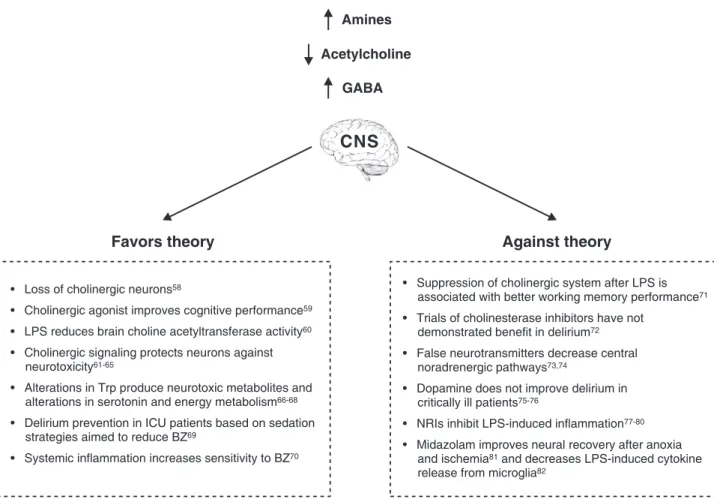
Evidence linking systemic inflammation and
deregulation of neurotransmitters
Since inflammation and alterations in
neurotransmit-ters are the major theories trying to explain brain
dysfunction we explore the evidences that links
in-flammation and major neurotransmitters system
dereg-ulation (Figure 3).
Acetylcholine (Ach)
A widely postulated mechanism to explain delirium is
cholinergic failure.
83The first evidence for this hypothesis
Figure 2
Cytokines produced in the infection site activate afferent signals to the brain, and the subsequent vagal activation
inhibits cytokine synthesis through the ‘‘inflammatory reflex’’ of the cholinergic pathway. Inflammation changes the structure
and function of the blood-brain barrier (BBB), increasing microvascular permeability, impairing microcirculatory blood flow, and
producing brain inflammation. During sepsis, alterations in the coagulation system results in microthrombus formation and
microinfarcts. Endothelial activation also impairs the microcirculation and worsens brain inflammation, which in turn is related
to brain dysfunction. Ach = acetylcholine; BBB = blood-brain barrier; SAE = sepsis-associated encephalopathy.
came from case reports linking delirium to acute
poison-ing with anticholinergic drugs and the reversal of delirium
with cholinergic drugs. Cholinergic signaling by both
nicotinic and muscarinic receptors modulates cognitive
function, arousal, learning, and memory, the major brain
functions affected in delirium. Thus, sepsis-induced
inflammation is presumed to affect cholinergic signaling
and contribute to the genesis of SE.
In an animal model of LPS-induced long-term cognitive
deficits, neuronal loss in the hippocampus and the
prefrontal cortex occurs mainly due to reduced
choliner-gic innervation at postrolandic cortical areas. This is
consistent with the fact that the hippocampus is
particu-larly sensitive to systemic inflammation.
84We
demon-strated that the use of cholinergic agonists improves
cognitive performance in septic animals,
58and that
endotoxin is able to reduce brain choline
acetyltransfer-ase activity.
59Thus, it is possible that cholinergic neurons
are particularly sensitive to systemic inflammation. This is
the major theory behind SE, but to date there is no direct
evidence to support it.
Endotoxin administration to healthy individuals
in-creases plasma acetylcholinesterase (AChE) activity,
which is associated with better performance in evocative
memory tasks, but worse performance in working
memory.
60In addition, patients that respond to endotoxin
by suppressing the cholinergic system have a better
working memory performance as compared with patients
that enhance cholinergic activity, indicating that limited
cholinergic activation may be beneficial for cognition.
60To date, human trials of cholinesterase inhibitors have not
demonstrated benefit to prevent or treat delirium.
71The cholinergic pathway may be involved indirectly in
the pathogenesis of delirium. The cholinergic pathway
acts as a predictor of individual variation in systemic
inflammatory response to infection; thus, by modulating
systemic inflammation, the cholinergic system can
indirectly affect brain function.
60High plasma levels of
an alpha-7 nicotinic Ach receptor agonist correlated with
lower cytokine levels in endotoxin-treated volunteers.
72Cholinergic signaling protects striatal, hippocampal, and
cortical neurons against neurotoxicity induced by
excito-toxic amino acids as well as other excito-toxic insults. Several
mechanisms have been postulated to explain these
effects, from the production of growth factors
61,85to a
decrease in superoxide anion generation
62to antioxidant
actions.
63,64In addition, resembling the peripheral
choli-nergic anti-inflammatory pathway, ACh and nicotine
65have been reported to modulate LPS-induced TNF-
a
release from microglia through activation of
a
-nAChR.
Thus, it is possible that the decrease in cholinergic
neurons during systemic inflammation decreases the
availability of an ‘‘anti-inflammatory’’ signal in the brain.
This is consistent with the decrease of cholinergic
neurons observed with aging
86that occurs in parallel to
microglia activation.
87Amines
Dopamine, norepinephrine, and serotonin have a role in
arousal and the sleep-wake cycle.
83In addition, the D2
dopamine receptor subtype has been associated with
hallucinations, stereotypic behavior, and thought
distur-bances,
88and norepinephrine plays an important role in
modulating attention, anxiety, and mood.
89Thus, amines
could be involved in several different symptoms
asso-ciated with brain dysfunction. In fact, excess dopamine
and norepinephrine has been associated with hyperactive
delirium.
89Experimentally, this is supported by the fact
that the administration of dopamine agonists results in
frontostriatal abnormalities that correlate with delirium,
and dopamine antagonists are classically used to treat
hyperactive delirium.
90,91Furthermore, elevated CNS
serotonin activity is postulated to occur in hepatic
encephalopathy, and serotonin syndrome secondary to
the withdrawal of serotonin reuptake inhibitors resembles
the clinical picture of SE.
92,93Brain serotonin synthesis depends on the availability of
tryptophan (Trp), and dopamine and norepinephrine
production requires tyrosine (Tyr) and phenylalanine
(Phe).
94Despite the fact that most delirium theories
suggest that an increase in amines drives delirium, in
healthy volunteers the administration of LPS increases
the cerebral delivery and influx of Phe.
95This can be
associated with the synthesis of ‘‘false’’
neurotransmit-ters, such as phenylethanolamine, which in turn can
decrease central noradrenergic pathways.
73,96An
ele-vated Phe/large neutral amino acids (LNAA) ratio during
acute febrile illness is associated with delirium in
hos-pitalized elderly patients.
74The systemic inflammatory
response is associated with a decrease in the ratio of
branched-chain (valine and isoleucine) and aromatic
amino acids (mainly phenylalanine). This is associated
with an increase in the cerebral delivery and
unidirec-tional cerebral influx of phenylalanine, an abolished influx
of leucine and isoleucine, and an ammonia-independent
cerebral efflux of glutamine.
95In this context, both low
and high levels of Trp/LNAA are associated with delirium
in the general ICU patient.
94Alterations in Trp
concentra-tions could lead to delirium due to the production of
neurotoxic metabolites or alterations in
serotonin/melato-nin synthesis. In fact, increased activation of the
kynurenine pathway (a neurotoxic metabolite of Trp) is
associated with mortality and brain dysfunction in ICU
patients.
97Besides its neurotoxic effect, the accumulation
of kynurenine or quinolinic acid can compromise immune
functions.
66In addition, the excessive degradation of
tryptophan, as seen in septic patients, could lead to
NAD+ depletion.
67In this context, neurons may become
functionally hypoxic (due to Krebs cycle impairment) even
in the presence of oxygen, and cellular hibernation may,
in part, reflect an underlying tryptophan shortage.
Plasma levels of Tyr/LNAA are also associated with
delirium. Patients with high levels of tyrosine could have
excess dopamine or norepinephrine, which is consistent
with a neurotoxic effect of norepinephrine.
68Despite this,
the use of dopamine antagonists in critically ill patients
does not consistently improve delirium severity and
duration,
75,98and the use of vasoactive drugs is not
independently associated with increased incidence of
delirium.
76Furthermore, as described with Ach,
norepi-nephrine seems to have anti-inflammatory properties.
The administration of norepinephrine reuptake inhibitors
(NRIs) inhibits LPS-induced expression of cytokines,
chemokines
and
endothelial
activation,
99probably
increasing norepinephrine availability to glial cells. In
addition,
a
2-adrenoceptor stimulation decreases vascular
endothelial cell permeability
77and reduces production of
inflammatory mediators.
78Supporting this view,
dopex-amine, an
a
2-adrenoceptor and dopamine 1 and 2
receptor agonist, protects against cerebral edema
induced by sepsis, and the co-administration of an
a
2-adrenoceptor antagonist blunted this effect.
79In humans,
there is preliminary evidence that dexmedetomidine, an
a
-agonist, exerts protective effects in septic patients,
80but this is not supported by animal models of primary
brain injury.
100The lack of a consistent effect is also
observed with the
b
-adrenergic receptor;
b
2-adrenocep-tor activation can induce
101or protect against brain
inflammation.
102,103Gamma-aminobutyric acid (GABA)
The most compelling evidence about delirium prevention
in ICU patients comes from sedation strategies aimed to
reduce benzodiazepine (BZ) use.
104Despite this, little is
known about GABA neurotransmission under
inflamma-tory conditions, or of the exact mechanisms whereby
increased GABA signaling drives delirium. The cortical
type A GABA (GABA-A) and corticotrophin-releasing
factor systems are major regulatory factors of the
behavioral response to stress.
69Acute stressors such
as restraint, infection, hypoxia or combined mild stressors
influence the GABA-A complex at two different levels: by
altering BZ-1 binding sites and modulating the expression
of selective GABA-A receptor subunits.
105Inflammatory
mediators increase the insertion of GABA-A receptors in
the neuron membrane,
106and an increase in GABA-A
receptor activity has been observed in septic rats.
107,108Thus, it could be presumed that increased sensitivity to
BZ occurs during systemic inflammation. In fact, GABA-A
agonists worsen postoperative pain only in the presence
of
inflammation.
109Cerebral
synaptic
activity
is
decreased in SE, and because GABA-A receptor
regulates synaptic transmission in most cerebral
inhibi-tory synapses, it is possible that GABA-A could be a
target for new therapeutic strategies aimed to treat or
prevent delirium. Indeed, increased GABAergic
neuro-transmission has been reported in patients with hepatic
encephalopathy.
70On the other hand, while the expression of GABA-A
receptors is found normally in glial cells, BZ receptor
non-associated with GABA-A expression, which is low in
normal glial cells, is increased during inflammatory
conditions.
110In this context, the BZ midazolam improves
neural recovery after anoxia and ischemia.
111Midazolam
decreases LPS-induced cytokine release from microglia
via non-GABA-A BZ receptors.
81This seems to be a
specific effect, as propofol has no such protective effect
in
vitro
.
82Thus, if systemic and brain inflammation leads to
delirium, it is expected that BZ could improve delirium in
the critically ill patient.
Concluding remarks
Despite the fact that SE and brain dysfunction are highly
prevalent in ICU patients and are associated with worse
prognosis, surprisingly little is known about their
patho-physiology. The most cited theory –
– neurotransmitter
deregulation –
– lacks solid evidence to be widely
accepted, and this may partly account for the lack of
effect of clinical interventions designed to treat acute
brain dysfunction, mainly strategies based on cholinergic
drugs. Thus, the hypothesis that neurotransmission and
inflammation are connected and are major players in
brain dysfunction pathophysiology requires further critical
assessment in the future.
Acknowledgements
This study received financial support from the Center of
Excellence in Applied Neurosciences of Santa Catarina
(NENASC), Program of Support to Centers of Excellence
(PRONEX), Conselho Nacional de Desenvolvimento
Cientı´fico e Tecnolo´gico (CNPq) and Fundac¸a˜o de
Amparo a` Pesquisa e Inovac¸a˜o do Estado de Santa
Catarina (FAPESC); from the National Science and
Technology Institute for Translational Medicine
(INCT-TM); and from Programa de Cooperac¸a˜o Acadeˆmica
(PROCAD) –
– Sepse.
Disclosure
The authors report no conflicts of interest.
References
1 Perl TM, Dvorak L, Hwang T, Wenzel RP. Long-term survival and function after suspected gram-negative sepsis. JAMA. 1995; 274:338-45.
2 Quartin AA, Schein RM, Kett DH, Peduzzi PN. Magnitude and duration of the effect of sepsis on survival. Department of Veterans Affairs Systemic Sepsis Cooperative Studies Group. JAMA. 1997;277:1058-63.
3 Angus DC, Linde-Zwirble WT, Lidicker J, Clermont G, Carcillo J, Pinsky MR. Epidemiology of severe sepsis in the United States: analysis of incidence, outcome, and associated costs of care. Crit Care Med. 2001;29:1303-10.
4 Iwashyna TJ, Ely EW, Smith DM, Langa KM. Long-term cognitive impairment and functional disability among survivors of severe sepsis. JAMA. 2010;304:1787-94.
5 Vincent JL, Opal SM, Marshall JC, Tracey K. Sepsis definitions: time for change. Lancet. 2013;381:774-5.
6 Tsung A, McCoy SL, Klune JR, Geller DA, Billiar TR, Hefeneider SH. A novel inhibitory peptide of Toll-like receptor signaling limitslipopolysaccharide-induced production of inflammatory med-iators andenhances survival in mice. Shock. 2007;27:364-9. 7 Song DH, Lee JO. Sensing of microbial molecular patterns by
toll-like receptors. Immunol Rev. 2012;250:216-29.
8 Sprung CL, Peduzzi PN, Shatney CH, Schein RM, Wilson MF, Sheagren JN, et al. Impact of encephalopathy on mortality in the sepsis syndrome. The Veterans Administration Systemic Sepsis Cooperative Study Group. Crit Care Med. 1990;18:801-6. 9 Ebersoldt M, Sharshar T, Annane D. Sepsis-associated delirium.
Intensive Care Med. 2007;33:941-50.
10 Kafa IM, Bakirci S, Uysal M, Kurt MA. Alterations in the brain electrical activity in a rat model of sepsis-associated encephalo-pathy. Brain Res. 2010;1354:217-26.
11 Papadopoulos MC, Davies DC, Moss RF, Tighe D, Bennett ED. Pathophysiology of septic encephalopathy: a review. Crit Care Med. 2000;28:3019-24.
12 Wilson JX, Young GB. Progress in clinical neurosciences: sepsis-associated encephalopathy: evolving concepts. Can J Neurol Sci. 2003;30:98-105.
13 Barichello T, Martins MR, Reinke A, Feier G, Ritter C, Quevedo J, et al. Cognitive impairment in sepsis survivors from cecal ligation and perforation. Crit Care Med. 2005;33:221-3.
14 Hopkins RO, Weaver LK, Pope D, Orme JF, Bigler ED, Larson-LOHR V. Neuropsychological sequelae and impaired health status in survivors of severe acute respiratory distress syndrome. Am J Respir Crit Care Med. 1999;160:50-6.
15 Heyland DK, Hopman W, Coo H, Tranmer J, McColl MA. Long-term healthrelated quality of life in survivors of sepsis. Short Form 36: a valid and reliable measure of health-related quality of life. Crit Care Med. 2000;28:3599-605.
16 Angus DC, Musthafa AA, Clermont G, Griffin MF, Linde-Zwirble WT, Dremsizov TT, et al. Quality adjusted survival in the first year after the acute respiratory distress syndrome. Am J Respir Crit Care Med. 2001;163:1389-94.
17 Granja C, Dias C, Costa-Pereira A, Sarmento A. Quality of life of survivors from severe sepsis and septic shock may be similar to that of others who survive critical illness. Crit Care. 2004;8: R91-8.
18 Jackson JC, Gordon SM, Ely EW, Burger C, Hopkins RO. Research issues in the evaluation of cognitive impairment in intensive care unit survivors. Intensive Care Med. 2004;30:2009-16.
19 Granja C, Lopes A, Moreira S, Dias C, Costa-Pereira A, Carneiro A, et al. Patients’ recollectionsof experiences in the intensive care unit may affect their quality of life. Crit Care. 2005;9:R96-109. 20 Hopkins RO, Weaver LK, Collingridge D, Parkinson RB, Chan KJ,
Orme JF Jr. Two-year cognitive, emotional, and quality-of-life outcomes in acute respiratory distress syndrome. Am J Respir Crit Care Med. 2005;171:340-7.
21 Hough CL, Curtis JR. Long-term sequelae of critical illness: memories and health-related quality of life. Crit Care. 2005;9:145-6. 22 Hopkins RO. Sepsis, oxidative stress, and brain injury. Crit Care
Med. 2007;35:2233-4.
23 Gordon SM, Jackson JC, Ely EW, Burger C, Hopkins RO. Clinical identification of cognitive impairment in ICU survivors: insights for intensivists. Intensive Care Med. 2004;30:1997-2008.
24 van Gool WA, van de Beek D, Eikelenboom P. Systemic infection and delirium: when cytokines and acetylcholine collide. Lancet. 2010;375:773-5.
25 Licinio J, Mastronardi C, Wong M. Pharmacogenomics of neuroim-mune interactions in human psychiatric disorders. Exp Physiol. 2008;92:807-11.
26 Comim CM, Vilela MC, Constantino LS, Petronilho F, Vuolo F, Lacerda-Queiroz N, et al. Traffic of leukocytes and cytokine up-regulation in the central nervous system in sepsis. Intensive Care Med. 2011;37:711-8.
28 Weberpals M, Hermes M, Hermann S, Kummer MP, Terwel D, Semmler A, et al. NOS2 gene deficiency protects from sepsis-induced long-term cognitive deficits. J Neurosci. 2009;29:14177-84.
29 Teeling JL, Perry VH. Systemic infection and inflammation in acute cns injury and chronic neurodegeneration: underlying mechanisms. Neuroscience. 2009;158:1062-73.
30 Dantzer R, Bluthe RM, Gheusi G, Cremona S, Laye S, Parnet P, et al. Molecular basis of sickness behavior. Ann N Y Acad Sci. 1998;856:132-8.
31 Dantzer R, Aubert A, Bluthe RM, Gheusi G, Cremona S, Laye S, et al. Mechanisms of the behavioural effects of cytokines. Adv Exp Med Biol. 1999;461:83-105.
32 Dantzer R, Konsman JP, Bluthe RM, Kelley KW. Neural and humoral pathways of communication from the immune system to the brain: parallel or convergent? Auton Neurosci. 2000;85:60-5. 33 Khurana V, Gambhir IS, Kishore D. Evaluation of delirium in elderly:
a hospital-based study. Geriatr Gerontol Int. 2011;11:467-73. 34 Ek M, Kurosawa M, Lundeberg T, Ericsson A. Activation of vagal
afferents after intravenous injection of interleukin-1beta: role of endogenous prostaglandins. J Neurosci. 1998;18:9471-79. 35 Bluthe RM, Laye S, Michaud B, Combe C, Dantzer R, Parnet P.
Role of interleukin-1beta and tumour necrosis factor-alpha in lipopolysaccharide-induced sickness behaviour: a study with inter-leukin-1 type I receptor-deficient mice. Eur J Neurosci. 2000; 12:4447-56.
36 Bluthe RM, Michaud B, Poli V, Dantzer R. Role of IL-6 in cytokine-induced sickness behavior: a study with IL-6 deficient mice. Physiol Behav. 2000;70:367-73.
37 Cartmell T, Poole S, Turnbull AV, Rothwell NJ, Luheshi GN. Circulating interleukin-6 mediates the febrile response to localized inflammation in rats. J Physiol. 2000;526:653-61.
38 Konsman JP, Blond D, Vigues S. Neurobiology of interleukin-1 receptors: getting the message. Eur Cytokine Netw. 2000;11: 699-702.
39 Reichenberg A, Yirmiya R, Schuld A, Kraus T, Haack M, Morag A, et al. Cytokine-associated emotional and cognitive disturbances in humans. Arch Gen Psychiatry. 2001;58:445-52.
40 Cohen O, Reichenberg A, Perry C, Ginzberg D, Pollmacher T, Soreq H, et al. Endotoxin-induced changes in human working and declarative memory associate with cleavage of plasma ‘‘read-through’’ acetylcholinesterase. J Mol Neurosci. 2003;21:199-212. 41 Krabbe KS, Reichenberg A, Yirmiya R, Smed A, Pedersen BK,
Bruunsgaard H. Low-dose endotoxemia and human neuropsycho-logical functions. Brain Behav Immun. 2005;19:453-60.
42 Munster BC, Aronica E, Zwinderman AH, Eikelenboom P, Cunningham C, Rooij SE. Neuroinflammation in delirium: a postmortem case-control study. Rejuvenation Res. 2011;14: 615-22.
43 McGrane S, Girard TD, Thompson JL, Shintani AK, Woodworth A, Ely W, et al. Procalcitonin and C-reactive protein levels at admission as predictors of duration of acute brain dysfunction in critically ill patients. Crit Care. 2011;15:R78.
44 Girard TD, Ware LB, Bernard GR, Pandharipande PP, Thompson JL, Shintani AK, et al. Associations of markers of inflammation and coagulation with delirium during critical illness. Intensive Care Med. 2012;38:1965-73.
45 Pfister D, Siegemund M, Dell-Kuster S, Smielewiski P, Ru¨egg S, Strebel SP, et al. Cerebral perfusion in sepsis-associated delirium. Crit Care. 2008;12:R63.
46 Szatma´ri S, Ve´gh T, Csomo´s A, Hallay J, Taka´cs I, Molna´r C, et al. Impaired cerebrovascular reactivity in sepsis-associated encepha-lopathy studied by acetazolamide test. Crit Care. 2010;14:R50. 47 Hughes CG, Morandi A, Girard TD, Riedel B, Thompson JL,
Shintani AK, et al. Association between endothelial dysfunction and acute brain dysfunction during critical illness. Anesthesiology. 2013;118:631-9.
48 Møller K, Strauss GI, Qvist J, Fonsmark L, Knudsen GM, Larsen FS, et al. Cerebral Blood Flow and Oxidative Metabolism During Human Endotoxemia. J Cereb Blood Flow Metab. 2002;22: 1262-70.
49 Abbott NJ, Ro¨nnba¨ck L, Hansson E. Astrocyte-endothelial interac-tions at the blood-brain barrier. Nat Rev Neurosci. 2006;7:41-53.
50 Gavins F, Yilmaz G, Granger DN. The evolving paradigm for blood cell-endothelial cell interactions in the cerebral microcirculation. Microcirculation. 2007;14:667-81.
51 Taccone FS, Su F, Pierrakos C, He X, James S, Dewitte O, et al. Cerebral microcirculation is impaired during sepsis: an experimental study. Crit Care. 2010;14:R140.
52 He F, Peng J, Deng XL, Yang LF, Wu LW, Zhang CL, et al. RhoA and NF-kB are involved in lipopolysaccharideinduced brain micro-vascular cell line hyperpermeability. Neuroscience. 2011;188: 35-47.
53 Wilson JX, Young GB. Sepsis-associated encephalopathy: evolving concepts. Can J Neurol Sci. 2003;30:98-105.
54 Vincent JL. Microvascular endothelial dysfunction: a renewed appreciation of sepsis pathophysiology. Crit Care. 2001;5:S1-5. 55 Dal-Pizzol F, Rojas HA, dos Santos EM, Vuolo F, Constantino L,
Feier G, et al. Matrix Metalloproteinase-2 and Metalloproteinase-9 Activities are Associated with Blood-Brain Barrier Dysfunction in an Animal Model of Severe Sepsis. Mol Neurobiol. 2013;48:62-70. 56 van den Boogaard M, Ramakers B, van Alfen N, van der Werf SP,
Fick WF, Hoedemaekers CW, et al. Endotoxemia-induced inflam-mation and the effect on the human brain. Crit Care. 2010;14:R81. 57 Grandi C, Tomasi CD, Fernandes K, Stertz L, Kapczinski F, Quevedo J, et al. Brain-derived neurotrophic factor and neuron-specific enolase, but not S100b, levels are associated to the occurrence of delirium in intensive care unit patients. J Crit Care. 2011;26:133-7.
58 Comim CM, Pereira JG, Steckert A, Petronilho F, Barichello T, Quevedo J, et al. Rivastigmine reverses habituation memory impairment observed in sepsis survivors rats. Shock. 2009; 32:270-1.
59 Willard LB, Hauss-Wegrzyniak B, Wenk GL. Pathological and biochemical consequences of acute and chronic neuroinflammation within the basal forebrain cholinergic system of rats. Neuroscience. 1999;88:193-200.
60 Ofek K, Krabbe KS, Evron T, Debecco M, Nielsen AR, Brunnsgaad H, et al. Cholinergic status modulations in human volunteers under acute inflammation. J Mol Med (Berl). 2007;85:1239-51.
61 Belluardo N, Mudo` G, Blum M, Fuxe K. Central nicotinic receptors, neurotrophic factors and neuroprotection. Behav Brain Res. 2000;113:21-34.
62 Cormier A, Morin C, Zini R, Tillement JP, Lagrue G. Nicotine protects rat brain mitochondria against experimental injuries. Neuropharmacology. 2003:44:642-52.
63 Guan ZZ, Yu WF, Nordberg A. Dual effects of nicotine on oxidative stress and neuroprotection in PC12 cells. Neurochem Int. 2003;43:243-9.
64 Newman MB, Arendash GW, Shytle RD, Bickford PC, Tighe T, Sanberg PR. Nicotine’s oxidative and antioxidant properties in CNS. Life Sci. 2002;1:71:2807-20.
65 Thomsen MS, Mikkelsen JD. Thea7 nicotinic acetylcholine receptor ligands methyllycaconitine, NS6740 and GTS-21 reduce lipopoly-saccharide-induced TNF-arelease from microglia. J Neuroimmunol. 2012;251:65-72.
66 Romani L, Fallarino F, De Luca A, Montagnoli C, D’Angelo C, Zelante T, et al. Defective tryptophan catabolism underlies inflamma-tion in mouse chronic granulomatous disease. Nature. 2008;451:211-5. 67 Zeden JP, Fusch G, Holtfreter B, Schefold JC, Reinke P, Domanska G, et al. Excessive tryptophan catabolism along the kynurenine pathway precedesongoing sepsis in critically ill patients. Anaesth Intensive Care. 2010;38:307-16.
68 Klotz L, Sastre M, Kreutz A, Gavrilyuk V, Klockgether T, Feinstein DL, et al. Noradrenaline induces expression of peroxisome proliferator activated receptor gamma (PPARgamma) in murine primary astrocytes and neurons. J Neurochem. 2003;86:907-16. 69 Shekhar A, Truitt W, Rainnie D, Sajdyk T. Role of stress,
corticotrophin releasing factor and amygdala plasticity in chronic anxiety. Stress. 2005;8:209-19.
70 Palomero-Gallagher N, Zilles K. Neurotransmitter receptor altera-tions in hepatic encephalopathy: a review. Arch Biochem Biophys 2013;536:109-21.
71 van Eijk MM, Roes KC, Honing ML, Kuiper MA, Karakus A, van der Jagt M, et al. Effect of rivastigmine as an adjunct to usual care with haloperidol on duration of delirium and mortality in critically ill
patients: a multicentre, double-blind, placebo-controlled rando-mized trial. Lancet. 2010;27:376:1829-37.
72 Kox M, Pompe JC, Gordinou de Gouberville MC, van der Hoeven JG, Hoedemaekers CW, Pickkers P. Effects of thea7 nicotinic acetylcholine receptor agonist gts-21 on the innate immune response in humans. Shock. 2011;36:5-11.
73 Freund H, Atamian S, Holroyde J, Fischer JE. Plasma amino acids as predictors of the severity and outcome of sepsis. Ann Surg. 1979;190:571-6.
74 Flacker JM, Lipsitz LA. Large neutral amino acid changes and delirium in febrile elderly medical patients. J Gerontol A Biol Sci Med Sci. 2000;55:B249-52.
75 van den Boogaard M, Schoonhoven L, van Achterberg T, van der Hoeven JG, Pickkers P. Haloperidol prophylaxis in critically ill patients with a high risk for delirium. Crit Care. 2013;17:R9. 76 van den Boogaard M, Pickkers P, Slooter AJ, Kuiper MA, Spronk
PE, van der Voort PH, et al. Development and validation of PRE-DELIRIC (PREdiction of DELIRium in ICu patients) delirium prediction model for intensive care patients: observational multi-centre study. BMJ. 2012;344:e420.
77 Gourdin M, Dubois P, Mullier F, Chatelain B, Dogne´ JM, Marchandise B, et al. The effect of clonidine, as alpha-2 adrenergic receptor agonist, on inflammatory response and postischemic endothelium function during early reperfusion in healthy volunteers. J Cardiovasc Pharmacol. 2012;60:553-60.
78 Juge´ M, Grimaud N, Petit JY. Involvement of alpha-2 adrenergic mechanisms in experimental analgesic and anti-inflammatory activities of a benzamide derivative. Pharmacol Res. 1997;36:179-85. 79 Moss RF, Parmar NK, Tihe D, Davies DC. Adrenergic agents modify cerebral edema and microvessel ultrastructure in porcine sepsis. Crit Care Med. 2004;32:1916-21.
80 Pandharipande PP, Sanders RD, Girard TD, McGrane S, Thompson JL, Shintani AK, et al. Effect of dexmdetomidine versus lorazepam on outcome outcome in patients with sepsis: an a priori-designed analysis of the MENDS randomized controlled trial. Crit Care. 2010;14:R38.
81 Wilms H, Claasen J, Ro¨hl C, Sievers J, Deuschl G, Lucius R. Involvement of benzodiazepine receptors in neuroinflammatory and neurodegenerative diseases: evidence from activated microglial cells in vitro. Neurobiol Dis. 2003;14:417-24.
82 Tanabe K, Kozawa O, Iida H. Midazolam suppresses interleukin-1b-induced interleukin-6 release from rat glial cells. J Neuroinflammation. 2011;8:68.
83 Hshieh TT, Fong TG, Marcantonio ER, Inouye SK. Cholinergic deficiency hypotesis in delirium: a synthesis of current evidence. J Gerontol A Biol Sci Med Sci. 2008;63:764-72.
84 Semmler A, Okulla T, Sastre M, Dumitrescu-Ozimek L, Heneka MT. Systemic inflammation induces apoptosis with variable vulnerability of different brain regions. J Chem Neuroanat. 2005;30:144-57. 85 Belluardo N, Mudo G, Blum M, Amato G, Fuxe K. Neurotrophic
effects of central nicotinic receptor activation. J Neural Transm Suppl. 2000;(60):227-45.
86 Terry AV Jr, Buccafusco JJ. The cholinergic hypothesis of age and Alzheimer’s disease-related cognitive deficits: recent challenges and their implications for novel drug development. J Pharmacol Exp Ther. 2003;306:821-7.
87 Streit WJ, Sparks DL. Activation of microglia in the brains of humans with heart disease and hypercholesterolemic rabbits. J Mol Med (Berl). 1997;75:130-8.
88 Mrzljak L, Goldman-Rakic PS. Acetylcholinesterase reactivity in the frontal cortex of human and monkey: contribution of AChE-rich pyramidal neurons. J Comp Neurol. 1992;324:261-81.
89 Hirano H, Day J, Fibiger HC. Serotonergic regulation of acetylcho-line release in rat frontal cortex. J Neurochem. 1995;65:1139-45. 90 Platt MM, Breitbart W, Smith M, Marotta R, Weisman H, Jacobsen
PB. Efficacy of neuroleptics for hypoactive delirium. J Neuropsychiatry Clin Neurosci. 1994;6:66-7.
91 Wilkinson LS. The nature of interactions involving prefrontal and striatal dopamine systems. J Psychopharmacol. 1997;11:143-50. 92 van der Mast RC, van den Broek WW, Fekkes D, Pepplinkhuizen L,
Habbema JD. Is delirium after cardiac surgery related to plasma amino acids and physical condition? J Neuropsychiatry Clin Neurosci. 2000;12:57-63.
93 Boyer EW, Shannon M. The serotonin syndrome. N Engl J Med. 2005;352:1112-20.
94 Pandharipande PP, Morandi A, Adams JR, Girard TD, Thompson JL, Shintani AK, et al. Plasma tryptophan and tyrosine levels are independent risk factors for delirium in critically ill patients. Intensive Care Med. 2009;35:1886-92.
95 Berg RM, Taudorf S, Bailey DM, Lundby C, Larsen FS, Pedersen BK, et al. Cerebral net exchange of large neutral amino acids afterlipopolysaccharide infusion in healthy humans. Crit Care. 2010;14:R16.
96 Freund HR, Ryan JA Jr, Fischer JE. Amino acid derangements in patients with sepsis: treatment with branched chain amino acid rich infusions. Ann Surg. 1978;188:423-30.
97 Adams Wilson JR, Morandi A, Girard TD, Thompson JL, Boomershine CS, Shintani AK, et al. The association of the kynurenine pathway of tryptophan metabolism with acute brain dysfunction during critical illness. Crit Care Med. 2012;40:835-41. 98 Girard TD, Pandharipande PP, Carson SS, Schmidt GA, Wright PE,
Canonico AE, et al. Feasibility, efficacy, and safety of antipsycho-tics for intensive care unit delirium: the MIND randomized, placebo-controlled trial. Crit Care Med. 2010;38:428-37.
99 O’Sullivan JB, Ryan KM, Curtin NM, Harkin A, Connor TJ. Noradrenaline reuptake inhibitors limit neuroinflammation in rat cortex following a systemic inflammatory challenge: implications for depression and neurodegeneration. Int J Neuropsychopharmacol. 2009;12:687-99.
100 Brede M, Braeuninger S, Langhauser F, Hein L, Roewer N, Stoll G, et al. alpha2-adrenoceptors do not mediate neuroprotection in acute ischemic stroke in mice. J Cereb Blood Flow Metab. 2011;31:e1-7.
101 Wohleb ES, Hanke ML, Corona AW, Powell ND, Stiner LM, Bailey MT, et al.b-adrenergic receptor antagonism prevents anxiety-like behavior and microglial reactivity induced by repeated social defeat. J Neurosci. 2011;31:6277-88.
102 Markus T, Hansson SR, Cronberg T, Cilio C, Wieloch T, Ley D. b-adrenoceptor activation depresses brain inflammation and is neuroprotective in lipopolysaccharide-induced sensitization to oxy-gen-glucose deprivation in organotypic hippocampal slices. J Neuroinflammation. 2010;7:94.
103 Gleeson LC, Ryan KJ, Griffin EW, Connor TJ, Harkin A. The b2-adrenoceptor agonist clenbuterol elicits neuroprotective, anti-inflammatory and neurotrophic actions in the kainic acid model of excitotoxicity. Brain Behav Immun. 2010;24:1354-61.
104 Price LH, Goddard AW, Barr LC, Godman WK. Anxiety disorders: pharmacological challenges in anxiety disorders. In: Bloom FE, Kupfer DJ. Psychopharmacology: the fourth generation of pro-gress. An official publication of the American College of Neuropsychopharmacology. New York: Raven Press; 1995. p. 1287-359.
105 Mora F, Segovia G, Del Arco A, de Blas M, Garrido P. Stress, neurotransmitters, corticosterone and body-brain integration. Brain Res. 2012;1476:71-85.
106 Wang DS, Zurek AA, Lecker I, Yu J, Abramian AM, Avramescu S, et al. Memory deficits induced by inflammation are regulated by a5-subunit-containing GABAA receptors. Cell Rep. 2012;2: 488-96.
107 Kadoi Y, Saito S. An alteration in the gamma-aminobutyric acid receptor system in experimentally induced septic shock in rats. Crit Care Med. 1996;24:298-305.
108 Komatsubara T, Kadoi Y, Saito S. Augmented sensitivity to benzodia-zepine in septic shock rats. Can J Anaesth. 1995;42:937-43. 109 Boegel K, Gyulai FE, Moore KK, Gold MS. Deleterious impact of a
c-aminobutyric acid type A receptor preferring general anesthetic when used in the presence of persistent inflammation. Anesthesiology. 2011;115:782-90.
110 Matsumoto T, Ogata M, Koga K, Shigematsu A. Effect of peripheral benzodiazepine receptor ligands on lipopolysaccharide-induced tumor necrosis factor activity in thioglycolate-treated mice. Antimicrob Agents Chemother. 1994;38:812-6.