Prognostic value of ventricular diastolic
dysfunction in patients with severe sepsis and
septic shock
INTRODUCTION
Myocardial dysfunction is one of the mechanisms involved in the pathophysiology of septic shock. Septic myocardial dysfunction is usually deined as global (systolic and diastolic) but reversible biventricular dysfunction.(1) An incidence ranging from 20 to 60% has been reported in the irst 3 days after the onset of septic shock.(2) Ventricular function generally returns to normal within 7 - 10 days.(3)
Left ventricular systolic function has been identiied as a major prognostic factor for most cardiac diseases.(4-7) he prognostic value of systolic dysfunction
in sepsis, however, remains controversial.(8) A study of 20 patients performed
using radioisotopes and pulmonary artery catheterization has shown that systolic dysfunction and ventricular dilation occur in 50% of septic shock patients. Paradoxically, patients with systolic dysfunction have been demonstrated to have lower mortality.(9,10) However, these indings have not been conirmed.(11,12) A recent meta-analysis of 14 studies with a total of 887 patients has concluded that Gustavo Rolando1,2, Emilio Daniel Valenzuela
Espinoza2, Emelin Avid2, Sebastián Welsh2, Juan Del Pozo2, Alejandro Risso Vazquez2, Yanina Arzani2, Fabio Daniel Masevicius2, Arnaldo Dubin2
1. Instituto Médico de Alta Complejidad (IMAC) - Buenos Aires, Argentina.
2. Sanatorio Otamendi - Buenos Aires, Argentina. Objectives: To evaluate the
prevalence of myocardial dysfunction and its prognostic value in patients with severe sepsis and septic shock.
Methods: Adult septic patients
admitted to an intensive care unit were prospectively studied using transthoracic echocardiography within the irst 48 hours after admission and thereafter on the 7th-10th days. Echocardiographic variables of biventricular function, including the E/e’ ratio, were compared between survivors and non-survivors.
Results: A total of 99
echocardiograms (53 at admission and 46 between days 7 - 10) were performed on 53 patients with a mean age of 74 (SD 13) years. Systolic and diastolic dysfunction was present in 14 (26%)
Conflicts of interest: None.
Corresponding author:
Emilio Daniel Valenzuela Espinoza Intensive Care Unit, Sanatorio Otamendi Azcuénaga 870
C1115AAB Buenos Aires, Argentina E-mail: [email protected]
Responsible editor: Luciano César Pontes de Azevedo
Valor prognóstico da disfunção ventricular diastólica em
pacientes com sepse grave e choque séptico
ABSTRACT
Keywords: Sepsis; Shock, septic; Echocardiography; Ventricular dysfunction; Aged
and 42 (83%) patients, respectively, and both types of dysfunction were present in 12 (23%) patients. he E/e’ ratio, an index of diastolic dysfunction, was the best predictor of hospital mortality according to the area under the ROC curve (0.71) and was an independent predictor of outcome, as determined by multivariate analysis (OR = 1.36 [1.05 - 1.76], p = 0.02).
Conclusion: In septic patients
admitted to an intensive care unit, echocardiographic systolic dysfunction is not associated with increased mortality. In contrast, diastolic dysfunction is an independent predictor of outcome.
there are no signiicant diferences in the ejection fraction or indexed ventricular dimensions between survivors
and non-survivors.(13) Another recent meta-analysis and
systematic review that included seven studies with a total of 585 patients has found that a low ventricular ejection fraction is neither a sensitive nor a speciic predictor of mortality in sepsis.(14) Although it has been less studied, diastolic dysfunction is a common inding in septic patients, and it has recently been associated with a poor outcome.(15-17)
he aim of this study was to evaluate cardiac function using echocardiography in patients admitted to an intensive care unit (ICU) with severe sepsis and septic shock. Our objectives were to assess the following: 1) the prevalence of echocardiographic myocardial dysfunction; 2) the persistence of such abnormalities after the irst 7 - 10 days following ICU admission; and 3) the prognostic value of left ventricular systolic and diastolic dysfunction for ICU mortality.
METHODS
his is a single center, prospective observational cohort study conducted from July 2009 to April 2011 at a mixed medical-surgical ICU of a private hospital located in the inner metropolitan area of Buenos Aires, Argentina. his hospital has 200 beds and admits approximately 20,000 patients per year. he ICU has 28 beds and admits approximately 1300 patients per year. Was obtained
approval from the Ethics Committee of Instituto Médico de
Alta Complejidad (IMAC), resolution Nº 10609. Written informed consent was obtained from the next of kin of the patients. Anonymity was maintained by providing each patient with a unique identiier.
We included adult (aged 18 years or older) patients with a diagnosis of severe sepsis or septic shock(18) who had been admitted to the ICU. We excluded patients with moderate-to-severe mitral and/or aortic valve disease and pre-existing severe impairment of the left or right ventricular ejection fraction (LVEF or RVEF, respectively) and those who had undergone myocardial revascularization surgery, received prosthetic valves, or had severe pulmonary hypertension.
he data were collected using a structured data collection form. We recorded patient demographic data, as well as cardiac echocardiography indings and patient outcomes. In addition, the Sequential Organ Failure Assessment (SOFA) score(19) was calculated at the time of
the irst echocardiography, and the Acute Physiology and
Chronic Health Evaluation (APACHE II) score(20) was
determined based on the irst 24 h of the ICU stay. All patients underwent transthoracic echocardiography examinations (Vivid 7, GE Medical Systems, Milwaukee, WI, USA). he irst examination was performed within the irst 48 hours of ICU admission, and the second was conducted between the 7th and 10th day of the ICU stay. A single cardiologist who specialized in echocardiography performed M-mode, 2-dimension and Doppler echocardiography. For these procedures, parasternal long- and short-axis, apical 4- and 2-chamber long-axis and subcostal views were utilized. Left ventricular end-diastolic and end-systolic volumes (LVEDV and LVESV, respectively) were measured using biplane modiied Simpson’s rule. hen, stroke volume (SV) and LVEF were calculated. he systolic and diastolic ventricular diameters and ejection and shortening fractions of both ventricles were measured. Ventricular diameters were adjusted for body surface area. he peak mitral and tricuspid inlow E and A velocity waves on pulsed-wave Doppler, E/A ratio, E-wave deceleration time, isovolumic relaxation time and color M-mode inlow velocity of propagation were measured from the apical four-chamber view. Tricuspid annular plane systolic excursion (TAPSE) was measured
by M-mode in the apical four-chamber view.(21) For
diastolic assessment using tissue Doppler imaging (TDI), the sector width was narrowed stepwise to visualize the lateral myocardial wall to obtain good alignment between the wall and ultrasound beam and to reach a frame rate of at least 98 frames/second. At least 3 consecutive beats were recorded at end expiratory breathing in the non-ventilated patients. he images were digitally stored for of-line analysis. hereafter, the e’/a’ ratio and ventricular illing index E/e’ ratio were calculated.
Data analysis
he distribution of continuous variables was explored using the Kolmogorov-Smirnov test. he irst and the second set of echocardiographic measurements were
compared with Student’s t test or the Mann-Whitney U
test. he chi square test or Fisher’s exact test was used to assess qualitative variables. Odds ratios and 95% conidence interval (95%CI) were calculated. Logistic regression analysis was performed to determine the independent associations between the echocardiographic variables and outcome. Diastolic dysfunction parameters were included in multivariable regression analysis as continuous variables. he primary measure of outcome was ICU mortality. he predictive abilities of the echocardiographic variables were evaluated by receiver operating characteristic (ROC) analysis.
RESULTS
Over the 21 month study period, 210 patients were deemed eligible for this study, 100 were excluded, and 50 were lost. Ultimately, 60 patients were included in the study. Seven patients were excluded because of diiculties with obtaining adequate echocardiogram images.
Twenty-nine (55%) of the patients were male, with a mean age of 74 (standard deviation - SD 13) years and a mean APACHE II score of 19 (SD 5). hirty (57%) patients had a diagnosis of severe sepsis, and 23 (43%) had septic shock. A total of 58% of the patients were taking broad-spectrum antibiotics at admission, including 9 and 22 of the survivors and non-survivors, respectively (p = 0.61). heir clinical and epidemiological characteristics are shown in table 1.
Overall, 99 echocardiograms were performed. All patients received baseline echocardiogram within the irst 48 hours after ICU admission. Seven patients died prior to undergoing echocardiogram between days 7-10.
here were no signiicant diferences in the echocardiographic variables between the irst and second measurements except for an increase in the left ventricular systolic volume and a decrease in the E/A ratio (Table 1S in electronic supplementary materials).
On admission, 14 patients (26%) had an LVEF ≤ 50%, 7 (13%) had an RVEF ≤ 50%, 42 (84%) had left ventricular diastolic dysfunction, 36 (83%) had right ventricular diastolic dysfunction, and 12 (23%) had systolic and diastolic dysfunction.
he echocardiographic indings on admission for the survivors and non-survivors are shown in table 2.
he patients with right ventricular diastolic dysfunction had a longer duration of mechanical ventilation compared to those without ventricular dysfunction (20 ± 16 versus 11 ± 5 days, p = 0.01). he patients with left and right ventricular diastolic dysfunction were older (Table 2S in electronic supplementary materials).
A total of 85% of the patients were on mechanical ventilation. he tidal volume (ml/Kg) means were 7.6 ± 1 and 7.5 ± 1, the positive end-expiratory pressure (PEEP)
means were 6 ± 2 and 7 ± 3cmH2O, and the PO2/FiO2
ratios (partial pressure of oxygen/inspiratory oxygen fraction) were 280 ± 80 and 250 ± 90mmHg for the survivors and non-survivors, respectively. None of these diferences were statistically signiicant.
Overall, the ICU mortality rate was 66%. Multivariate regression analysis of ICU mortality showed that only the E/e’ ratio and SOFA score were independent predictors of mortality (Table 3). he areas under the ROC curves of the SOFA score and E/e’ ratio for predicting lCU mortality were 0.67 (95%CI 0.52 - 0.82) and 0.71 (95%CI 0.56 - 0.86), respectively. he pooled sensitivities and speciicities of E/e’ ratios > 11.0 and > 8.0 for predicting ICU mortality were 50% (95%CI 0.27 - 0.73) and 94% (95%CI 0.79 - 0.99) and 54% (95%CI 0.37 - 0.70) and 77% (95%CI 0.52 - 0.93), respectively.
DISCUSSION
he main inding of our study was that old septic patients had a high prevalence of diastolic dysfunction. his dysfunction persisted over the course of 7 - 10 days. Moreover, it was associated with a poor outcome, and the E/e’ ratio was the best echocardiographic parameter for predicting mortality. Finally, systolic dysfunction was not associated with survival.
We found prevalence rates of 84% and 83% for left and right ventricular diastolic dysfunction, respectively, in the group of septic patients. hese igures are higher than those previously described in other series of septic patients, for example, incidences of left ventricular diastolic
dysfunction of 37% and 38% have been reported.(11,17)
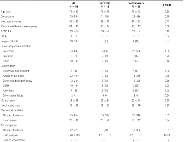
Table 1 - Clinical and epidemiological characteristics between survivors and non-survivors
All N = 53
Survivors N = 18
Nonsurvivors
N = 35 p value
Age (years) 74 ± 13 71 ± 15 76 ± 12 0.30
Gender, male 29 (55) 31 (58) 22 (42) 0.19
Heart rate (beats/min) 86 ± 20 83 ± 15 87 ± 22 0.47
Mean arterial blood pressure (mmHg) 84 ± 13 85 ± 14 83 ± 12 0.59
APACHE II 19 ± 5 18 ± 4 20 ± 5 0.10
SOFA 7 ± 3 6 ± 3 8 ± 3 0.04
Surgical patients 10 (19) 6 (33) 4 (11) 0.07
Primary diagnosis of infection
Pneumonia 34 (64) 12(66) 22 (63) 1.00
Peritonitis 8 (15) 2 (11) 6 (17) 0.70
Other 10 (19) 2 (11) 8 (23) 0.46
Comorbidities
Cerebrovascular accident 6 (11) 2 (11) 4 (11) 1.00
Arterial hypertension 22 (42) 9 (50) 13 (37) 0.39
Chronic cardiac insufficiency 12 (23) 2 (11) 10 (29) 0.18
COPD 10 (19) 3 (17) 7 (20) 1.00
Arrhythmia 7 (13) 2 (11) 5 (14) 1.00
Chronic renal failure 2 (4) 0 (0) 2 (6) 0.54
ICU stay (days) 23 ± 19 29 ± 24 20 ± 14 0.10
Hospital stay (days) 23 ± 18 29 ± 23 22 ± 16 0.20
Mechanical ventilation
Number of patients 45 (85) 15 (33) 30 (64) 0.82
Duration (days) 20 ± 16 23 ± 22 18 ± 12 0.94
Norepinephrine
Number of patients 22 (42) 3 (14) 19 (86) 0.01
Dose (µg/Kg/min) 0.28 ± 0.3 0.20 ± 0.05 0.29 ± 0.31 0.311
Days on vasopressors 5 ± 6 2 ± 5 7 ± 6 0.02
APACHE II - Acute Physiology and Chronic Health Evaluation; SOFA - Sequential Organ Failure Assessment; COPD - chronic obstructive pulmonary disease; ICU - intensive care unit. Results are expressed as a number (%) and the mean ± standard deviation.
that diastolic dysfunction was associated with increased mortality. his relationship was still present after adjusting for age and the SOFA and APACHE scores. he inding of an association of decreased ventricular compliance with poor outcome may be consistent with the results of a previous study demonstrating a lack of ventricular dilation in non-survivors with septic shock.(10)
Although we used a thorough approach for the assessment of diastolic function, only the E/e’ ratio was independently related to outcome. his ratio represents the relationship between early mitral inlow velocity and mitral annulus movement. herefore, it is an estimate of cardiac illing pressure. Accordingly, an observational study of patients with septic shock has found a strong
correlation between pulmonary artery wedge pressure and the E/e’ ratio.(25) An elevated E/e’ ratio indicates poor ventricular compliance. Because this ratio was found to be the only independent predictor of mortality in this study, it may be a better prognostic indicator than the other variables associated with diastolic failure. An alteration in the E/e’ ratio has been described previously in patients
with septic shock.(22) Although one study found that the
E/e’ ratio is an independent predictor of mortality,(17)
another investigation failed to ind this association.(11)
Table 2 - Echocardiographic characteristics in survivors and nonsurvivors
Survivors N = 18
Nonsurvivors N = 35
Odds ratio
(95%CI)* p value
LV diastolic diameter (mm) 47 ± 7 48 ± 6 1.01 (0.92 - 1.10) 0.40
LV systolic diameter (mm) 31 ± 8 33 ± 7 1.04 (0.95 - 1.14) 0.10
LVDV (mL) 88 ± 60 87 ± 38 0.99 (0.99 - 1.01) 0.90
LVSV (mL) 39 ±39 42 ± 26 1.00 (0.98 - 1.02) 0.80
LV ejection fraction 59 ± 10 53 ± 11 0.95 (0.88 - 1.02) 0.06
S wave (cm/s) 11 ± 3 10 ± 5 0.94 (0.88 - 1.00) 0.80
RVDV (mL) 67 ± 29 63 ± 22 0.17 (0 - 45961) 0.60
RVSV (mL) 27 ± 17 27 ± 11 0.99 (0.97 - 1.01) 0.90
RV ejection fraction 62 ± 7 58 ± 10 0.99 (0.96 - 1.04) 0.10
TAPSE (mm) 23 ± 5 21 ± 4 0.94 (0.87 - 1.01) 0.20
LVDD 12 (29) 30 (71) 0.92 (0.80 - 1.05) 0.01
LV E deceleration time (m/s2) 211 ± 42 211 ± 72 7.50 (1.32 - 42.50) 0.10
LV e’ (cm/s) 12 ± 4 13 ± 14 1 (0.99 - 1.00) 0.80
LV E/A ratio 1.0 ± 0.4 1.1 ± 0.5 1.78 (0.01 - 315) 0.40
LV E/e’ ratio 6.6 ± 2.5 8.8 ± 2.9 1.71 (0.43 - 6.93) 0.01
RVDD 9 (24) 29 (76) 1.36 (1.06 - 1.75) 0.02
LV E deceleration time (m/s2) 214 ± 64 194 ± 67 0.99 (0.98 - 1.00) 0.30
LV e’ (cm/s) 13 ± 3 14 ± 8 27.16 (0.00 - 53.30) 0.51
LV E/A ratio 0.97 ± 0.3 1.11 ± 0.4 3.40 (0.55 - 21.10) 0.18
LV E/e’ ratio 3.8 ± 1.2 4.3 ± 1.8 1.21 (0.83 - 1.78) 0.32
LVSDD 2 (11) 10 (29) 3.20 (0.62 - 16.54) 0.16
LV - left ventricle; RV - right ventricle; LVDV - left ventricular diastolic volume; LVSV - left ventricular systolic volume; TDI - tissue Doppler imaging; RVDV - right ventricular diastolic volume; RVSV - right ventricular systolic volume; TAPSE - tricuspid annular systolic plane excursion; LVDD - left ventricular diastolic dysfunction; LVSD - left ventricular systolic dysfunction; RVDD - right ventricular diastolic dysfunction; LVSDD - left ventricular systolic and diastolic dysfunction; * 95% confidence interval. Results are expressed as a number (%) and the mean ± standard deviation.
Table 3 - Multivariate logistic regression analysis of intensive care unit mortality
Odds ratio (95%CI) Wald p value
E/e’ ratio 1.36 (1.05 - 1.76) 5.74 0.02
SOFA 1.28 (0.99 - 1.65) 3.81 0.05
95%CI - 95% confidence interval; SOFA - Sequential Organ Failure Assessment.
of mechanical ventilation as well as inotropic drugs. Our indings suggest that an E/e’ ratio of greater than 8.0 might be a predictor of mortality. Nevertheless, the current E/e’ ratio reference limits are based on patients with cardiac disease.(22,23)
Left and right systolic dysfunction was present in 26% and 13% of the patients, respectively. As previously described, systolic dysfunction was not related to ICU mortality.(8,13,14) Similarly, changes in ventricular diameter or volume did not afect the outcome.
In addition to the ejection fraction, the S wave, as determined by spectral TDI, can be used to assess systolic function. he S wave may more accurately relect myocardial contractility than the ejection fraction because
it is less dependent on cardiac illing pressure.(26,27) Weng et al. have found that an S wave > 9cm/s is predictive of mortality in septic patients.(28) In our study, we did not ind diferences in the S wave value between the survivors and non-survivors.
is used to improve diastolic dysfunction in septic critically ill patients.
An important strength of this study is that the echocardiographic examination was repeated after a period of 7 - 10 days. he lack of resolution of the alterations in cardiac function might mean that these abnormalities were premorbid conditions. However, our study has some limitations: our sample size was small, and the study was performed at a single center. Furthermore, prior ventricular function was unknown. hus, it was not possible to discern whether alterations in diastolic function preceded or were a consequence of sepsis, However, the presence of diastolic dysfunction in patients appears to indicate an increased risk of death.
CONCLUSION
In conclusion, our prospective observational study has evaluated cardiac function via echocardiography in septic intensive care unit patients and has found that left ventricular diastolic dysfunction, as assessed according to the E/e’ ratio, is highly prevalent in old patients and is an independent predictor of mortality. Further investigations of the prognostic value and clinical usefulness of early echocardiography for septic patients appear warranted.
ACKNOWLEDGEMENTS
We are indebted to Dr. Glenn M. Eastwood and Professor Dr. Andrew Hilton for their meaningful comments.
Objetivo: Avaliar a prevalência de disfunção miocárdica e seu valor prognóstico em pacientes com sepse grave e choque séptico.
Métodos: Pacientes sépticos adultos, admitidos em uma unidade de terapia intensiva, foram estudados de forma prospectiva por meio de ecocardiograia transtorácica dentro das primeiras 48 horas após sua admissão e, então, entre o sétimo e o décimo dias. As variáveis ecográicas de função biventricular, inclusive a relação E/e’, foram comparadas entre sobreviventes e não sobreviventes.
Resultados: Foi realizado um total de 99 ecocardiogramas (53 na admissão e 46 entre os dias 7 e 10) em 53 pacientes com média de idade de 74 anos (desvio padrão de 13 anos). Estava presente disfunção sistólica em 14 (26%); disfunção diastólica
foi observada em 42 (83%) pacientes; e ambos os tipos de disfunção estavam presentes em 12 (23%) pacientes. A relação E/e’, ou índice de disfunção diastólica, foi o melhor preditor de mortalidade hospitalar segundo a área sob a curva ROC (0,71) e se constituiu em um preditor independente do desfecho, conforme determinado pela análise multivariada (odds ratio - OR = 1,36 [1,05 - 1,76]; p = 0,02).
Conclusão: Em pacientes sépticos admitidos em uma unidade de terapia intensiva, a disfunção sistólica determinada ecograicamente não se associa com aumento da mortalidade. Em contraste, a disfunção diastólica foi um preditor independente do desfecho.
RESUMO
Descritores: Sepse; Choque séptico; Ecocardiograia; Disfunção ventricular; Idoso
REFERENCES
1. Antonucci E, Fiaccadori E, Donadello K, Taccone FS, Franchi F, Scolletta S. Myocardial depression in sepsis: from pathogenesis to clinical manifestations and treatment. J Crit Care. 2014;29(4):500-11. Review. 2. Vieillard-Baron A. Septic cardiomyopathy. Ann Intensive Care. 2011;1(1):6. 3. Vieillard-Baron A, Caille V, Charron C, Belliard G, Page B, Jardin F. Actual
incidence of global left ventricular hypokinesia in adult septic shock. Crit Care Med. 2008;36(6):1701-6.
4. Verdecchia P, Angeli F, Gattobigio R, Sardone M, Porcellati C. Asymptomatic left ventricular systolic dysfunction in essential hypertension: prevalence, determinants and prognostic value. Hypertension. 2005;45(3):412-8. 5. Marchioli R, Avanzini F, Barzi F, Chieffo C, Di Castelnuovo A, Franzosi
MG, Geraci E, Maggioni AP, Marfisi RM, Mininni N, Nicolosi GL, Santini M, Schweiger C, Tavazzi L, Tognoni G, Valagussa F; GISSI-Prevenzione Investigators. Assessment of absolute risk of death after myocardial infarction by use of multiple-risk-factor assessment equations. GISSI-Prevenzione mortality risk chart. Eur Heart J. 2001;22(22):2085-103.
6. Solomon SD, Anavekar N, Skali H, McMurray JJ, Swedberg K, Yusuf S, Granger CB, Michelson EL, Wang D, Pocock S, Pfeffer MA; Candesartan in Heart Failure Reduction in Mortality (CHARM) Investigators. Influence of ejection fraction on cardiovascular outcomes in a broad spectrum of heart failure patients. Circulation. 2005;112(24):3738-44.
7. Carabello BA, Green LH, Grossman W, Cohn LH, Koster JK, Collins JJ Jr. Hemodynamic determinants of prognosis of aortic valve replacement in critical aortic stenosis and advanced congestive heart failure. Circulation. 1980;62(1):42-8.
8. Repessé X, Charron C, Vieillard-Baron A. Evaluation of left ventricular systolic function revisited in septic shock. Crit Care. 2013;17(4):164. 9. Parker MM, Shelhamer JH, Bacharach SL, Green MV, Natanson C,
Frederick TM, et al. Profound but reversible myocardial depression in patients with septic shock. Ann Intern Med. 1984;100(4):483-90. 10. Parker MM, McCarthy KE, Ognibene FP, Parrillo JE. Right ventricular
11. Pulido JN, Afessa B, Masaki M, Yuasa T, Gillespie S, Herasevich V, et al. Clinical spectrum, frequency and significance of myocardial dysfunction in severe sepsis and septic shock. Mayo Clin Proc. 2012;87(7):620-8. 12. Vincent JL, Gris P, Coffernils M, Leon M, Pinsky M, Reuse C, et al.
Myocardial depression characterizes the fatal course of septic shock. Surgery. 1992;111(6):660-7.
13. Huang SJ, Nalos M, McLean AS. Is early ventricular dysfunction or dilatation associated with lower mortality rate in adult severe sepsis and septic shock? A meta-analysis. Crit Care. 2013;17(3):R96.
14. Sevilla Berrios RA, O’Horo JC, Velagapudi V, Pulido JN. Correlation of left ventricular systolic dysfunction determined by low ejection fraction and 30-day mortality in patients with severe sepsis and septic shock: a systematic review and meta-analysis. J Crit Care. 2014;29(4):495-9. 15. Bouhemad B, Nicolas-Robin A, Arbelot C, Arthaud M, Féger F, Rouby JJ.
Acute left ventricular dilatation and shock-induced myocardial dysfunction. Crit Care Med. 2009;37(2):441-7.
16. Sturgess DJ, Marwick TH, Joyce C, Jenkins C, Jones M, Masci P, et al. Prediction of hospital outcome in septic shock: a prospective comparison of tissue Doppler and cardiac biomarkers. Crit Care. 2010;14(2):R44. 17. Landesberg G, Gilon D, Meroz Y, Georgieva M, Levin PD, Goodman S, et al.
Diastolic dysfunction and mortality in severe sepsis and septic shock. Eur Heart J. 2012;33(7):895-903.
18. Dellinger RP, Levy MM, Carlet JM, Bion J, Parker MM, Jaeschke R, Reinhart K, Angus DC, Brun-Buisson C, Beale R, Calandra T, Dhainaut JF, Gerlach H, Harvey M, Marini JJ, Marshall J, Ranieri M, Ramsay G, Sevransky J, Thompson BT, Townsend S, Vender JS, Zimmerman JL, Vincent JL; International Surviving Sepsis Campaign Guidelines Committee; American Association of Critical-Care Nurses; American College of Chest Physicians; American College of Emergency Physicians; Canadian Critical Care Society; European Society of Clinical Microbiology and Infectious Diseases; European Society of Intensive Care Medicine; European Respiratory Society; International Sepsis Forum; Japanese Association for Acute Medicine; Japanese Society of Intensive Care Medicine; Society of Critical Care Medicine; Society of Hospital Medicine; Surgical Infection Society; World Federation of Societies of Intensive and Critical Care Medicine. Surviving Sepsis Campaign: international guidelines for management of severe sepsis and septic shock: 2008. Crit Care Med. 2008;36(1):296-327. Erratum in: Crit Care Med. 2008;36(4):1394-6. 19. Vincent JL, de Mendonça A, Cantraine F, Moreno R, Takala J, Suter PM,
et al. Use of the SOFA score to assess the incidence of organ dysfunction/ failure in intensive care units: results of a multicenter prospective study. Working group on “sepsis-related problems” of the European Society of Intensive Care Medicine. Crit Care Med. 1998;26(11):1793-800.
20. Knaus WA, Draper EA, Wagner DP, Zimmerman JE. APACHE II: a severity of disease classification system. Crit Care Med. 1985;13(10):818-29. 21. Paulus WJ, Tschöpe C, Sanderson JE, Rusconi C, Flachskampf FA,
Rademakers FE, et al. How to diagnose diastolic heart failure: a consensus statement on the diagnosis of heart failure with normal left ventricular ejection fraction by the Heart Failure and Echocardiography Associations of the European Society of Cardiology. Eur Heart J. 2007;28(20):2539-50. 22. Nagueh SF, Appleton CP, Gillebert TC, Marino PN, Oh JK, Smiseth OA, et al.
Recommendations for the evaluation of left ventricular diastolic function by echocardiography. J Am Soc Echocardiogr. 2009;22(2):107-33. 23. Hill JC, Palma RA. Doppler tissue imaging for the assessment of left
ventricular diastolic function: a systematic approach for the sonographer. J Am Soc Echocardiogr. 2005;18(1):80-8; quiz 89.
24. Chang WT, Chen JS, Hung YK, Tsai WC, Juang JN, Liu PY. Characterization of aging-associated cardiac diastolic dysfunction. PLoS One. 2014;9(5):e97455.
25. Mousavi N, Czarnecki A, Ahmadie R, Tielan Fang, Kumar K, Lytwyn M, et al. The utility of tissue Doppler imaging for the noninvasive determination of left ventricular filling pressures in patients with septic shock. J Intensive Care Med. 2010;25(3):163-7.
26. Ho CY, Solomon SD. A clinician’s guide to tissue Doppler imaging. Circulation. 2006;113(10):e396-8.
27. A’roch R, Gustafsson U, Johansson G, Poelaert J, Haney M. Left ventricular strain and peak systolic velocity: responses to controlled changes in load and contractility. explored in a porcine model. Cardiovasc Ultrasound. 2012;10:22.
28. Weng L, Liu YT, Du B, Zhou JF, Guo XX, Peng JM, et al. The prognostic value of left ventricular systolic function measured by tissue Doppler imaging in septic shock. Crit Care. 2012;16(3):R71.
29. Sanfilippo F, Corredor C, Fletcher N, Landesberg G, Benedetto U, Foex P, et al. Diastolic dysfunction and mortality in septic patients: a systematic review and meta-analysis. Intensive Care Med. 2015;41(6):1004-13. Erratum in Intensive Care Med. 2015;41(6):1178-9.