Effect of a Gluten-Free Diet on Cortical
Excitability in Adults with Celiac Disease
Rita Bella1, Giuseppe Lanza2, Mariagiovanna Cantone2, Salvatore Giuffrida1,
Valentina Puglisi1, Luisa Vinciguerra1, Manuela Pennisi3, Riccardo Ricceri1, Carmela Cinzia D’Agate4, Giulia Malaguarnera5, Raffaele Ferri2, Giovanni Pennisi6*
1Department‘‘G.F. Ingrassia”, Section of Neurosciences, University of Catania, Catania, Italy,
2Department of Neurology I.C.,“Oasi”Institute for Research on Mental Retardation and Brain Aging (I.R.C. C.S.), Troina (EN), Italy,3Spinal Unit, Emergency Hospital“Cannizzaro”, Catania, Italy,4Gastroenterology and Endoscopy Unit, University of Catania, Catania, Italy,5Department of Biomedical Sciences, University of Catania, Catania, Italy,6Department“SpecialitàMedico-Chirurgiche”, University of Catania, Catania, Italy
Abstract
Introduction
An imbalance between excitatory and inhibitory synaptic excitability was observed in de novo patients with celiac disease (CD) in a previous study with Transcranial Magnetic Stim-ulation (TMS), suggesting a subclinical involvement of GABAergic and glutamatergic neuro-transmission in asymptomatic patients. The aim of this investigation was to monitor the eventual changes in the same cohort of patients, evaluated after a period of gluten-free diet.
Methods
Patients were re-evaluated after a median period of 16 months during which an adequate gluten-free diet was maintained. Clinical, cognitive and neuropsychiatric assessment was repeated, as well as cortical excitability by means of single- and paired-pulse TMS from the first dorsal interosseous muscle of the dominant hand.
Results
Compared to baseline, patients showed a significant decrease of the median resting motor threshold (from 35% to 33%,p<0.01). The other single-pulse (cortical silent period, motor
evoked potentials latency and amplitude, central motor conduction time) and paired-pulse TMS measures (intracortical inhibition and intracortical facilitation) did not change signifi-cantly after the follow-up period. Antibodies were still present in 7 subjects.
Discussion
In patients under a gluten-free diet, a global increase of cortical excitability was observed, suggesting a glutamate-mediated functional reorganization compensating for disease progression. We hypothesize that glutamate receptor activation, probably triggered by CD-related immune system dysregulation, might result in a long-lasting motor cortex hyperexcit-ability with increased excitatory post-synaptic potentials, probably related to phenomena of OPEN ACCESS
Citation:Bella R, Lanza G, Cantone M, Giuffrida S, Puglisi V, Vinciguerra L, et al. (2015) Effect of a Gluten-Free Diet on Cortical Excitability in Adults with Celiac Disease. PLoS ONE 10(6): e0129218. doi:10.1371/journal.pone.0129218
Academic Editor:François Tremblay, University of Ottawa, CANADA
Received:February 25, 2015
Accepted:May 6, 2015
Published:June 8, 2015
Copyright:© 2015 Bella et al. This is an open access article distributed under the terms of the
Creative Commons Attribution License, which permits unrestricted use, distribution, and reproduction in any medium, provided the original author and source are credited.
Data Availability Statement:All relevant data are within the paper.
Funding:These authors have no support or funding to report.
long-term plasticity. The impact of the gluten-free diet on subclinical neurological abnormali-ties needs to be further explored.
Introduction
In recent years, a wide spectrum of neurological and psychiatric complications of Celiac Dis-ease (CD) has been described, although most of the studies focused on overt clinical manifesta-tions such as epilepsy, ataxia, peripheral neuropathy, headache, cognitive impairment,
depression, anxiety, and schizophrenia [1–3]. However, subtle neurological changes, that do not reach the clinical threshold, have also been reported [4] and might play a role in the patho-physiological mechanisms underlying neurological involvement in CD.
In a previous study aiming to evaluate the effects of the gluten-mediated immune disorder on intracortical circuits, we investigated the profile of cortical excitability of 20 neurologically asymptomatic de novo CD patients by means of Transcranial Magnetic Stimulation (TMS). Compared to healthy controls, a pattern toward a“disinhibition”and“hyperfacilitation”of the motor cortex was observed, suggesting a subclinical involvement of GABAergic and glutama-tergic neurotransmission in asymptomatic patients. In particular, it was suggested that the cross-reaction between anti-gliadin antibodies and neuronal antigens, as well as the altered ion levels related to transglutaminase 6-immunoglobulin deposition, might affect the normal bal-ance between excitatory and inhibitory synaptic excitability. Similarly, anti-glutamic acid de-carboxylase (GAD) antibodies synthesized within the nervous system might interfere with GABAergic interneurons activity [5].
In this context, most of the studies, although not all [6,7], highlight the potential therapeutic role of the gluten-free diet not only for the typical intestinal manifestations of CD but also for some of the psychiatric and neurological complications, such as depression, anxiety, beha-vioural symptoms, peripheral neuropathy and cerebellar ataxia [8–12].
The aim of this new study was to assess the impact of the gluten-free diet at the level of TMS, monitoring the neurophysiological changes observed in the same cohort from baseline. We hypothesized that the dietary regimen may modulate the balance between intracortical ex-citatory/inhibitory circuits.
Materials and Methods
Ethics Statement
The study was approved by the ethics committee of the Azienda Ospedaliero-Universitaria
“Policlinico-Vittorio Emanuele”, Catania (Italy). Written informed consent was obtained from all participants prior to the participation, in accordance with the Declaration of Helsinki. All assessments were performed in a controlled laboratory environment.
Subjects and assessment
University of Catania (Italy), fulfilled the criteria for CD according to the European Society for Paediatric Gastroenterology Hepatology and Nutrition (ESPGHAN) guidelines [13]. Accord-ing to the international“ACG Clinical Guidelines for the diagnosis and management of Celiac Disease”[14] patients enrolled in the present study underwent periodic visits with both CD specialist and skilled dietician as well as structured survey to evaluate adherence to the gluten-free diet [15]. Diet compliance was adequate in all patients.Table 1summarizes the main clini-cal data and the serologiclini-cal findings of CD patients at follow-up. Major neurologiclini-cal disorders, acute medical illness or organ failure, head trauma, epilepsy, Mini-Mental State Examination (MMSE) score<24 [16], alcohol or drug abuse, use of drugs affecting cortical excitability (i.e.
benzodiazepines, mood stabilizers, antipsychotics) and any condition precluding TMS execu-tion were exclusion criteria.
The patients underwent the same assessment performed at the entry of the previous study [5], including clinical-demographic evaluation (age, gender, education, handedness, social and living conditions, general and neurological examinations, co-morbidities), and neuropsycho-logical tests: Mini-mental state examination (MMSE), Structured Clinical Interview for DSM-IV Axis I Disorders (SCID-I) and 17-items Hamilton Depression Rating Scale (HDRS) [17]. Electroencephalogram (EEG) and brain computed tomography scan were not repeated given that the main objective was to screen CD patients for the presence of cerebral calcifica-tions and to rule out evident neuroradiological lesions; at baseline, all the patients had normal scans. Moreover, none of the participants complained of neurological symptoms and general and neurological exams did not show other findings with respect to the first examination.
Transcranial magnetic stimulation
TMS was performed using a High-power Magstim 200 magnetic stimulator (Magstim Co., Whitland, Dyfed, UK). A 70 mm figure-of-eight coil was held over the motor cortex at the opti-mum scalp position to elicit Motor Evoked Potentials (MEPs) in the contralateral First Dorsal Interosseous (FDI) muscle of the dominant hand, according to the Edinburgh Handedness In-ventory [18]. Electromyographic (EMG) activity was amplified using a Medelec Synergy Table 1. Clinical-serological features of CD-patients before and after the gluten-free diet.
Baseline Follow-up Weightchange (kg)
Ab Comorbidities Ab Comorbidities
1 EMA, tTG iron-deficiency anemia - none 0
2 EMA, tTG iron-deficiency anemia - none +7
3 EMA, tTG thyroiditis - thyroiditis +4
4 EMA, tTG asthma - none -3
5 EMA, tTG thyroiditis - thyroiditis 0
6 EMA, tTG thyroiditis, iron-deficiency anemia EMA, tTG thyroiditis, iron-deficiency anemia +1
7 EMA, tTG vitiligo EMA, tTG vitiligo +4
8 EMA, tTG iron-deficiency anemia, thyroiditis, infertility EMA, tTG iron-deficiency anemia, thyroiditis, osteopenia 0
9 - osteopenia - iron-deficiency anemia, osteopenia 0
10 EMA, tTG none EMA, tTG none +8
11 EMA, tTG none EMA, tTG none +3
12 EMA, tTG none EMA, tTG none +1
13 EMA, tTG thyroiditis, iron-deficiency anemia EMA, tTG thyroiditis, iron-deficiency anemia 0
Ab: antibodies; CD: celiac disease; tTG: tissue transglutaminase antibodies; EMA: endomysial antibodies;—: negative.
system (Oxford Instruments) and recorded from silver/silver-chloride surface active electrode, placed over the muscular belly of the target muscle (FDI), with the reference electrode placed distally at the metacarpo-phalangeal joint of the index finger and the ground on the dorsal face of the wrist. Motor responses were amplified and filtered (bandwidth 3–3000 Hz) with gains of
100μV and 5 mV/div. For the motor nerve conduction study (M and F waves from the FDI
muscle), a bipolar nerve stimulation electrode (Medelec Synergy system, Oxford Instruments) with 6-mm diameter felt pads and an inter-electrode separation of 25 mm was used and ap-plied to the ulnar nerve at wrist. Resting motor threshold (rMT) was defined as the lowest stim-ulus intensity able to elicit MEPs at rest of an amplitude 50μV in at least 5 of 10 trials,
according to the IFCN recommendation [19]. When determining the rMT, TMS started with a subthreshold intensity of stimulation, with the coil placed over the optimal site of stimulation; then, stimulus intensity was gradually increased in steps of 5% of the maximal stimulator out-put (MSO) until TMS consistently evoked MEPs with peak-to-peak amplitude of more than
50μV in each trial. Thereafter, stimulus intensity was gradually lowered in steps of 1% MSO
until 5 positive responses out of 10 trials were recorded [20]. According to the IFCN practical guidelines, an intertrial interval over 3 seconds (4–6 sec) was used in MEPs measurements [21]. Central motor conduction time (CMCT) was calculated by subtracting the conduction time in peripheral nerves, estimated by F wave techniques, from MEP latency obtained during moderate active muscle contraction, with a stimulus intensity set at 130% of the rMT. The clear negative waveform from the baseline was used to determine the MEPs onset latency. M and F waves are elicited by giving supramaximal electrical stimulation to the ulnar nerve at wrist. The size of the MEPs was expressed as a percentage of supramaximal M wave amplitude (A ratio). The cortical silent period (CSP) was determined with an approximately 50% of maxi-mum tonic voluntary contraction of the FDI muscles, induced by single TMS pulses delivered at 130% of rMT. The mean CSP duration of 10 rectified trials was calculated.
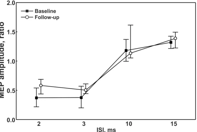
Curves of intracortical excitability were obtained using the paired-pulse TMS protocol. Intracortical inhibition (ICI) and Intracortical facilitation (ICF) were studied using the condi-tioning-test paradigm applying two magnetic stimuli in rapid succession [22], and the effect of the first (conditioning) stimulus on the second (test) stimulus was investigated. The procedure was performed over the primary motor cortex through the same stimulating coil deriving pulses from two Magstim 200 Stimulators, connected via a Bistim module (The Magstim Com-pany, Whitland, Dyfed, UK) connected to a CED micro 1401 interface (Cambridge Electronic Design, Cambridge, UK). The conditioning stimulus was set at 80% of the subjects rMT where-as the test stimulus at 130% of the rMT. The interstimulus intervals (ISIs) tested were 2, 3, 10 and 15 ms. Ten trials for each ISI were recorded in a random way. The responses were express-ed as the ratio between the MEP amplitude producexpress-ed by pairexpress-ed stimulation and that producexpress-ed by test stimulus alone.
The subjects were seated in a comfortable chair with continuous EMG monitoring to ensure either a constant level of EMG activity during tonic contraction or complete relaxation at rest. Data were collected on a computer and stored with an ad hoc software for off-line analysis [23]. All TMS procedures were performed in the same laboratory and experimental conditions, at the same time of the day and by the same operators, thus minimizing the inter-subject variability.
Statistical analysis
comparisons were performed by means of the non-parametric Wilcoxon test for paired data-sets. Differences were considered significant a p<0.05 level. However, because of the relatively
limited number of subjects available and to rule out possible type II errors, we also calculated effect sizes using the Cohen’s d value [24]. Cohen’s d is defined as the difference between two means divided by their pooled standard deviation. According to Cohen, 0.2 is indicative of a small effect, 0.5 of a medium and 0.8 of a large effect size.
Results
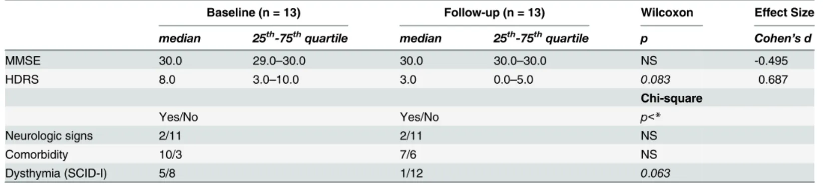
As shown inTable 2, no difference was found for clinical features between the initial and the follow-up evaluation. Global cognitive functioning as assessed by MMSE did not change signif-icantly over time. The HDRS score showed a trend towards an improvement at follow-up (with a moderate effect size); a dysthymic disorder, evaluated by the SCID-I, was found in only one patient at follow-up, vs. 5 at baseline.
Compared to baseline, treated patients showed a statistically significant decrease of the me-dian rMT values (from 35% down to 33%, p = 0.0042). The other single-pulse TMS measures (Table 3), as well as the paired-pulse TMS parameters (ICI and ICF), did not change signifi-cantly after the follow-up period (Fig 1), thus showing persistent disinhibition and hyperfacili-tation. Maximal M-waves were comparable between sessions. Examination of the individual Table 2. Clinical features of the CD subjects at follow-up.
Baseline (n = 13) Follow-up (n = 13) Wilcoxon Effect Size
median 25th-75thquartile median 25th-75thquartile p Cohen
’s d
MMSE 30.0 29.0–30.0 30.0 30.0–30.0 NS -0.495
HDRS 8.0 3.0–10.0 3.0 0.0–5.0 0.083 0.687
Chi-square
Yes/No Yes/No p<*
Neurologic signs 2/11 2/11 NS
Comorbidity 10/3 7/6 NS
Dysthymia (SCID-I) 5/8 1/12 0.063
CD = celiac disease; MMSE = Mini Mental State Examination; HDRS = 17 item-Hamilton Depression Rating Scale; SCID-I = Structured Clinical Interview for DSM-IV Axis I.
doi:10.1371/journal.pone.0129218.t002
Table 3. Comparison of electrophysiological data.
Baseline (n = 13) Follow-up (n = 13) Wilcoxon Effect Size
Median 25th-75thquartile Median 25th-75thquartile p Cohen’s d
rMT, % 35.0 34.0–41.0 33.0 29.0–37.0 0.0042 0.472
CSP, ms 100.0 76.0–112.0 83.0 57.0–128.0 NS -0.090
MEP latency, ms 18.4 18.3–20.2 18.9 17.9–20.6 NS -0.118
CMCT, ms 5.6 4.3–6.3 5.2 5.0–5.7 NS 0.199
CMCTF, ms 4.8 4.5–5.5 5.2 4.7–5.6 NS -0.161
A ratio 0.3 0.2–0.6 0.3 0.2–0.4 NS -0.234
F amplitude,μV 0.1 0.1–0.2 0.2 0.1–0.2 NS -0.192
rMT: resting motor threshold; CPS: cortical silent period; MEP: motor evoked potentials; CMCT: central motor conduction time; CMCTF: central motor conduction time estimated by F wave; A ratio: amplitude ratio.
data taking into account the differences of the rMT values from baseline (ΔrMT) and the length of the follow-up showed additional information. Specifically, after a longer follow-up period, 4 out of 6 patients with serological conversion had the largestΔrMT whereas, at shorter time of follow-up, 5 out of 7 patients with antibodies persistence exhibited the smallestΔrMT. The small number of patients did not allow a more detailed statistical analysis of this aspect.
Discussion
To our knowledge, this is the first prospective study exploring the impact of the gluten-free diet on cortical excitability to TMS in CD patients. The main result is that, after the gluten-free diet, a global increase of cortical excitability, as indexed by a reduction of the median rMT, was observed, without any significant changes of the other TMS measures. As known, the rMT is a global measure of motor system excitability and is thought to reflect mainly the membrane ex-citability of the cortical-spinal neurons and the glutamatergic excitatory interneurons that proj-ect into these neurons [25,26].
Although the reason underlying this change is rather complex, it is known that the net effect of increased excitation could be the result of prevailing activation and/or weaker inhibition, phenomena involving different neurochemical pathways. In this context, it is worth to note that both the CSP shortening and the reduced ICI did not change over time, suggesting the per-sistence of a GABA-mediated intracortical dysfunction despite the dietary restriction [5]. Ac-cordingly, the hyperexcitability might be the result of an immune-mediated dysregulation of inhibitory GABAergic interneurons or a parallel glutamate-mediated cortical functional Fig 1. Intracortical excitability at different interstimuls intervals at baseline and follow-up.ISI = interstimuls interval; MEP = Motor Evoked Potential.
reorganization compensating for disease progression despite the adoption of the gluten free-diet. In particular, the AMPA type ionotropic glutamate receptor is known to be crucial for plasticity and excitatory synaptic transmission at the level of many post-synaptic membranes [27], including the motor cortex [28]. Thus, it cannot be excluded that receptor activation, probably triggered by CD-related immune system dysregulation, might result in a long-lasting increase of excitatory post-synaptic potentials underlying the observed motor cortex hyperex-citability and probably related to phenomena of long-term plasticity. Although obtained from a small number of patients, the analysis of individual data seems to reveal a correlation between theΔrMT, the serological status and the length of the gluten-free diet. In particular, even if a median decrease of the rMT was globally observed in all participants, a relatively greater hyper-excitability occurred in those with seroconversion and a longer gluten restriction. This might suggest a more powerful involvement of hyperexcitability-induced cortical plasticity in these patients, as demonstrated in other neurological disorders by specific TMS mapping techniques [29,30].
The lack of studies assessing TMS parameters in newly diagnosed CD does not allow to compare our data with other cohorts and very few studies have explored subclinical neuro-physiology of CD after alimentary therapy. In 27 asymptomatic children on gluten-free diet, Cakir et al. found 2 of them with EMG-documented peripheral polyneuropathy, one with pro-longed central conduction time at somatosensory evoked potentials and 3 with a single seizure but normal EEG, indicating that subclinical neurological abnormalities may persist despite the diet [31]. More recently, Parisi et al. revealed subclinical EEG abnormalities in 48% of children, which disappeared in 78% of them after 6 months of dietary restriction, suggesting that cortical excitability in asymptomatic children with newly diagnosed CD is modified by the introduction of the diet [32].
Notably, the role of the gluten-free diet in subclinical neurological abnormalities has not been assessed and the impact on the histologic course needs to be further investigated. It is well established that the gluten-free diet represents the most important aspect of the management of CD patients and it is the only treatment that allows the prevention of several associated ma-lignant and non-mama-lignant complications, including neurological diseases. Histologic recovery of the small intestinal mucosa is assumed to occur within 6–12 months after starting a gluten restriction, simultaneously with clinical remission. However, follow-up data on intestinal re-covery in CD patients are scarce and contradictory, even in patients with a good diet compli-ance [33–36]. In a long-term follow-up study of 158 patients, the histologic recovery in gluten-sensitive sprue after starting a gluten-free diet took more than 2 years in 35.4% of patients and was incomplete or absent in a substantial subgroup of patients [37].Other studies reported that neurological damage or antibodies persisted despite the strict adherence to the diet, showing the ineffectiveness of a gluten-free diet in obtaining neurological remission in CD [38–41]. It is noteworthy that 7 of our patients did not achieve serological conversion. Furthermore, neuro-logical deficits may even develop despite strict adherence to a gluten-free diet, as shown in dif-ferent studies [38–42].
related to the duration of neurological disorders as well as to the age of the patient at the start of the gluten-free diet [32,43].
Finally, in this study, a certain degree of improvement of depressive symptoms was also ob-served, supporting the role of the gluten-free diet in the amelioration of psychiatric CD-related disorders. Recognizing depression as a risk factor for a poor adherence to the gluten-free diet is of great importance. Consequently, it would be advisable to consider, both at the diagnosis and at follow-up, not only clinical and serological features, but also the presence of depression, so that psychological support or pharmacological therapy might be started [4,44].
There are some limitations to take into account when interpreting the findings of this study. First, as usual in TMS research, the relatively small number of patients requires further inde-pendent investigations with larger series of participants. In addition, future studies on CD pa-tients with a long-term dietary restriction will further clarify the meaning of the detected changes and their temporal relation with the gluten-free diet duration and compliance. Second, the reliability of the rMT measurements over time is of crucial importance given that there was no untreated control group. For instance, the hypothesis that the observed increase of motor cortex excitability could be related to aging should be considered. However, most TMS studies have not found differences in the primary motor cortex excitability threshold between young and elderly [45–48], whereas others have shown an increase in motor threshold in healthy sub-jects with aging [49,50]. Conversely, our patients showed a significant decrease of the median rMT value at follow-up, indicating a higher excitability. Regarding the test-retest reliability of rMT measured at baseline and repeated at follow-up, most TMS studies converge on the evi-dence that rMT is reliable when measured over time, in both healthy subjects [51,52] and pa-tients with neurological disorders such as stroke [53–55]. However, as the effect of the gluten-free diet on the rMT was relatively small (35%vs33%), the observed change needs to be
consid-ered in view of a number of issues pertaining to reliability of repeated cortical-motor threshold measurements (such as the TMS coil placement, the arm and electrode positioning). Therefore, this finding might not be large enough to uphold the conclusion of increased cortical excitabil-ity, although multiple comparisons have been made increasing the likelihood of a significant result. Finally, the timing of testing after starting the gluten-free diet was not uniform. Never-theless, given that, as known, complete healing of the small intestine after the adoption of the gluten-free diet may take longer than 12 months (even more than 24 months in some cases), we established to re-evaluate these patients after at least one year of adequate gluten restriction.
In conclusion, in this study we showed that the gluten-free diet modulated the TMS indexes of cortical function in adult CD patients. TMS, integrated with clinical, immunological and ad-vanced neuroimaging data, might be considered as an additional tool for evaluating the pro-gression of neurological involvement in CD and the response to the dietary regimen.
Author Contributions
Conceived and designed the experiments: GP SG RB. Performed the experiments: LV VP RR. Analyzed the data: RF MP. Contributed reagents/materials/analysis tools: CCD GM. Wrote the paper: MC GL RB.
References
1. Hadjivassiliou M, Sanders DS, Grünewald RA, Woodroofe N, Boscolo S, Aeschlimann D. Gluten sensi-tivity: from gut to brain. Lancet Neurol. 2010; 9: 318–330. doi:10.1016/S1474-4422(09)70290-XPMID: 20170845
3. Jackson JR, Eaton WW, Cascella NG, Fasano A, Kelly DL. Neurologic and psychiatric manifestations of celiac disease and gluten sensitivity. Psychiatr Q. 2012; 83: 91–102. doi: 10.1007/s11126-011-9186-yPMID:21877216
4. Abenavoli L. Subclinical neurological abnormalities and gluten-free diet. J Am Diet Assoc. 2008; 108: 1995. doi:10.1016/j.jada.2008.10.027PMID:19027400
5. Pennisi G, Lanza G, Giuffrida S, Vinciguerra L, Puglisi V, Cantone M, et al. Excitability of the motor cor-tex in de novo patients with celiac disease. PLoS One. 2014; 9: e102790. doi:10.1371/journal.pone. 0102790PMID:25062250
6. Chin RL, Sander HW, Brannagan TH, Green PH, Hays AP, Alaedini A, et al. Celiac neuropathy. Neurol-ogy. 2003; 60: 1581–1585. PMID:12771245
7. Luostarinen L, Himanen S- L, Luostarinen M, Collin P, Pirttilä T. Neuromuscular and sensory distur-bances in patients with well treated coeliac disease. J Neurol Neurosurg Psychiatry. 2003; 74: 490–
494. PMID:12640070
8. Pynnönen PA, Isometsä ET, Aronen ET, Verkasalo MA, Savilahti E, Aalberg VA. Mental disorders in adolescents with celiac disease. Psychosomatics. 2004; 45: 325–335. PMID:15232047
9. Addolorato G. Anxiety but not depression decreases in coeliac patients after one-year gluten-free diet: a longitudinal study. Scand J Gastroenterol. 2001 36: 502–506. PMID:11346203
10. Hallert C, Sedvall G. Improvement in central monoamine metabolism in adult coeliac patients starting a gluten-free diet. Psychol Med. 1983; 13: 267–271. PMID:6192458
11. Hadjivassiliou M, Kandler RH, Chattopadhyay AK, Davies-Jones AG, Jarratt JA, Sanders DS, et al. Die-tary treatment of gluten neuropathy. Muscle Nerve. 2006; 34: 762–766. PMID:17013890
12. Pellecchia MT, Scala R, Perretti A, De Michele G, Santoro L, Filla A, et al. Cerebellar ataxia associated with subclinical celiac disease responding to gluten-free diet. Neurology. 1999; 53: 1606–1608. PMID: 10534283
13. Husby S, Koletzko S, Korponay-Szabó IR, Mearin ML, Phillips A, Shamir R, et al. European Society for Pediatric Gastroenterology, Hepatology, and Nutrition guidelines for the diagnosis of coeliac disease. J Pediatr Gastroenterol Nutr. 2012; 54: 136–160. doi:10.1097/MPG.0b013e31821a23d0PMID: 22197856
14. Rubio-Tapia A, Hill ID, Kelly CP, Calderwood AH, Murray JA. ACG clinical guidelines: diagnosis and management of celiac disease. Am J Gastroenterol. 2013; 108: 656–676. doi:10.1038/ajg.2013.79 PMID:23609613
15. Biagi F, Bianchi PI, Marchese A, Trotta L, Vattiato C, Balduzzi D, et al. A score that verifies adherence to a gluten-free diet: a cross-sectional, multicentre validation in real clinical life. Br J Nutr. 2012; 108: 1884–1888. doi:10.1017/S0007114511007367PMID:22321199
16. Folstein MF, Folstein SE, McHugh PR. "Mini-mental state". A practical method for grading the cognitive state of patients for the clinician. J Psychiatr Res. 1975; 12: 189–198. PMID:1202204
17. Hamilton M. A rating scale for depression. J Neurol Neurosurg Psychiatry. 1960; 23: 56. PMID: 14399272
18. Oldfield RC. The assessment and analysis of handedness: the Edinburgh inventory. Neuropsycholo-gia. 1971; 9: 97–113. PMID:5146491
19. Rossini P, Barker A, Berardelli A, Caramia M, Caruso G, Cracco RQ, et al. Non-invasive electrical and magnetic stimulation of the brain, spinal cord and roots: basic principles and procedures for routine clin-ical application. Report of an IFCN committee. Electroencephalogr Clin Neurophysiol. 1994; 91: 79–
92. PMID:7519144
20. Groppa S, Oliviero A, Eisen A, Quartarone A, Cohen L, Mall V, et al. A practical guide to diagnostic tran-scranial magnetic stimulation: report of an IFCN committee. Clin Neurophysiol. 2012; 123: 858–882. doi:10.1016/j.clinph.2012.01.010PMID:22349304
21. Rothwell J, Hallett M, Berardelli A, Eisen A, Rossini P, Paulus W. Magnetic stimulation: motor evoked potentials. Electroencephalogr Clin Neurophysiol Suppl. 1999; 52: 97–103. PMID:10590980
22. Kujirai T, Caramia M, Rothwell JC, Day B, Thompson P, Ferbert A, et al. Corticocortical inhibition in human motor cortex. J Physiol. 1993; 471: 501–519. PMID:8120818
23. Giordano D, Kavasidis I, Spampinato C, Bella R, Pennisi G, Pennisi M. An integrated computer-con-trolled system for assisting researchers in cortical excitability studies by using transcranial magnetic stimulation. Comput Methods Programs Biomed. 2012; 107: 4–15. doi:10.1016/j.cmpb.2011.10.008 PMID:22172294
24. Cohen J. Statistical power analysis. Curr Dir Psychol Sci. 1992; 1: 98–101.
26. Paulus W, Classen J, Cohen LG, Large CH, Di Lazzaro V, Nitsche M, et al. State of the art: pharmaco-logic effects on cortical excitability measures tested by transcranial magnetic stimulation. Brain Stimul. 2008; 1: 151–163. doi:10.1016/j.brs.2008.06.002PMID:20633382
27. Levite M. Glutamate receptor antibodies in neurological diseases: AMPA-GluR3 antibodies, NMDA-NR1 antibodies, NMDA-NR2A/B antibodies, mGluR1 antibodies or mGluR5 anti-bodies are present in subpopulations of patients with either: epilepsy, encephalitis, cerebellar ataxia, systemic lupus erythematosus (SLE) and neuropsychiatric SLE, Sjogren's syndrome, schizophrenia, mania or stroke. These autoimmune anti-glutamate receptor antibodies can bind neurons in few brain regions, activate glutamate receptors, decrease glutamate receptor's expression, impair glutamate-in-duced signaling and function, activate blood brain barrier endothelial cells, kill neurons, damage the brain, induce behavioral/psychiatric/cognitive abnormalities and ataxia in animal models, and can be removed or silenced in some patients by immunotherapy. J Neural Transm. 2014; 121: 1029–1075. doi:10.1007/s00702-014-1193-3PMID:25081016
28. Ganor Y, Gottlieb M, Eilam R, Otmy H, Teichberg VI, Levite M. Immunization with the glutamate recep-tor-derived peptide GluR3B induces neuronal death and reactive gliosis, but confers partial protection from pentylenetetrazole-induced seizures. Exp Neurol. 2005; 195: 92–102. PMID:15907325
29. Guerra A, Petrichella S, Vollero L, Ponzo D, Pasqualetti P, Määttä S, et al. Neurophysiological features of motor cortex excitability and plasticity in Subcortical Ischemic Vascular Dementia: A TMS mapping study. Clin Neurophysiol. 2015; 126: 906–913 doi:10.1016/j.clinph.2014.07.036PMID:25262646
30. Pennisi G, Bella R, Lanza G. Motor cortex plasticity in subcortical ischemic vascular dementia: What can TMS say? Clin Neurophysiol. 2015; 126: 851–852. doi:10.1016/j.clinph.2014.09.001PMID: 25270240
31. Cakir D, Tosun A, Polat M, Celebisoy N, Gokben S, Aydogdu S, et al. Subclinical neurological abnor-malities in children with celiac disease receiving a gluten-free diet. J Pediatr Gastroenterol Nutr. 2007; 45: 366–369. PMID:17873753
32. Parisi P, Pietropaoli N, Ferretti A, Nenna R, Mastrogiorgio G, Del Pozzo M, et al. Role of the gluten-free diet on neurological-EEG findings and sleep disordered breathing in children with celiac disease. Sei-zure. 2015; 25: 181–183. doi:10.1016/j.seizure.2014.09.016PMID:25457448
33. Shmerling DH, Franckx J. Childhood celiac disease: a long-term analysis of relapses in 91 patients. J Pediatr Gastroenterol Nutr. 1986; 5: 560–569. PMID:3735006
34. Congdon P, Mason MK, Smith S, Crollick A, Steel A, Littlewood J. Small-bowel mucosa in asymptomat-ic children with celiac disease: Mucosal changes with gluten-free diets. Am J Dis Child. 1981; 135: 118–121. PMID:7468543
35. Grefte JM, Bouman JG, Grond J, Jansen W, Kleibeuker JH. Slow and incomplete histological and func-tional recovery in adult gluten sensitive enteropathy. J Clin Pathol. 1988; 41: 886–891. PMID:3170777
36. Selby W, Painter D, Collins A, Faulkner-Hogg K, Loblay R. Persistent mucosal abnormalities in coeliac disease are not related to the ingestion of trace amounts of gluten. Scand J Gastroenterol. 1999; 34: 909–914. PMID:10522611
37. Wahab PJ, Meijer JW, Mulder CJ. Histologic follow-up of people with celiac disease on a gluten-free diet: slow and incomplete recovery.Am J Clin Pathol. 2002; 118: 459–463. PMID:12219789
38. Tursi A, Giorgetti GM, Iani C, Arciprete F, Brandimarte G, Capria A, et al. Peripheral neurological distur-bances, autonomic dysfunction, and antineuronal antibodies in adult celiac disease before and after a gluten-free diet. Dig Dis Sci. 2006; 51: 1869–1874. PMID:16967315
39. Luostarinen L, Himanen S, Luostarinen M, Collin P, Pirttilä T. Neuromuscular and sensory disturbances in patients with well treated coeliac disease. J Neurol Neurosurg Psychiatry. 2003; 74: 490–494. PMID:12640070
40. Volta U, De Giorgio R, Petrolini N, Stangbellini V, Barbara G, Granito A, et al. Clinical findings and anti-neuronal antibodies in coeliac disease with neurological disorders. Scand J Gastroenterol. 2002; 37: 1276–1281. PMID:12465725
41. Briani C, Ruggero S, Zara G, Toffanin E, Ermani M, Betterle C, et al. Anti‐ganglioside antibodies in chil-dren with coeliac disease: correlation with gluten‐free diet and neurological complications. Aliment Pharmacol Ther. 2004; 20: 231–235. PMID:15233704
42. Bürk K, Farecki ML, Lamprecht G, Roth G, Decker P, Weller M, et al. Neurological symptoms in patients with biopsy proven celiac disease. Mov Disord. 2009; 24: 2358–2362. doi:10.1002/mds.22821PMID: 19845007
43. Mavroudi A, Karatza E, Papastavrou T, Panteliadis C, Spiroglou K. Successful treatment of epilepsy and celiac disease with a gluten-free diet. Pediatr Neurol. 2005; 33: 292–295. PMID:16194732
45. Peinemann A, Lehner C, Conrad B, Siebner HR. Age-related decrease in paired-pulse intracortical inhi-bition in the human primary motor cortex. Neurosci Lett. 2001; 313: 33–36. PMID:11684333
46. Oliviero A, Profice P, Tonali PA, Pilato F, Saturno E, Dileone M, et al. Effects of aging on motor cortex excitability. Neurosci Res. 2006; 55: 74–77. PMID:16584795
47. Fujiyama H, Hinder MR, Schmidt MW, Tandonnet C, Garry MI, Garry MI, et al. Age-related differences in corticomotor excitability and inhibitory processes during a visuomotor RT task. J Cogn Neurosci. 2012; 24: 1253–1263. doi:10.1162/jocn_a_00201PMID:22288391
48. Coppi E, Houdayer E, Chieffo R, Spagnolo F, Inuggi A, Straffi L, et al. Age-related changes in motor cortical representation and interhemispheric interactions: a transcranial magnetic stimulation study. Front Aging Neurosci. 2014; 6: 209. doi:10.3389/fnagi.2014.00209PMID:25157232
49. Rossini PM, Desiato MT, Caramia MD. Age-related changes of motor evoked potentials in healthy hu-mans: non-invasive evaluation of central and peripheral motor tracts excitability and conductivity. Brain Res. 1992; 593: 14–19. PMID:1458317
50. Petitjean M, Ko JY. An age-related change in the ipsilateral silent period of a small hand muscle. Clin Neurophysiol. 2013; 124: 346–353. doi:10.1016/j.clinph.2012.07.006PMID:22883478
51. Christie A, Fling B, Crews RT, Mulwitz LA, Kamen G. Reliability of motor-evoked potentials in the ADM muscle of older adults. J Neurosci Methods. 2007; 164: 320–324. PMID:17588673
52. Cacchio A, Cimini N, Alosi P, Santilli V, Marrelli A. Reliability of transcranial magnetic stimulation-relat-ed measurements of tibialis anterior muscle in healthy subjects. Clin Neurophysiol. 2009; 120: 414–
419. doi:10.1016/j.clinph.2008.11.019PMID:19135412
53. Koski L, Lin JC- H, Wu AD, Winstein CJ. Reliability of intracortical and corticomotor excitability esti-mates obtained from the upper extremities in chronic stroke. Neurosci Res. 2007; 58: 19–31. PMID: 17303273
54. Cacchio A, Paoloni M, Cimini N, Mangone M, Liris G, Aloisi P, et al. Reliability of TMS-related measures of tibialis anterior muscle in patients with chronic stroke and healthy subjects. J Neurol Sci. 2011; 303: 90–94. doi:10.1016/j.jns.2011.01.004PMID:21262510