Beneficial Effects of Evogliptin, a Novel
Dipeptidyl Peptidase 4 Inhibitor, on Adiposity
with Increased
Ppargc1a
in White Adipose
Tissue in Obese Mice
Yu-Na Chae☯, Tae-Hyoung Kim☯, Mi-Kyung Kim, Chang-Yell Shin, Il-Hoon Jung, Yong
Sung Sohn, Moon-Ho Son*
Research Institute of Dong-A ST Co., Ltd., Yongin-si, Gyeonggi-do, 446–905, Republic of Korea
☯These authors contributed equally to this work. *[email protected]
Abstract
Although dipeptidyl peptidase 4 (DPP4) is an adipokine known to positively correlate with adiposity, the effects of pharmacological DPP4 inhibition on body composition have not been fully understood. This study was aimed to assess the effects of DPP4 inhibitors on adi-posity for the first time in the established obese mice model. The weight loss effects of multi-ple DPP4 inhibitors were compared after a 4 week treatment in diet-induced obese mice. In addition, a 2 week study was performed to explore and compare the acute effects of evo-gliptin, a novel DPP4 inhibitor, and exenatide, a glucagon-like peptide-1 (GLP-1) analogue, on whole body composition, energy consumption, various plasma adipokines and gene expression in white adipose tissue (WAT). After the 4 week treatment, weight loss and blood glucose reductions were consistently observed with multiple DPP4 inhibitors. More-over, after 2-week treatment, evogliptin dose-dependently reduced whole body fat mass while increasing the proportion of smaller adipocytes. However, insulin sensitivity or plasma lipid levels were not significantly altered. In addition to increased active GLP-1 levels by plasma DPP4 inhibition, evogliptin also enhanced basal metabolic rate without reduction in caloric intake, in contrast to exenatide; this finding suggested evogliptin's effects may be mediated by pathways other than via GLP-1. Evogliptin treatment also differentially
increasedPpargc1aexpression, a key metabolic regulator, in WAT, but not in skeletal mus-cle and brown adipose tissue. The increased expression of the downstream mitochondrial gene,Cox4i1, was also suggestive of the potential metabolic alteration in WAT by DPP4 inhibitors. We are the first to demonstrate that pharmacological DPP4 inhibition by evoglip-tin directly causes fat loss in established obese mice. In contradisevoglip-tinction to exenatide, the fat-loss effect of DPP4 inhibitor is partly attributed to enhanced energy expenditure along with metabolic changes in WAT. These results provide insight into the regulation of energy storage in WAT caused by DPP4 inhibition.
OPEN ACCESS
Citation:Chae Y-N, Kim T-H, Kim M-K, Shin C-Y,
Jung I-H, Sohn YS, et al. (2015) Beneficial Effects of Evogliptin, a Novel Dipeptidyl Peptidase 4 Inhibitor, on Adiposity with IncreasedPpargc1ain White Adipose Tissue in Obese Mice. PLoS ONE 10(12): e0144064. doi:10.1371/journal.pone.0144064
Editor:Zane Andrews, Monash University,
AUSTRALIA
Received:July 6, 2015
Accepted:November 12, 2015
Published:December 3, 2015
Copyright:© 2015 Chae et al. This is an open
access article distributed under the terms of the
Creative Commons Attribution License, which permits unrestricted use, distribution, and reproduction in any medium, provided the original author and source are credited.
Data Availability Statement:All relevant data are
within the paper and its Supporting Information files.
Funding:This research was funded by Dong-A ST
Introduction
Obesity is closely related to insulin resistance and type 2 diabetes because adipose tissue, which secretes various adipokines with autocrine and paracrine action, is central to the control of energy storage and whole body insulin sensitivity [1]. Recently, dipeptidyl peptidase 4 (DPP4; also known as CD26, EC3.4.14.5) was identified as a novel adipokine potentially link-ing obesity to metabolic syndrome [2]. DPP4 has gained considerable interest as an antidia-betic target for lowering blood glucose levels; inhibition of DPP4 enzyme activity increases endogenous intact glucagon-like peptide-1 (GLP-1), thereby stimulating insulin secretion which subsequently lowers blood glucose. Therefore, multiple DPP4 inhibitors have been developed for treating type 2 diabetes [3–4]. Although these DPP4 inhibitors have been reportedly neutral with regards to their impact on body weight in humans [3,5], their effects on body composition have not yet been reported. Animal studies have revealed conflicting results on the effect of DPP4 inhibitors on body weight. In one study, DPP4 inhibitor reduced fat pad weight and adipocyte size in weight gaining mice [6], whereas other studies showed no changes in body weight [7–10]. Meanwhile, there are no reports, to our knowledge, investigat-ing the effects of DPP4 inhibitors on the weight loss and whole body composition in estab-lished obese mice.
GLP-1 receptors also exist on the surface of adipose tissue and their activation can cause adipogenesis [11] but enhance lipolysis [12]. GLP-1 analogues have shown to reduce body weight by restricting caloric intake [13–15]. In contrast to GLP-1 analogues and genetic defi-ciency of DPP4 [16], DPP4 inhibitors do not affect food consumption [8,17], suggesting its effects in energy homeostasis may be governed by different mechanisms of action. Increased GLP-1 action by pharmacological DPP4 inhibition partly accounts for reduced adiposity in weight-gaining mice through improving lipid metabolism [18]. Due to the limited secretion of GLP-1, plasma levels of endogenous GLP-1 enhanced by inhibiting DPP4 are also limited, yet a much higher plasma level of exenatide, a GLP-1 analogue, is needed for body fat loss than for glucose control. Thus it has been unclear whether the effect of DPP4 inhibitors on reducing adiposity can be fully explained by increased circulating GLP-1 levels.
There is very limited information about the direct effects of DPP4 inhibitors on the adipo-kine action of DPP4 [2], independent of GLP-1 action. DPP4 is highly expressed in kidneys and also exists in various types of adipose tissue such as visceral and to a lesser extent, subcuta-neous fat tissue [2,18]. Adipocytic DPP4 expression increases according to adipocyte differen-tiation [19] with a positive correlation between plasma DPP4 and body weight [20–21]. This evidence provides a potential strong linkage between DPP4 and adiposity. Although DPP4-de-ficient mice with at least 6-fold higher ambient plasma GLP-1 levels had less body weight and fat mass due to the increased energy expenditure and reduced food intake when fed on a high-fat diet [16], GLP-1 dependent or independent effects on adiposity have not been dissected yet. Fukuda and colleagues thereafter hypothesized that energy dissipation in muscle can be the potential underlying mechanism of reduced weight gain by DPP4 inhibition after a 10-week long-term treatment [22]. However, because observed effects on fat tissue in long-term studies can be confounded by multiple factors by way of both direct and indirect actions, our understanding of the direct effects of DPP4 inhibitors on fat tissue has remained
nebulous.
Herein we investigated the direct effects of our novel DPP4 inhibitor, evogliptin [17,23–
24], on body composition and adiposity in an established obese mice model. Additionally, the effects of evogliptin were compared with those of exenatide, a GLP-1 analogue.
Competing Interests:All the authors are employers
of Dong-A ST Co., Ltd. However, this does not alter the authors' adherence to PLOS ONE policies on sharing data and materials.
Abbreviations:AMC, 7-amido-4-methylcoumarin;
Materials and Methods
Materials
Evogliptin ((R)-4-[(R)-3-amino-4-(2, 4, 5-trifluorophenyl)butanoyl]-3-(t-butoxymethyl) piperazin-2-one, purity>97.0%) L-tartrate salt was synthesized in house [24]. Sitagliptin
phosphate and vildagliptin were purchased from Jiacheng-Chem (Hangzhou, China). Saxaglip-tin hydrate and linaglipSaxaglip-tin were products of APIchem (Shanghai, China) and Nanjing Chemlin (Nanjing, China), respectively. Gly-Pro-7-amido-4-methylcoumarin (Gly-Pro-AMC) as a sub-strate for the DPP4 enzyme and exenatide were obtained from Merck (Frankfurt, Germany) and American Peptide (Sunnyvale, CA), respectively. All other chemicals unless otherwise specified, were purchased from Sigma-Aldrich (St. Louis, MO).
Ethics statement
All experiments were performed in compliance with Korean legislation under the Laboratory Animal Act 2009 and were approved by the Institutional Animal Care and Use Committee of Dong-A ST Research Institute. Male C57BL/6J mice at age of 6 weeks were obtained from Dae Han Biolink (Eumseong, Korea). Mice were maintained at 23 ± 2°C on a 12:12-h light-dark cycle (lights on 0700–1900h) and housed 2 animals per cage. Mice were allowed free access to food and water.
High-fat diet-induced obese (HF-DIO) mice studies
Seven-week-old mice were fed a high-fat diet (60% fat and 20% carbohydrates [kcal 100 g-1], D12492, Research Diets, New Brunswick, NJ)ad libitum. After 14 weeks on a high-fat diet, the mice were housed two animals per cage and were individually acclimated in metabolic cages for 1 or 2 weeks. As a lean control, age-matched male C57BL/6J mice were kept on a normal chow diet. Each repeated dosing study is summarized inTable 1.
Study 1. HF-DIO mice were allocated to each group (8 animals/group) according to body weight and blood glucose levels. We selected preliminary doses of evogliptin, sitagliptin, vilda-gliptin, saxavilda-gliptin, and linagliptin in HF-DIO mice based on the doses used elsewhere [17,
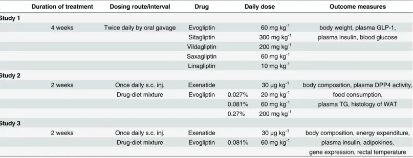
Table 1. Study design.
Duration of treatment Dosing route/interval Drug Daily dose Outcome measures
Study 1
4 weeks Twice daily by oral gavage Evogliptin 60 mg kg-1 body weight, plasma GLP-1,
Sitagliptin 300 mg kg-1 plasma insulin, blood glucose
Vildagliptin 200 mg kg-1
Saxagliptin 60 mg kg-1
Linagliptin 10 mg kg-1 Study 2
2 weeks Once daily s.c. inj. Exenatide 30μg kg-1 body composition, plasma DPP4 activity,
Drug-diet mixture Evogliptin 0.027% 20 mg kg-1 food consumption,
0.081% 60 mg kg-1 plasma TG, histology of WAT 0.27% 200 mg kg-1
Study 3
2 weeks Once daily s.c. inj. Exenatide 30μg kg-1 body composition, energy expenditure,
Drug-diet mixture Evogliptin 0.081% 60 mg kg-1 plasma insulin, adipokines, gene expression, rectal temperature
25–27,10]. To confirm their pharmacodynamic effects, plasma DPP4 inhibition was assessed 12 h after single oral administration of each drug. Due to the observed short half lives of sita-gliptin and evosita-gliptin in mice (unpublished data), all the drugs were administered twice daily by oral gavage and given for 4 weeks using 0.5% (w/v) methylcellulose as a vehicle. Body weight and diet consumption were measured weekly. Before the last administration, fed blood glucose levels were measured from the tail vein blood using a glucometer (AccuChek Active, Roche Diagnostics, Mannheim, Germany). Twelve hours after the last administration, mice were euthanized in the non-fasted state and fed GLP-1 and insulin levels were determined. Blood for determining active GLP-1 was collected in heparinized tubes with sitagliptin (final 1 mM) to minimize the degradation of GLP-1.
Study 2. HF-DIO mice were allocated to each group (8 animals/group) according to body weight and whole body fat mass. To determine the acute effects on body composition, treat-ment period was shortened to 2 weeks. In this study, evogliptin at three doses targeting 20, 60, and 200 mg kg-1day-1for determining dose response and exenatide at the moderate dose were compared. Considering the half-life (2–3 h) of exenatide in mice and the minimum effective dose of 1μg kg-1based on oral glucose tolerance test in mice after single subcutaneous injection
of exenatide (unpublished data), we designated 30μg kg-1day-1as the moderate dose. To
reduce the stress caused by animal handling, evogliptin was given to mice as a drug-diet admix-ture (0.027, 0.081, or 0.27% (w/w)) and exenatide was subcutaneously given to mice once daily. Mice which did not receive exenatide received subcutaneous injections of saline. Control and exenatide-treated mice were fed on a repelleted diet without evogliptin. Diet consumption and body weight were measured daily for 2 weeks. Body composition was measured before and after treatment. After euthanasia in the non-fasted state, plasma was obtained for determining plasma DPP4 activity and triglycerides levels. Because epididymal fat mass was highly corre-lated with whole body fat mass (unpublished data; Pearson’s correlation coefficient = 0.949,
P<0.0001, n = 60), epididymal white adipose fat pad (WAT) was isolated and immediately
fro-zen for histology.
Study 3. HF-DIO mice were allocated to each group (8 animals/group) according to body weight and whole body fat mass. To explore the underlying mechanisms of fat loss, HF-DIO mice received either daily subcutaneous injections of 30μg kg-1day-1of exenatide or saline
with a high-fat diet with/without 0.081% (w/w) evogliptin. Two weeks after treatment, rectal temperature was determined and indirect calorimetry was performed. Body composition was measured before and after treatment. After euthanasia in the non-fasted state, plasma was obtained for determining plasma insulin and adipokine panel. Given that soleus muscle mass directly reflects metabolic change [28] and interscapular fat is representative of brown adipose tissue (BAT) and widely used to investigate the metabolic effects on BAT [29–30], soleus mus-cle, interscapular BAT, and epididymal WAT were isolated and immediately frozen for evalua-tion of gene expression.
Body composition analysis
Body composition was measured under conscious state using a Minispec LF90II NMR spec-trometer (Bruker Optics, Ettlingen, Germany) before and after treatment. Lean body mass was obtained by subtracting fat mass from body weight.
Histological evaluation of adipose tissue
Epididymal adipose tissues were removed, prepared for 4μm-thick microtome sections, and
(http://rsbweb.nih.gov/ij). After processing background of images, the cellular boundary line was automatically detected and manually compared to the original image. Then the area of each cell in the same area was automatically determined except for cells appearing partly at the edge of image frame. Three sections per animal were analyzed.
Determination of plasma parameters
Plasma DPP4 activity was measured using Gly-Pro-AMC as previously described [15]. Plasma triglycerides were determined using an automatic analyzer (Reflotron1
Plus System, Roche, Mannheim, Germany). Plasma insulin was measured using a Milliplex Mouse Adipokine Panel assay service (Millipore, St. Charles, MO). Plasma active GLP-1 and leptin levels were mea-sured using ELISA kits from LINCO Research (St. Charles, MO). An ELISA kit for high molec-ular weight adiponectin from Shibayaki (Shibukawa, Japan) was used for detecting biologically active form of adiponectin.
Indirect calorimetry
After the 2-week treatment, mice were transferred to a new cage to evaluate metabolic rate and accommodated under free access to food and water. Data were obtained for 21 h using a com-puter-controlled open-circuit indirect calorimetry system (Oxylet system; Panlab Harvard Apparatus, Barcelona, Spain) with airflow of 0.35~0.37 l min-1, and metabolic parameters such as oxygen consumption, carbon dioxide production and mean energy expenditure were auto-matically assessed according to manufacturer’s instructions. Respiratory quotient (Rq) was computed by dividing carbon dioxide production by oxygen consumption.
RT-PCR
Information of primers and probes used is listed inS1 Table. For mouse peroxisome prolifera-tor-activated receptor gamma coactivator 1-alpha (Ppargc1a) and hypoxia inducible factor 1-alpha (Hif1a), SYBR Green I Master Mix was used in a LightCycler 480 (Roche Applied Sci-ence, Indianapolis, IN). TaqMan probe method was used for mouse uncoupling protein 1 (Ucp1) and cytochrome c oxidase subunit 4 isoform 1 (Cox4i1). Data analysis was performed using standard curve method. Relative gene expression was presented after normalization by mouse ribosomal protein S3 (Rps3) expression (when fold difference = 2-ΔCt, an average fold change of<2 and a maximal variability of<6-fold between samples) which was validated as
described elsewhere [31].
Statistical analysis
All data was expressed as means ± SE. Except for ANCOVA, statistical analyses were per-formed using SigmaStat1
2.0 (SPSS, Chicago, IL). Comparisons with baseline values and com-parisons between two groups were analyzed using Student’s pairedt-test and Student’st-test, respectively. For comparisons of data involving more than three groups at the same time point, one-way analysis of variance (ANOVA) was used. For comparisons of data at different time points from the same animals among more than three groups, we used repeated measures (RM) two-way ANOVA. When ANOVA indicated a significant difference, the differences were evaluated further using Dunnett’s multiple comparison test. The adipocyte size distribu-tion was analyzed using the Mann—Whitney rank sum test between two groups due to data skewness.Pvalues<0.05 were considered significant. One-way analysis of covariance
Results
Weight-loss effect of DPP4 inhibitors in HF-DIO mice
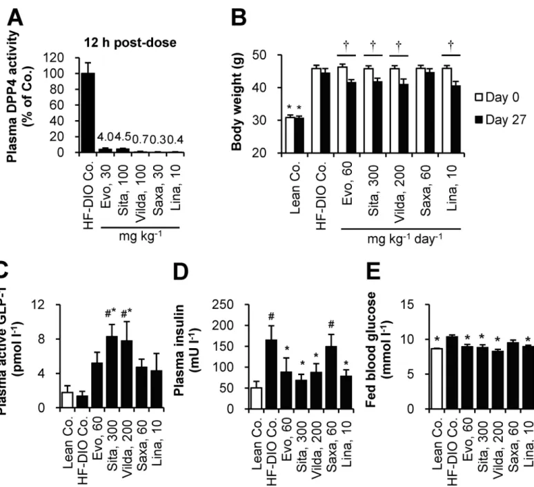
For dose selection, plasma DPP4 activity was assessed 12 h after single oral administration of each drug at a preselected dose in HF-DIO mice. All the DPP4 inhibitors showed over 90% inhibition of plasma DPP4 activity at the doses tested (Fig 1A). Given that DPP4
Fig 1. Body weight-loss effect of DPP4 inhibitors in established obese mice after 4-week treatment.(A) To select each dosage for twice daily administration, plasma DPP4 inhibition was assessed 12 h after a single oral administration in HF-DIO mice. (B-E) After 4 week-administration of evogliptin, sitagliptin, vildagliptin, saxagliptin, or linagliptin twice daily by oral gavage in HF-DIO mice, (B) body weight, (C) fed plasma levels of active GLP-1, (D) insulin, and (E) fed blood glucose levels were determined (n = 5/group). #,P<0.05 vs. lean control;*,P<0.05 vs. HF-DIO control by one-way ANOVA;†,P<0.05 vs. the baseline by RM two-way ANOVA.
inhibition over 80% was considered clinically meaningful [32], those tested doses were selected for the next study phase except for sitagliptin whose dose was increased from 100 mg kg-1to 150 mg kg-1twice daily for full efficacy based on a reported target daily dose of 280 mg kg-1[25].
After four weeks of treatment to HF-DIO mice, body weight-loss effects were observed and statistically equivalent among four DPP4 inhibitors except for saxagliptin (Fig 1B). Cumulative diet consumption did not differ among treatment groups (data not shown). As expected, plasma DPP4 inhibition caused an increase of active GLP-1 levels, with the highest levels observed in sitagliptin or vildagliptin treatment groups at the doses tested (Fig 1C). However, basal insulin levels similarly declined in evogliptin, sitagliptin, vildagliptin and linagliptin-treated mice, indicative of improved insulin sensitivity. These results supported the significant reduction in blood glucose levels by four DPP4 inhibitors (Fig 1E). The changes in basal insulin and blood glucose were concordant with body weight loss across all drugs except saxagliptin whose effects on weight were not significant despite similar plasma DPP4 inhibition and increase of active GLP-1 levels.
Acute effect of evogliptin on whole body composition in HF-DIO mice
Next, we compared treatment response to evogliptin and exenatide to examine whether the observed weight loss was directly due to DPP4 inhibition. To confirm a dose response, evoglip-tin, as a drug-diet mixture, was given to mice at three doses including lower and higher dosages than the dose of evogliptin (60 mg kg-1day-1) which was tested in the 4-week study. Mean administered doses were 21.0±1.3, 62.5±2.0, and 202.7±6.2 mg kg-1day-1for 0.027, 0.081, and 0.27% (w/w), respectively.
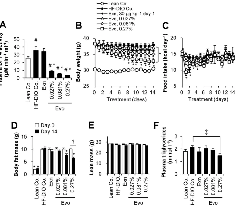
After 2-week treatment, evogliptin caused dose-dependent inhibition of plasma DPP4 activ-ity (74.0%, 84.5%, and 90.7% at doses of 0.027%, 0.081%, and 0.27%, respectively)(Fig 2A). Evogliptin treatment at 0.27% continuously decreased body weight with a significant reduction of 13.3% vs. HF-DIO control (Fig 2B;P<0.05 by RM two-way ANOVA). However, evogliptin
at 0.081% and exenatide treatments did not show significant weight reductions (-7.74% and -4.77% vs. HF-DIO control, respectively). Furthermore, a return to a lean mouse phenotype was not observed with any of the treatments.
Daily energy intake acutely decreased on day 1 by 0.27% evogliptin and exenatide alone and then recovered 3–5 days afterwards (Fig 2C). However the pattern of energy intake of the same animals was unchanged and neither was cumulative diet consumption (data not shown). The body weight in 0.081% and 0.27% evogliptin-treated mice continu-ously declined throughout the treatment period regardless of energy intake; meanwhile, weight-loss by exenatide treatment was maintained after a rapid drop on the second day of administration, implying the effects of either drug were driven by different
pharmacodynamics.
As for whole body fat mass, evogliptin treatment dose-dependently lowered fat mass with the greatest reduction achieved at the dose of 0.27% (-38.5% vs. HF-DIO Co.,P<0.05 by RM
two-way ANOVA)(Fig 2D,S2 Table). Although there was no statistically significant difference in fat-loss trend (P>0.05 by RM two-way ANOVA), evogliptin 0.081% treatment exhibited a
Acute effects on adiposity in HF-DIO mice
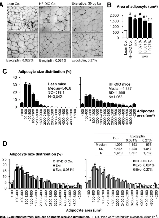
We histologically analyzed adipocyte size distribution in epididymal fat section. HF-DIO mice typically exhibited prominent adipocyte hypertrophy (Fig 3A and 3B) and the size distribution was significantly right-shifted in HF-DIO mice compared to that in lean mice (P<0.05, Mann —Whitney rank sum test), indicating an increased proportion of large adipocytes (Fig 3C). Evogliptin treatment dose-dependently reduced mean adipocyte size and left-shifted its size distribution (Fig 3D). It was a noteworthy finding that the percentage of large size
adipocytes>3 600μm2decreased significantly in a dose-dependent manner. Intriguingly,
Fig 2. Two-week treatment of evogliptin decreased whole body fat mass in obese mice.After HF-DIO mice were treated with exenatide (30μg kg-1,
once daily, s.c.) or evogliptin at 0.027%, 0.081%, or 0.27% (w/w) for 2 weeks, (A) plasma DPP4 activity, (B) body weight, (C) energy intake, (D) whole body fat mass, (E) lean body mass, and (F) plasma triglycerides levels were determined (n = 8/group). #,P<0.05 vs. lean control and*,P<0.05 vs. HF-DIO control by one-way ANOVA;†,P<0.05 vs. the baseline by RM two-way ANOVA;‡,P<0.05 vs. HF-DIO control by Student’st-test.
Fig 3. Evogliptin treatment reduced adipocyte size and distribution.HF-DIO mice were treated with exenatide (30μg kg-1, once daily, s.c.) or evogliptin
at 0.027%, 0.081%, or 0.27% (w/w) for 2 weeks. The effects on adipocytes from epididymal fat tissues were histologically determined (three sections/ animals, eight animals/group). (A) Representative images (original magnification, ×200), (B) mean adipocyte areas and (C-D) percentile distribution of adipocyte size were presented. #,P<0.05 vs. lean control and*,P<0.05 vs. HF-DIO control by one-way ANOVA.
when the 2-week evogliptin treatment significantly reduced whole body fat mass at 0.081%, mean adipocyte size and the median value of adipocyte size distribution was significantly improved compared to that of the HF-DIO control (P<0.05, Mann—Whitney rank sum test).
The adipocyte size distribution was comparable between exenatide- and 0.081% evogliptin-treated groups. Compared to exenatide, evogliptin treatment at 0.27% showed a similar reduc-tion in initial diet consumpreduc-tion but was superior in ameliorareduc-tion of adiposity to exenatide (P<0.05, Mann—Whitney rank sum test), which also suggested an additional fat-loss
induc-ing mechanism of action by evogliptin treatment.
Evogliptin increased energy expenditure unlike exenatide
The effects on glucose metabolism were compared at the end of 2-week treatment. Both evo-gliptin at 0.081% and exenatide did not significantly improve the insulin sensitivity (11–13%,
P>0.09 by One-way ANOVA) (S1A and S1B Fig). Notably, while fat-loss effects by both
treat-ments were comparable, reduction of 6 h-fasted blood glucose was doubled in exenatide-treated mice compared to 0.081% evogliptin (-29.5% vs. -11.9%, respectively;P<0.05)(S1C
Fig). Endogenous active GLP-1 levels were not altered by exenatide treatment (S1D Fig). Because restricted energy intake was not a cause of fat-loss by 0.081% evogliptin, we exam-ined the acute effects on energy expenditure. In Study 3, when the changes in body composi-tion of Study 2 were recapitulated (S2 Fig), 0.081% evogliptin treatment enhanced oxygen consumption, carbon dioxide production (Fig 4A) and energy expenditure especially at night-time (Fig 4B). Meanwhile, exenatide treatment had no effect on energy expenditure. Neither evogliptin nor exenatide treatment altered the respiratory quotient (Fig 4C). Because energy expenditure can be altered by both lean body mass and fat mass, mean total energy expenditure was examined according to body composition (S3 Fig). When the effect of treatment on energy expenditure was comprehensively analyzed by ANCOVA using [lean mass+0.2×fat mass] as a covariate as previously described [33], the equal slope assumption passed indicating no interac-tions between each treatment and covariates. Interestingly, adjusted means of energy expendi-ture were greater in evogliptin-treated mice than HF-DIO control or exenatide-treated mice. Moreover, in this equal slope model (R2= 0.717, adjusted R2= 0.675), the y-intercept of evo-gliptin treatment was greater than those of HF-DIO control and exenatide (-35.8 vs. -47.6 and -50.2, respectively;P<0.001, ANCOVA) (Fig 4D), indicative of higher energy expenditure in
evogliptin treated-mice. However, there were no differences in core body temperature (S4 Fig). This result suggested that part of evogliptin-induced fat loss may be attributed to the enhanced metabolic rate which may not be mediated by increased active GLP-1.
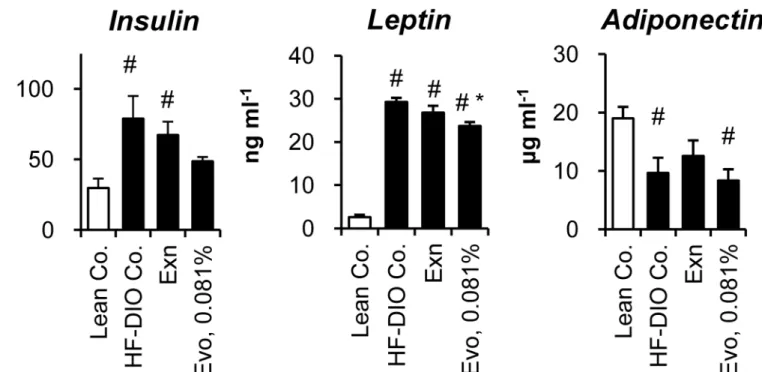
Effects on plasma parameters
Adipose tissue is an endocrine organ secreting various cytokines that regulate whole body metabolism, as well as a highly active metabolic organ for storing surplus energy. Therefore, we evaluated the effects of each treatment on the plasma levels of insulin and five cytokines. Increased plasma levels of insulin and leptin denoted insulin resistance and leptin resistance in HF-DIO mice (Fig 5). Both exenatide and 0.081% evogliptin alone did not significantly improve hyperinsulinemia in HF-DIO mice (P= 0.535 andP= 0.071 by One-way ANOVA, respectively), which corroborated the results of the insulin tolerance test. Meanwhile, only 0.081% evogliptin treatment led to a significant reduction of plasma leptin. The physiologically active form of adiponectin stayed lower in obese mice than lean mice, while neither exenatide (P= 0.690) nor 0.081% evogliptin altered adiponectin levels.
Fig 4. Evogliptin increased energy expenditure unlike exenatide.HF-DIO mice were treated with exenatide (30μg kg-1, once daily, s.c.) or evogliptin at
0.081% (w/w) for 2 weeks. Metabolic parameters were measured using an indirect calorimetry system; (A) Oxygen consumption (VO2) and carbon dioxide
production (VCO2), (B) mean energy expenditure (EE), (C) respiratory quotient (Rq), and (D) a plot of individual data of total mean EE vs. (lean mass
+ 0.2 × fat mass) as a covariate (n = 8/group). #,P<0.05 vs. lean control and*,P<0.05 vs. HF-DIO control by one-way ANOVA.
were not significantly altered by any treatments (S5 Fig). Plasma levels of tumor necrosis factor αand monocyte chemoattractant protein-1 simultaneously analyzed using a Milliplex kit were under the minimum detection limit of 2.5 pg ml-1and 7.8 pg ml-1, respectively (data not shown).
Taken together, these results suggested that reduced adiposity after 2-week treatment of evogliptin was not attributed to alteration of adipokines levels or inflammation.
Evogliptin increased
Ppargc1a
gene expression in WAT
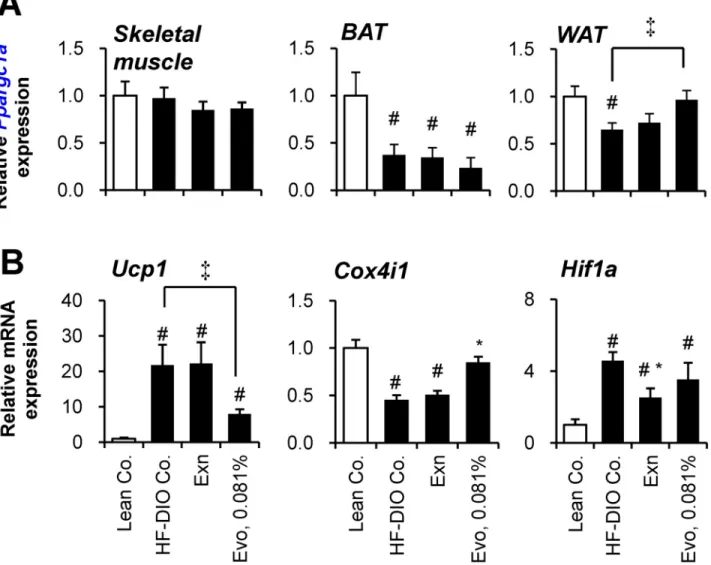
Increased energy expenditure can be explained by enhanced metabolic rate in skeletal muscle [22] or adipose tissues [34]. Given that PGC1α(Ppargc1a) is a key regulator involved in lipid metabolism in various peripheral tissues including muscle [35–36] and adipocyte [37–38], we presumed that evogliptin regulates PGC1αexpression in its target organ.
To determine the target organ of evogliptin, we evaluated the gene expression levels of
Ppargc1ain skeletal muscle, BAT and WAT with regard to energy homeostasis.Ppargc1a
expression in skeletal muscle was not altered by a high-fat diet or any treatments (Fig 6A). In BAT,Ppargc1aexpression was significantly decreased by a high-fat diet, but not affected by any treatments. However, in WAT, evogliptin treatment significantly restoredPpargc1agene expression which was normally suppressed by feeding a high-fat diet, suggesting evogliptin’s influence on metabolic changes in WAT.
Given that PGC1αstimulates thermogenesis via inducing UCP1 expression, gene expres-sion of UCP1 (Ucp1) was first examined. Unexpectedly, evogliptin treatment reduced gene expression ofUcp1in WAT (Fig 6B), suggesting an alternative pathway may be responsible. Because PGC1αenhances metabolism through up-regulating mitochondrial biogenesis in adi-pose tissue, the effect of evogliptin on the expression levels of a putative mitochondrial gene, Fig 5. Effects on plasma insulin and adipokines.After 2-week treatment of exenatide (30μg kg-1, once daily, s.c.) or evogliptin at 0.081% (w/w) in HF-DIO mice, fed plasma concentrations of (A) insulin, (B) leptin and (D) high molecular weight (HMW) adiponectin were assessed (n = 8/group). #,P<0.05 vs. lean control and*,P<0.05 vs. HF-DIO control by one-way ANOVA.
Cox4i1, was also examined. High-fat feeding reduced mitochondrialCox4i1gene expression, but evogliptin treatment, unlike exenatide, restored gene expression ofCox4i1. Conversely, exenatide, unlike evogliptin, reducedHif1agene expression enhanced by a high-fat diet. Evo-gliptin showed a differential reduction in adipocyte gene expression ofPparg2contrary to
Srebp1candDpp4(S6 Fig). Given that PPARγ2 is a typical adipogenic transcription factor, we examined whether evogliptin directly inhibited the differentiation of human adipocytes. In this assay, evogliptin neither induced adipogenesis per se nor antagonized the adipocyte differentia-tion induced by rosiglitazone, a typical PPARγagonist, along with no direct activation of human and mouse PPARα/γ(S6 Fig).
These results indicated that, compared to exenatide, 0.081% evogliptin treatment differen-tially influenced the lipid metabolism in WAT which may serve the potential target for evogliptin.
Fig 6. Effect of evogliptin treatment on the gene expression in muscle and adipose tissues.After 2–week administration of exenatide (30μg kg-1, once
daily, s.c.) or evogliptin at 0.081% (w/w) in HF-DIO mice, (A) PGC-1α(Ppargc1a) gene expression in soleus muscle and two types of fat tissues
(interscapular fat and epididymal fat for brown adipose tissue and white adipose tissue, respectively), and gene expression levels of (B) UCP1 (Ucp1), (C) COXIV (Cox4i1) and HIF-1α(Hif1a) were assessed in white adipose tissue by realtime qPCR method (n = 8/group). #,P<0.05 vs. lean control and*, P<0.05 vs. HF-DIO control by one-way ANOVA;‡,P<0.05 by Student’s t-test.
Discussion
Although various gliptins are known to be neutral on body weight in type 2 diabetic patients, the effect on body fat mass has not been fully elucidated in humans and animals yet. To the best of our knowledge, this is the first report on the body weight-loss effect through pharmaco-logically inhibiting DPP4 activity in established obese mice. We also demonstrated that our novel DPP4 inhibitor, evogliptin, in contrast to exenatide, directly reduced whole body fat mass by enhanced energy expenditure at a clinically relevant dose achieving over 80% plasma DPP4 inhibition for 24 hours. Our findings proposed the possibility that the target organ of evogliptin treatment may be WAT.
An imbalance between energy intake and expenditure causes weight gain or loss. Exenatide produced an anorexic effect at three- to ten-fold higher doses than that for the glucose-lower-ing effect [6,39]. On the other hand, the maximum increase in endogenous intact GLP-1 levels enhanced through inhibiting DPP4 activity was limited due to restricted secretion. While chronic treatment with sitagliptin led to peak active GLP-1 levels of around 50 pM after a meal challenge which rapidly declined within 2–3 h in HF-DIO mice [40], evogliptin 0.081% led to mean ambient GLP-1 levels of 10.3±1.01 pM (3.7 fold greater compared to HF-DIO control). This evidence supports the notion that the endogenous GLP-1 levels increased by DPP4 inhibi-tion are insufficient to produce the anorexic effect of GLP-1 signaling.
In this study, the glucose-lowering effect of exenatide at 30μg kg-1was superior to 0.081%
evogliptin, suggesting that the fat-loss effect of evogliptin may not be a result of improvement in glucose metabolism. Among adipokines, 2-week evogliptin treatment significantly lowered only plasma leptin levels. Leptin acts as a negative regulator of energy intake on hypothalamus [41] and plasma leptin levels were lowered by 0.081% evogliptin at the end of treatment. Fur-thermore, evogliptin at 0.081% did not alter daily diet consumption, but enhanced energy expenditure. These results cannot be fully explained by the improvement of leptin resistance [42], although we did not determine levels of leptin receptors. Thus we concluded that reduc-tion in plasma leptin may be a result of reduced adiposity rather than direct causality with DPP4 inhibition.
We suggested that one possible mechanism in evogliptin-induced fat loss is increased energy dissipation. While it is clear that exenatide decreases food intake by increasing satiety and anorexic effect, the effect of exenatide on energy expenditure was controversial. It was reported that exenatide reduced physical activity, oxygen consumption, and energy expendi-ture in rodents [43–44]. While some clinical researchers have suggested the possibility of increased energy expenditure [45–47], a recent clinical study, which investigated energy expen-diture in obese subjects after exenatide administration, disproved this theory [48]. Goldsmith et al. reported that sitagliptin induced energy expenditure after 10 week-treatment in diabetic mice, which was partially sustained even in GLP-1 receptor-deficient mice [49]. Therefore, this data supports the notion that increased energy expenditure by evogliptin may be partly GLP-1 independent.
discovered that the key metabolic factor was altered in WAT along with alteration of its down-stream target gene, but not in muscle or BAT. Fat tissue accounts for a large proportion of body mass (15–30% in healthy subjects and more than 40% in obese subjects) [54]. Moreover, the evidence that reduced gene expression of AMPKα, PGC1αand PPARαwas associated with decreased oxidative capacity in WAT [55] and that chronic activation of adipocyte AMP-acti-vated protein kinase (AMPK) increased energy dissipation through enhancing fatty acid oxida-tion in adipose tissue [56], supports the hypothesis that metabolic change in WAT can
contribute to energy expenditure. Nevertheless, further studies are needed to explain how evo-gliptin up-regulatesPpargc1aexpression in adipose tissue and whether evogliptin affects PGC1αactivation state.
PGC-1αregulates thermogenesis and mitochondrial biogenesis through increasing UCPs expression and augmenting mitochondrial DNA replication in adipocytes [57]. Unexpect-edly, evogliptin caused a significant decrease ofUcp1expression in WAT, but this result was partly in line with the unaffected rectal temperature in the evogliptin-treated mice. On the other hand, one of mitochondrial components,Cox4i1expression, which correlates with increasedPpargc1aexpression and was not seen with exenatide, was increased by evogliptin treatment.
The 2-week evogliptin treatment significantly ameliorated adipocyte size distribution. Adi-pocyte size can reflect the metabolic function of adiAdi-pocytes [58]. It is yet unknown which abso-lute range of adipocytes sizes is metabolically harmful and no feasible method exists to directly compare adipocyte size from different studies due to discrepancies in sample preparation methods [59]. However, larger adipocytes are known to be metabolically more harmful than smaller ones [58]. In our data, the area of greater than 99% of the adipocytes from lean mice were<2,400μm2. Interestingly, evogliptin treatment significantly reduced the portion of large
adipocytes (>3 600μm2), implying the metabolic improvement.
Our study has some limitations. The maximum fat-loss effect of evogliptin has not yet been verified. We previously reported the weight-loss effect of cannabinoid (CB) 1 receptor antago-nist (SR 141716A also known as rimonabant which had an approval for anti-obesity drug) in a similar mice model [60]. Rimonabant achieved maximum efficacy 2 to 3 weeks post-dose according to the doses tested. However, the appropriate time for maximum efficacy can differ based on the mode of action involved in the fat-loss effects. We speculate that the fat-loss effect of evogliptin along with improvement in glucose and lipid metabolism, could be amplified with longer-term treatment. Additionally, we used a high-fat diet containing relatively lower carbohydrate, supplying 60% calories from fat. In mice fed on a high-fat diet with lower caloric contribution from fat (45%) with increased sucrose, the contribution of glucose metabolism to fat storage may be more significant.
In conclusion, our findings suggest the beneficial effects of evogliptin at a clinically relevant dose, on whole body composition, particularly whole body fat loss in an established obese mice model. This study verified fat loss by DPP4 inhibition which is likely mediated by increased energy expenditure and alteration in white adipose tissue metabolism. Our findings provide new insight into the role of DPP4 inhibitors in adiposity.
Supporting Information
S1 Fig. Effects on glucose metabolism after 2-week treatment. (PDF)
S3 Fig. Analysis of treatment effects on total energy expenditure according to the body composition.
(PDF)
S4 Fig. Effect on rectal temperature. (PDF)
S5 Fig. Effects on other cytokine levels. (PDF)
S6 Fig. Effects on gene expression in white adipose tissue after 2-week treatment in HF-DIO mice.
(PDF)
S7 Fig. Effects of on adipogenesis and PPARα/γactivation. (PDF)
S1 Table. Primers and probe for mouse genes used. (PDF)
S2 Table. Body composition changes in Study 2 after 2-week treatment. (PDF)
Acknowledgments
We would like to thank Mr. Young E. Lee, M.D., M.B.A. (UC Irvine Medical Center, CA, USA) for his careful proofreading and language help during the revision of the present paper.
Author Contributions
Conceived and designed the experiments: MKK CYS MHS. Performed the experiments: YNC THK CYS IHJ YSS. Analyzed the data: YNC THK MKK CYS MHS. Wrote the paper: MKK MHS.
References
1. Guillet C, Masgrau A, Walrand S, Boirie Y. Impaired protein metabolism: interlinks between obesity, insulin resistance and inflammation. Obes Rev. 2012; 13 Suppl 2: 51–57. PMID:23107259doi:10. 1111/j.1467-789X.2012.01037.x
2. Lamers D, Famulla S, Wronkowitz N, Hartwig S, Lehr S, Ouwens DM, et al. Dipeptidyl peptidase 4 is a novel adipokine potentially linking obesity to the metabolic syndrome. Diabetes. 2011; 60(7): 1917– 1925. PMID:21593202doi:10.2337/db10-1707
3. Scheen AJ. A review of gliptins in 2011. Expert Opin Pharmacother. 2012; 13(1): 81–99. PMID: 22149369doi:10.1517/14656566.2012.642866
4. Cahn A, Raz I. Emerging gliptins for type 2 diabetes. Expert Opin Emerg Drugs. 2013; 18(2): 245–258. PMID:23725569doi:10.1517/14728214.2013.807796
5. Amori RE, Lau J, Pittas AG. Efficacy and safety of incretin therapy in type 2 diabetes: systematic review and meta-analysis. JAMA. 2007; 298(2): 194–206. PMID:17622601
6. Lamont BJ, Drucker DJ. Differential antidiabetic efficacy of incretin agonists versus DPP-4 inhibition in high fat fed mice. Diabetes. 2008; 57(1): 190–198. PMID:17928394
7. Hamamoto S, Kanda Y, Shimoda M, Tatsumi F, Kohara K, Tawaramoto K, et al. Vildagliptin preserves the mass and function of pancreatic beta cells via the developmental regulation and suppression of oxi-dative and endoplasmic reticulum stress in a mouse model of diabetes. Diabetes Obes Metab. 2013; 15(2): 153–163. PMID:22950702doi:10.1111/dom.12005
methyl)-3,7-dihydro-purine-2,6-dione] increases basal glucagon-like peptide-1 and improves glycemic control in diabetic rodent models. J Pharmacol Exp Ther. 2009; 328(2): 556–563. doi:10.1124/jpet.108.143966
9. Flock G, Baggio LL, Longuet C, Drucker DJ. Incretin receptors for glucagon-like peptide 1 and glucose-dependent insulinotropic polypeptide are essential for the sustained metabolic actions of vildagliptin in mice. Diabetes. 2007; 56(12): 3006–3013. PMID:17717280
10. Kern M, Klöting N, Niessen HG, Thomas L, Stiller D, Mark M, et al. Linagliptin improves insulin sensitiv-ity and hepatic steatosis in diet-induced obessensitiv-ity. PLoS One. 2012; 7(6): e38744. PMID:22761701doi: 10.1371/journal.pone.0038744
11. Challa TD, Beaton N, Arnold M, Rudofsky G, Langhans W, Wolfrum C. Regulation of adipocyte forma-tion by GLP-1/GLP-1R signaling. J Biol Chem. 2012; 287(9): 6421–6430. PMID:22207759doi:10. 1074/jbc.M111.310342
12. Vendrell J, El Bekay R, Peral B, Garcia-Fuentes E, Megia A, Macias-Gonzalez M, et al. Study of the potential association of adipose tissue GLP-1 receptor with obesity and insulin resistance. Endocrinol-ogy. 2011; 152(11): 4072–4079. PMID:21862620doi:10.1210/en.2011-1070
13. Ostenson CG, Matthaei S, Reaney M, Krarup T, Guerci B, Kiljanski J, et al. Treatment outcomes after initiation of exenatide twice daily or insulin in clinical practice: 12-month results from CHOICE in six European countries. Diabetes Metab Syndr Obes. 2013; 6: 171–185. PMID:23667315doi:10.2147/ DMSO.S41827
14. Flint A, Kapitza C, Zdravkovic M. The once-daily human GLP-1 analogue liraglutide impacts appetite and energy intake in patients with type 2 diabetes after short-term treatment. Diabetes Obes Metab. 2013; 15(10): 958–962. PMID:23551925doi:10.1111/dom.12108
15. Elfers CT, Simmons JH, Roth CL. Glucagon-like peptide-1 agonist exendin-4 leads to reduction of weight and caloric intake in a rat model of hypothalamic obesity. Horm Res Paediatr. 2012; 78(1): 47– 53. PMID:22831885doi:10.1159/000338464
16. Conarello SL, Li Z, Ronan J, Roy RS, Zhu L, Jiang G, et al. Mice lacking dipeptidyl peptidase IV are pro-tected against obesity and insulin resistance. Proc Natl Acad Sci U S A. 2003; 100(11):6825–6830. PMID:12748388
17. Kim MK, Chae YN, Kim HD, Yang EK, Cho EJ, Choi SH, et al. DA-1229, a novel and potent DPP4 inhib-itor, improves insulin resistance and delays the onset of diabetes. Life Sci. 2012; 90(1–2): 21–29. PMID:22056373doi:10.1016/j.lfs.2011.10.007
18. Kos K, Baker AR, Jernas M, Harte AL, Clapham JC, O'Hare JP, et al. DPP-IV inhibition enhances the antilipolytic action of NPY in human adipose tissue. Diabetes Obes Metab. 2009; 11(4): 285–92. PMID:19175376doi:10.1111/j.1463-1326.2008.00909.x
19. Rosmaninho-Salgado J, Marques AP, Estrada M, Santana M, Cortez V, Grouzmann E, et al. Dipepti-dyl-peptidase-IV by cleaving neuropeptide Y induces lipid accumulation and PPAR-gamma expression. Peptides. 2012; 37(1): 49–54. PMID:22819773doi:10.1016/j.peptides.2012.06.014
20. Kirino Y, Kamimoto T, Sato Y, Kawazoe K, Minakuchi K, Nakahori Y. Increased plasma dipeptidyl pepti-dase IV (DPP IV) activity and decreased DPP IV activity of visceral but not subcutaneous adipose tis-sue in impaired glucose tolerance rats induced by high-fat or high-sucrose diet. Biol Pharm Bull. 2009; 32(3): 463–467. PMID:19252296
21. Kirino Y, Sato Y, Kamimoto T, Kawazoe K, Minakuchi K. Altered dipeptidyl peptidase-4 activity during the progression of hyperinsulinemic obesity and islet atrophy in spontaneously late-stage type 2 dia-betic rats. Am J Physiol Endocrinol Metab. 2011; 300(2): E372–E379. PMID:21139073doi:10.1152/ ajpendo.00319.2010
22. Fukuda-Tsuru S, Kakimoto T, Utsumi H, Kiuchi S, Ishii S. The novel dipeptidyl peptidase-4 inhibitor teneligliptin prevents high-fat diet-induced obesity accompanied with increased energy expenditure in mice. Eur J Pharmacol. 2014; 723: 207–215. PMID:24309217doi:10.1016/j.ejphar.2013.11.030
23. Cho JM, Jang HW, Cheon H, Jeong YT, Kim DH, Lim YM, et al. A novel dipeptidyl peptidase IV inhibitor DA-1229 ameliorates streptozotocin-induced diabetes by increasing beta-cell replication and neogen-esis. Diabetes Res Clin Pract. 2011; 91: 72–79. PMID:21093089doi:10.1016/j.diabres.2010.10.012
24. Kim HJ, Kwak WY, Min JP, Lee JY, Yoon TH, Kim HD, et al. Discovery of DA-1229: a potent, long acting dipeptidyl peptidase-4 inhibitor for the treatment of type 2 diabetes. Bioorg Med Chem Lett. 2011; 21: 3809–3812. PMID:21570283doi:10.1016/j.bmcl.2011.04.029
25. Mu J, Petrov A, Eiermann GJ, Woods J, Zhou YP, Li Z, et al. Inhibition of DPP-4 with sitagliptin improves glycemic control and restores islet cell mass and function in a rodent model of type 2 diabetes. Eur J Pharmacol. 2009; 623:148–154. PMID:19765579doi:10.1016/j.ejphar.2009.09.027
27. Vyas AK, Aerni-Flessner LB, Payne MA, Kovacs A, Jay PY, Hruz PW. Saxagliptin Improves Glucose Tolerance but not Survival in a Murine Model of Dilated Cardiomyopathy. Cardiovasc Endocrinol. 2012; 1(14): 74–82. PMID:23795310
28. Cannavino J, Brocca L, Sandri M, Bottinelli R, Pellegrino MA. PGC1-αover-expression prevents
meta-bolic alterations and soleus muscle atrophy in hindlimb unloaded mice. J Physiol. 2014; 592(Pt 20): 4575–4589. PMID:25128574doi:10.1113/jphysiol.2014.275545
29. Yo K, Yu YM, Zhao G, Bonab AA, Aikawa N, Tompkins RG, et al. Brown adipose tissue and its modula-tion by a mitochondria-targeted peptide in rat burn injury-induced hypermetabolism. Am J Physiol Endocrinol Metab. 2013; 304(4): E331–E341. PMID:23169784doi:10.1152/ajpendo.00098.2012
30. Bartelt A, Heeren J. Adipose tissue browning and metabolic health. Nat Rev Endocrinol. 2014; 10(1): 24–36. PMID:24146030doi:10.1038/nrendo.2013.204
31. Dheda K, Huggett JF, Bustin SA, Johnson MA, Rook G, Zumla A. Validation of housekeeping genes for normalizing RNA expression in real-time PCR. Biotechniques. 2004; 37(1): 112–114, 116, 118–119. PMID:15283208
32. Bergman AJ, Stevens C, Zhou Y, Yi B, Laethem M, De Smet M, et al. Pharmacokinetic and pharmaco-dynamic properties of multiple oral doses of sitagliptin, a dipeptidyl peptidase-IV inhibitor: a double-blind, randomized, placebo-controlled study in healthy male volunteers. Clin Ther. 2006; 28(1):55–72. PMID:16490580
33. Even PC, Nadkarni NA. Indirect calorimetry in laboratory mice and rats: principles, practical consider-ations, interpretation and perspectives. Am J Physiol Regul Integr Comp Physiol. 2012; 303(5): R459– R476. PMID:22718809doi:10.1152/ajpregu.00137.2012
34. Wendel AA, Purushotham A, Liu LF, Belury MA. Conjugated linoleic acid induces uncoupling protein 1 in white adipose tissue of ob/ob mice. Lipids. 2009; 44(11): 975–982. PMID:19779754doi:10.1007/ s11745-009-3348-9
35. Patti ME, Butte AJ, Crunkhorn S, Cusi K, Berria R, Kashyap S, et al. Coordinated reduction of genes of oxidative metabolism in humans with insulin resistance and diabetes: Potential role of PGC1 and NRF1. Proc Natl Acad Sci U S A. 2003; 100(14): 8466–8471. PMID:12832613
36. Lin J, Wu H, Tarr PT, Zhang CY, Wu Z, Boss O, et al. Transcriptional co-activator PGC-1 alpha drives the formation of slow-twitch muscle fibres. Nature. 2002; 418(6899): 797–801. PMID:12181572
37. Liang H, Ward WF. PGC-1alpha: a key regulator of energy metabolism. Adv Physiol Educ. 2006; 30 (4): 145–151. PMID:17108241
38. Bogacka I, Xie H, Bray GA, Smith SR. Pioglitazone induces mitochondrial biogenesis in human subcu-taneous adipose tissue in vivo. Diabetes. 2005; 54(5): 1392–1399. PMID:15855325
39. Talsania T, Anini Y, Sinus S, Drucker DJ, Brubaker PL. Peripheral exendin-4 and peptide YY(3–36) synergistically reduce food intake through different mechanisms in mice. Endocrinology. 2005; 146(9): 3748–3756. PMID:15932924
40. Zambrowicz B, Ding ZM, Ogbaa I, Frazier K, Banks P, Turnage A, et al. Effects of LX4211, a dual SGLT1/SGLT2 inhibitor, plus sitagliptin on postprandial active GLP-1 and glycemic control in type 2 dia-betes. Clin Ther. 2013; 35(3): 273–285.e7. PMID:23433601doi:10.1016/j.clinthera.2013.01.010
41. Campfield LA, Smith FJ, Guisez Y, Devos R, Burn P. Recombinant mouse OB protein: evidence for a peripheral signal linking adiposity and central neural networks. Science. 1995; 269(5223): 546–549. PMID:7624778
42. Ceddia RB. Direct metabolic regulation in skeletal muscle and fat tissue by leptin: implications for glu-cose and fatty acids homeostasis. Int J Obes (Lond). 2005; 29(10):1175–1183. PMID:16030519
43. Mack CM, Moore CX, Jodka CM, Bhavsar S, Wilson JK, Hoyt JA, et al. Antiobesity action of peripheral exenatide (exendin-4) in rodents: effects on food intake, body weight, metabolic status and side-effect measures. Int J Obes (Lond). 2006; 30(9): 1332–1340. PMID:16534527
44. Baggio LL, Huang Q, Brown TJ, Drucker DJ. Oxyntomodulin and glucagon-like peptide-1 differentially regulate murine food intake and energy expenditure. Gastroenterology. 2004; 127(2): 546–558.PMID: 15300587
45. Horowitz M, Flint A, Jones KL, Hindsberger C, Rasmussen MF, Kapitza C, et al. Effect of the once-daily human GLP-1 analogue liraglutide on appetite, energy intake, energy expenditure and gastric emptying in type 2 diabetes. Diabetes Res Clin Pract. 2012; 97(2): 258–266. PMID:22446097doi:10.1016/j. diabres.2012.02.016
47. Pannacciulli N, Bunt JC, Koska J, Bogardus C, Krakoff J. Higher fasting plasma concentrations of glu-cagon-like peptide 1 are associated with higher resting energy expenditure and fat oxidation rates in humans. Am J Clin Nutr. 2006; 84(3): 556–560. PMID:16960169
48. Bradley DP, Kulstad R, Racine N, Shenker Y, Meredith M, Schoeller DA. Alterations in energy balance following exenatide administration. Appl Physiol Nutr Metab. 2012; 37: 893–899. PMID:22735035doi: 10.1139/h2012-068
49. Goldsmith F, Keenan MJ, Raggio AM, Ye X, Hao Z, Durham H, et al. Induction of Energy Expenditure by Sitagliptin Is Dependent on GLP-1 Receptor. PLoS One. 2015; 10(5): e0126177. PMID:25938560
50. Boschmann M, Engeli S, Dobberstein K, Budziarek P, Strauss A, Boehnke J, et al. Dipeptidyl-pepti-dase-IV inhibition augments postprandial lipid mobilization and oxidation in type 2 diabetic patients. J Clin Endocrinol Metab. 2009; 94(3):846–852. PMID:19088168doi:10.1210/jc.2008-1400
51. Dobrian AD, Ma Q, Lindsay JW, Leone KA, Ma K, Coben J, et al. Dipeptidyl peptidase IV inhibitor sita-gliptin reduces local inflammation in adipose tissue and in pancreatic islets of obese mice. Am J Physiol Endocrinol Metab. 2011; 300(2):E410–E421. PMID:21081706doi:10.1152/ajpendo.00463.2010
52. Souza-Mello V, Gregório BM, Cardoso-de-Lemos FS, de Carvalho L, Aguila MB, Mandarim-de-Lacerda CA. Comparative effects of telmisartan, sitagliptin and metformin alone or in combination on obesity, insulin resistance, and liver and pancreas remodelling in C57BL/6 mice fed on a very high-fat diet. Clin Sci (Lond). 2010; 119(6):239–250. PMID:20415664doi:10.1042/CS20100061
53. Sawada T, Miyoshi H, Shimada K, Suzuki A, Okamatsu-Ogura Y, Perfield JW 2nd, et al. Perilipin over-expression in white adipose tissue induces a brown fat-like phenotype. PLoS One. 2010; 5(11): e14006. PMID:21103377doi:10.1371/journal.pone.0014006
54. Abernathy RP, Black DR. Healthy body weights: an alternative perspective. Am J Clin Nutr. 1996; 63(3 Suppl):448S–451S. PMID:8615340
55. Gaidhu MP, Anthony NM, Patel P, Hawke TJ, Ceddia RB. Dysregulation of lipolysis and lipid metabo-lism in visceral and subcutaneous adipocytes by high-fat diet: role of ATGL, HSL, and AMPK. Am J Physiol Cell Physiol. 2010; 298(4):C961–971. PMID:20107043doi:10.1152/ajpcell.00547.2009
56. Gaidhu MP, Fediuc S, Anthony NM, So M, Mirpourian M, Perry RL, et al. Prolonged AICAR-induced AMP-kinase activation promotes energy dissipation in white adipocytes: novel mechanisms integrating HSL and ATGL. J Lipid Res. 2009; 50(4):704–715. PMID:19050316doi:10.1194/jlr.M800480-JLR200
57. Finck BN, Kelly DP. PGC-1 coactivators: inducible regulators of energy metabolism in health and dis-ease. J Clin Invest. 2006; 116(3): 615–622. PMID:16511594
58. Skurk T, Alberti-Huber C, Herder C, Hauner H. Relationship between adipocyte size and adipokine expression and secretion. J Clin Endocrinol Metab. 2007; 92(3): 1023–1033. PMID:17164304
59. Verhoef SPM, van Dijk P, Westerterp KR. Relative shrinkage of adipocytes by paraffin in proportion to plastic embedding in human adipose tissue before and after weight loss. Obes Res Clin Pract. 2013; 7: e8–13. PMID:24331678doi:10.1016/j.orcp.2012.03.001