*Corresponding author: Mohammad Taghi Mohammadi, Tel/Fax: +98 21 82483419, Email: Mohammadimohammadt@bmsu.ac.ir ©
2016 The Authors. This is an Open Access article distributed under the terms of the Creative Commons Attribution (CC BY), which permits Adv Pharm Bull, 2016, 6(4), 531-539
doi: 10.15171/apb.2016.067 http://apb.tbzmed.ac.ir
Advanced
Pharmaceutical
Bulletin
Captopril and Valsartan May Improve Cognitive Function Through
Potentiation of the Brain Antioxidant Defense System and Attenuation
of Oxidative/Nitrosative Damage in STZ-Induced Dementia in Rat
Yasaman Arjmand Abbassi1, Mohammad Taghi Mohammadi2*, Mahsa Sarami Foroshani3, Javad Raouf Sarshoori4
1
Department of Biology, Science and Research Branch, Islamic Azad University, Tehran, Iran. 2
Department of Physiology and Biophysics, Faculty of Medicine, Baqiyatallah University of Medical Sciences, Tehran, Iran. 3 Department of Nanotechnology, School of new sciences and technology, Islamic Aazad University of Pharmaceutical Scinces
Branch, Tehran, Iran. 4
Department of Anatomy, Faculty of Medicine, Baqiyatallah University of Medical Sciences, Tehran, Iran.
Introduction
Alzheimer's disease (AD) is one of the most common forms of neurodegenerative disorders that leads to deficit in learning and memory.1,2 Also, it is associated with formation of senile plaques and neurofibrillary tangles in memory-related parts of the brain such as hippocampus.3,4 The main reason of this disease is not well understood, but based on recent studies the brain renin-angiotensin system (RAS) plays an important role in pathogenesis of AD.5,6 Angiotensin-II (Ang-II) is the main effector of the RAS and has two receptors in the brain (angiotensin type-1; AT1, and angiotensin type-2; AT2).7 Based on recent findings, activation of AT1 receptor induces several neurodegenerative pathways such as reactive oxygen specious (ROS),
inflammatory responses and apoptotic signals.8-10 Also, oxidative imbalance and significant increase of its by-products have been consistently reported in AD.9,11 The brain is highly susceptible to oxidative imbalance due to its high oxygen consumption, rich abundance of easily peroxidizable polyunsaturated fatty acids and feeble antioxidant defense system in comparison to the other tissues.12 Thus, either enhanced ROS production or impaired brain antioxidant system will affect the cellular redox balance to oxidative imbalance and cause ROS overproduction.13 It is no wonder that oxidative imbalance and subsequent oxidative stress mediated damage to biomolecules are extensively reported in Article History:
Received: 19 July 2016 Revised: 8 September 2016 Accepted: 10 September 2016 ePublished: 22 December 2016
Keywords: Alzheimer’s disease Antioxidant
Renin-angiotensin system Captopril
Valsartan
Abstract
Purpose:Previous findings have shown the crucial roles of brain renin-angiotensin system
(RAS) in pathogenesis of Alzheimer’s disease (AD). Since RAS inhibitors may have
beneficial effects on dementia and cognitive function in elderly people, the aim of present study was to examine the neuroprotective actions of captopril and valsartan on memory function and neuronal damage in experimental model of AD.
Methods: Adult forty male Wistar rats (220-280g) were randomly divided into 5 groups; Control, Vehicle, Alzheimer and treatment groups. AD was induced by the injections of streptozotocin (3mg/kg, bilateral intracerebroventricular) at days 1&3. Treated rats received orally captopril (50mg/kg/day) and valsartan (30mg/kg/day). Memory function and histological assessments were done at termination of experiment. Finally, superoxide dismutase (SOD) and catalase (CAT) activities as well as malondialdehyde (MDA) and NOx contents were determined.
Results: There was a significant increase in the mean value of latency in Alzheimer group (66%). Captopril and valsartan considerably decreased this value in both treatment groups
(45% and 72%, respectively). In Alzheimer group the activities of brain’s SOD and CAT reduced (40% and 47%, respectively) in accompany with an increase in MDA and NOx contents (49% and 50%, respectively). Captopril and valsartan significantly increased the
activities of brain’s SOD and CAT concomitant reduction in MDA and NOx contents.Also, histopathological damages noticeably decreased in both treatment groups.
Conclusion: Our findings indicate that RAS inhibition by using captopril and valsartan potentiates the antioxidant defense system of brain and reduces oxidative/nitrosative stress in accompany with neuronal damage during AD.
Arjmand Abbassi et al.
AD and increasing evidence suggests that oxidative imbalance plays a critical role in the disease.9,14 Additionally, enhancement of NO production in brain during AD induces nitrosative damage and combination of NO with ROS leads to formation of very toxic compound of peroxynitrite (ONOO-), which yields to the protein nitrotyrosination and cell death.10,14,15
Previous reports have demonstrated the possibility that treatment with antihypertensive RAS inhibitors prevent the impairment of cognitive performance.16,17 Preclinical and clinical studies confirm involvement of the brain RAS in memory dysfunction.16,18 However, the evidence is limited but treatment with antihypertensive RAS inhibitors has been associated with reduction of brain damage in different experimental and clinical models of neurodegenerative diseases.19-21 According to previous findings, AT1 is involved in the beginning and progression of several neurodegenerative disorders such as AD.22,23 These studies suggest some neuroprotective actions of AT1 receptor inhibition against many neurodegenerative conditions. The study of AbdAlla et al, indicated that certain ACE inhibitors such as captopril was able to reduce the ischemia-induced brain damage.24 Moreover, the findings of Inaba et al, suggest that the continuous activation of the RAS during neuro-pathophysiologic conditions impairs cognitive function via stimulation of AT1 receptor in accompany with a decrease in the cerebral blood flow and an increase in ROS production.25 Finally, Mogi et al, demonstrated a preventive effects of non-hypotensive dose of telmisartan, as a specific AT1 inhibitor, on cognitive impairment in mice model of AD.22 Therefore, it is appeared that RAS inhibition by AT1 receptor antagonists or angiotensin converting enzyme (ACE) inhibitors, which are widely used as antihypertensive drugs, will be able to prevent age-related neurodegenerative diseases.
According to mentioned studies, the aim of present study was to examine the neuroprotective effects of RAS inhibition on cognitive function and neuronal damage in experimental model of AD. Moreover, this study evaluated the probable neuroprotective actions of ACE inhibition and AT1 receptor blockage through inhibition of ROS overproduction and potentiation of the brain antioxidant defense system in AD.
Materials and Methods Animals
Adult forty male Wistar rats, weighing 220-250 g, were obtained from the experimental animal center of Baqiyatallah University of Medical Sciences, Iran. Animals were kept with free access to food and water, temperature of 23±2 °C, and 12 hours light/dark cycle throughout the study. All experimental protocols were in according to the Animal Care Committee of Baqiyatallah University of Medical Sciences Guidelines.
Drugs and chemicals
All of the drug solutions were freshly prepared before use. Captopril (Daru Lab, Iran) and Valsartan (Avicenna Lab, Iran) were suspended in distilled water and were being administered orally with the help of an oral tube. Captopril and valsartan were being taken at dosage of 50 and 30 mg/kg/day, respectively. Streptozotocin (STZ) was purchased from Sigma Aldrich (USA) and was dissolved in saline and delivered through intracerebroventricular (i.c.v.) injection.
Induction of experimental model of Alzheimer’s disease (AD)
AD was induced by using bilateral i.c.v. injection of STZ solution by stereotaxic apparatus.26,27 Briefly, animals were anaesthetized with a mixture of ketamine (80 mg/kg, Imalgene1000®, Merial) and xylazine (10 mg/kg, Rompun® 2%, Bayer) through intra-peritoneal injection. Rats were then placed into a stereotactic apparatus and received i.c.v. injection of STZ solution (stereotactic was being coordinated relative to bregma, duramater and inter-hemispheric scissure: antero-posterior: -0.8 mm; ventral: -3.6 mm, lateral: ±1.5 mm). Injections of STZ were performed into the right or left cerebral ventricles in two times (3 mg/kg) at days 1 and 3 (Figure 1). The concentration was adjusted so as to deliver a maximum of 5 μl in a single injection. The solutions were freshly prepared just before the injection to avoid decomposition of the drug.
Experimental design
In total, five groups were used in the present study and each group consisted of eight Wistar rats. The doses of drugs were selected according to the previous reports.28,29 Group I was used as control (Control). The animals in this group were normal rats without any administration of drugs or surgeries that were kept for 25 days in standard condition. Group II was used as sham (Vehicle) and the rats of this group received
bilateral i.c.v. injection of 0.9% saline (5 μl/ site in a
single injection) on days first and third. These rats were kept for 25 days in standard condition without any administration of drugs. Group III was used as AD (Alzheimer). AD was induced in these rats based on described protocol. The rats of this group were kept for 25 days in standard condition. Group IV was used as captopril treatment (AD+Captopril): In this group, after induction of AD same as group III, captopril (50 mg/kg/day) was orally taken for 25 days. Group V was used as valsartan treatment (AD+Valsartan): In these rats, after induction of AD same as group III and IV, valsartan (30 mg/kg/day) was orally taken for 25 days.
Assessment of learning and memory by using the T-Maze
RAS and Alzheimer’s disease
First preference test was taken and rats were asked to choose between two options. No reward was placed in each arm of the maze. The rats were allowed to explore the whole maze. Then, they were placed in the start location. Then the preference of each rat was recorded. This stage was repeated 5 times. Thereafter, memory test was taken. Rats were placed in the start location and were asked to choose between two options. Reward was placed in the opposite arm that each rat was selected according to preference test. The reward was food. The rats were allowed to explore the whole maze. Then each rat's choice and the amount of time spent during the test to the end of each arm (latency) were recorded.
Tissue preparation
After deep anesthesia, the brains were quickly removed, washed in ice-cold phosphate buffer saline (PBS) for nitrate (NOx), malondialdehyde (MDA), superoxide dismutase (SOD) and catalase (CAT) assays. Washed tissues were immediately immersed in liquid nitrogen and stored at -80 ◦C until biochemical analysis. On the day of use, frozen tissue samples were quickly weighed and homogenized 1:10 in ice-cold PBS. The homogenates were then centrifuged at 14000×g for 15 min at 4°C. The supernatants were separated and used for enzyme activity assays and protein determination.
SOD activity
The activity of SOD was determined by using the method described by Winterbourn et al, based on the ability of SOD to inhibit the reduction of NBT by superoxide.30 For assay, 0.067 M potassium phosphate buffer, pH 7.8 was added to 0.1 M EDTA containing 0.3 mM sodium cyanide, 1.5 mM NBT and 0.1 ml of sample. Then, 0.12 mM riboflavin was added to each sample to initiate the reaction and was incubated for 12 min. The absorbance of samples was read on a Genesys 10 UV spectrophotometer at 560 nm for 5 minutes. The amount of enzyme required to produce 50% inhibition was taken as 1 U and results were expressed as U/mg protein.
CAT activity
CAT activity was measured in tissues homogenates by the method of Aebi H.31 Reaction mixture containing 0.85 ml potassium phosphate buffer 50 mM, pH 7.0 and 0.1 ml homogenate was incubated at room temperature for 10 min. Reaction was initiated by addition of 0.05 ml H2O2 (30 mM prepared in potassium phosphate buffer 50 mM, pH 7.0) and the decrease in absorbance was recorded for 3 min at 240 nm. Specific activity is expressed as 1µmole H2O2 decomposed U/mg protein.
Lipid peroxidation assay
The end product of lipid peroxidation was estimated by measuring the level of MDA according to the method of Satoh K.32 0.5 ml of tissue homogenate was added to 1.5 ml of 10% TCA, vortexed and incubated for 10 min
at room temperature. 1.5 ml of supernatant and 2 ml of thiobarbituric acid (0.67%) were added and placed in a boiling water bath in sealed tubes for 30 min. The samples were allowed to cool at room temperature. 1.25 ml of n-butanol was added, vortexed and centrifuged at 2000 g for 5 min. The resulting supernatant was removed and measured at 532 nm on a spectrophotometer. MDA concentrations were determined by using 1,1,3,3-tetraethoxypropane as standard. MDA concentration was expressed as µmol/mg protein.
NOx assay
The nitrite-nitrate level (NOx) of brain was measured by the colorimetric reaction of the Griess reagent. 0.1 ml of homogenate solution was deproteinized by adding 0.2 ml of zinc sulfate solution and centrifuged for 20 min at 4000 rpm and 4 °C to separate supernatant for NOx assay. 0.1 ml of supernatant (as sample) or pure water (as blank) or sodium nitrate (as standard) was mixed with 0.1 ml vanadium III chloride to reduce nitrite to nitrate. 0.05 ml sulfanilamide (0.01 %) and 0.05 ml N-[1-naphthyl] ethylenediamindihydrochloride (NED, 0.01 %) were incubated for 30 min in dark place at 37 °C. Thereafter, the absorbance of solution was determined at wave length of 540 nm. Nitrate concentration was estimated from a standard curve generated from absorbance of each sodium nitrate solution. Finally, the nitrite-nitrate levels were expressed as µmol/mg protein.33
Protein assay
Protein concentration was estimated according to the method of Bradford using bovine serum albumin (BSA) as a standard.34
Histological assessment
At the end of the experiment, animals were sacrificed under deep anesthesia. The brains were removed and fixed in formalin (10%) for two weeks. After fixation and tissue processing, coronal serial sections (5μm in thickness) were prepared for conventional histological
examination. Paraffin embedded sectioning (each 50μm
intervals) was processed routinely for toluidine blue (TB) staining. After staining procedure, sections were dehydrated with administration of 70, 80, 96, 100, and 100% ethanol, respectively. The samples were placed in xylene solution for two times (each time was 15 min) owing to clearing. Due to mounting, the samples were covered with entelan sticker and then lamels were placed on them. The histological changes were observed through a light microscope (Nikon, Japan) connected to digital camera (CMEX, Holland) for capturing the photograph.
Statistical analysis
Arjmand Abbassi et al.
Data between groups were analyzed by one-way ANOVA in behavioral and biochemical tests. In the case of significant variation (p<0.05), the values were compared by Tukey Post-hoc test. P values < 0.05 were considered to be statistically significant.
Results
Assessment of memory function
The mean values of latency (the average time of the number of correct orientations) in control and vehicle groups were 73.4±15.5 and 67.8±14.4 sec on day 25, respectively. There was a significant increase in the mean value of latency in Alzheimer group compared with control and vehicle groups (216.2±77.9 sec, P<0.05). Captopril and valsartan meaningfully decreased the mean value of latency in both treatment groups (118.0±51.7, P<0.05 and 60.2±5.5, P<0.01 sec, respectively). The average of this time in both treatment groups was statistically significant (Figure 2).
Figure 1. The photograph shows the entrance of cannula into lateral ventricles of brain by using Evans Blue dye (2%) injection through cannula.
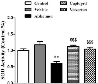
The activity of superoxide dismutase (SOD)
Figure 3 shows the SOD activity of brain tissue at the end of experiment (day 25). The activity of this enzyme significantly decreased by 40% in Alzheimer group compared with control and vehicle groups, (P<0.01). Captopril and valsartan significantly increased the activity of SOD in both treatment groups by 46% and 42%, respectively, compared with Alzheimer group (P<0.001).
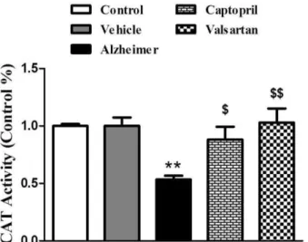
The activity of catalase (CAT)
Figure 4 denotes the quantitative CAT activity of brain tissue at the end of experiment (day 25). The activity of this enzyme significantly decreased by 47% in Alzheimer group compared with control and vehicle groups, (P<0.01). Captopril and valsartan significantly increased the activity of SOD in both treatment groups by 39% (P<0.05) and 48% (P<0.01), respectively, compared with Alzheimer group.
Figure 2. Test of memory function (Latency) at termination of the experiment. All values are presented as mean±SEM. *Significantly different from control and vehicle groups (P<0.05)
$
Significantly different from Alzheimer group (P<0.05)
$$
Significantly different from Alzheimer group (P<0.01)
Figure 3. Relative activity of superoxide dismutase (SOD) in the brain tissues at the end of experiment. The graph represents the relative changes quantified by normalization to the data of control rats. All values are Mean±SEM.
** Significantly different from control group (P<0.01)
$$$
Significantly different from Alzheimer group (P<0.001)
Parameter of oxidative stress (MDA)
RAS and Alzheimer’s disease
treatment groups (0.24±0.03 µmol/mg protein, P<0.05 and 0.17±0.01 µmol/mg protein, P<0.01, respectively).
Figure 4. Relative activity of catalase (CAT) in the brain tissues at the end of experiment. The graph represents the relative changes quantified by normalization to the data of control rats. All values are Mean±SEM.
** Significantly different from control group (P<0.01)
$
Significantly different from Alzheimer group (P<0.05)
$$
Significantly different from Alzheimer group (P<0.01)
Figure 5. The MDA content of brain (µmol/mg protein) at the end of experiment. All values are Mean±SEM.
* Significantly different from control group (P<0.05)
$
Significantly different from Alzheimer group (P<0.05)
$$
Significantly different from Alzheimer group (P<0.01)
Parameter of nitrosative stress (NOx)
Figure 6 shows the NOx content of brain tissue, as index of nitrosative stress, in different groups of experiment. Brain NOx level of control and vehicle groups was 0.43±0.06 and 0.57±0.09 µmol/mg protein, respectively. The content of NOx significantly increased in Alzheimer group in comparison to control and vehicle groups (0.86±0.16 µmol/mg protein, P<0.05). Captopril and valsartan significantly decreased the content of brain NOx in both treatment groups
(0.46±0.03 and 0.41±0.02 µmol/mg protein, respectively, P<0.05).
Figure 6. The NOx content of brain (µmol/mg protein) at the end of experiment. All values are Mean±SEM.
* Significantly different from control group (P<0.05)
$
Significantly different from Alzheimer group (P<0.05)
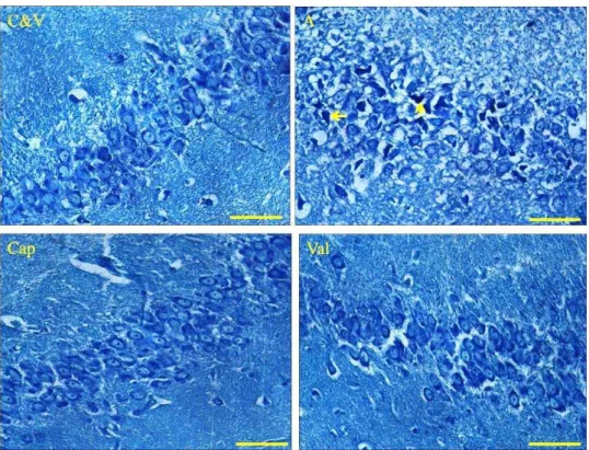
Histopathological assessment (TB staining)
Figure 7 shows the cells of hippocampus in CA1 region. The cells of CA1 area in control and vehicle groups are in normal states because the cells lined up regularly and the nuclei of cells are observed clearly in normal situation. The nuclei of cells in Alzheimer group are arranged disorderly and the numbers of intact neurons are decreased relative to control and vehicle. In addition, necrotic and apoptotic cells (pyknotic and shrinkage of nuclei), increased pericellular spaces of neuronal tissue were clearly observed during Alzheimer. In qualitative assessment, the level of neuronal damages considerably decreased in both treatment groups.
Discussion
Arjmand Abbassi et al.
activities (Figures 3 and 4). Furthermore, histological evaluations revealed the neuroprotective roles of
captopril and valsartan against the STZ-induced neurodegeneration (Figure 7).
Figure 7. Photographs show the histological changes (TB staining) of the cells of CA1 regions in the hippocampus of control (C), vehicle (V), Alzheimer (A) and Alzheimer-treated (Cap and Val) groups at the termination of experiment. Valsartan (Val) and captopril (Cap) considerably reduced the number of damaged neurons (necrotic and apoptotic neurons) in treated groups. (400X-scal bar 50 µm).
Memory dysfunction and cognitive deficiency are the common symptoms of AD that are deteriorated during the years.3 In the present study, induction of AD by i.c.v. injection of STZ generated cognitive and memory dysfunction like AD (Figure 2). It has been demonstrated that the antihypertensive drugs may have beneficial effects on cognitive functions in AD patients.18 According to epidemiological assessment using RAS inhibitors in hypertensive patients, which have cognitive deficit and dementia, has improved the cognitive dysfunctions.7 Moreover, administration of telmisartan was able to improve learning and memory functions of rodents, which had been damaged during AD.15 These findings confirm the role of brain RAS in pathogenesis of AD. Based on experimental evidence, RAS is one of the most important systems that are intensively activated in the brain of Alzheimer’s people.20,24 Therefore, brain RAS inhibition is expected to have potential therapeutic effect in treatment of AD. The findings of present study showed that using captopril and valsartan during the experiment has improved the cognitive and memory dysfunctions in STZ-induced dementia in rats (Figure 2). These findings indicate that antihypertensive drugs (ACE and AT1 receptor inhibitors) are useful for improvement of cognitive and memory functions in AD, which are clinically used as the antihypertensive agents. It is appeared that these beneficial effects of these drugs be
RAS and Alzheimer’s disease
plaques and neurofibrillary tangles.14,41 These subjects exhibit a significant oxidative imbalance.8,9,14 The findings of present study indicated that using captopril and valsartan during AD has attenuated the free radicals production of ROS and RNS, (Figures 5 and 6). Based on previous studies, activation of AT1 receptor is the most important origin of ROS/RNS production in different states of brain pathophysiology.15,20,21,42 Therefore, inhibition of brain RAS can be a probable pathway that production of ROS or RNS has been attenuated in treated rats by RAS inhibitors. According to previous findings, ACE inhibitors like captopril directly act as a scavenger of free radicals.43,44 Additionally, inhibition of RAS may diminish the activation of NADPH-oxidase, which is a key enzyme in production of oxygen free radicals.20 Since using captopril and valsartan in our study has enhanced the enzyme activities of SOD and CAT in both treated rats (Figures 3 and 4), it is concluded that these inhibitors of RAS has potentiated the brain antioxidant system during the STZ-induced dementia. Previous findings have also shown that ACE inhibition is able to improve the cellular antioxidant defense system in other pathological states.45 Therefore according to our findings, enhancement of SOD and CAT activities is another mechanism that RAS inhibition by captopril and valsartan results in reduction of brain free radicals and neuronal damage during STZ-induced dementia. AD is characterized by structural and histopathological alterations in the areas that are associated with learning and memory functions.2,4 These histopathological changes can be obviously seen in other model of dementia such as i.c.v. STZ-induced dementia.26,46 In histopathological evaluations based on TB staining, we showed the extensive damage to cells of hippocampus in Alzheimer group (Figure 7). Necrotic, apoptotic and pre-apoptotic neurons was clearly observed in these areas of brain. It has been demonstrated that oxidative and nitrosative stress could precede the pronounced AD neuro-pathological alterations.9,39,47 Enhanced overall protein peroxidation, as well as oxidative modification of specific proteins, has been also found in hippocampus and superior and middle temporal gyri from MCI and Alzheimer subjects.4,11,47 These facts strongly suggest that oxidative stress plays an important role in the pathogenesis of AD and progression of this disease. In the present study inhibition of brain RAS (using captopril and valsartan) considerably reduced the histopathological alterations of brain concomitant reduction of oxidative/nitrosative damage (Figure 7). Also in the study of Singh et al, administration of lisinopril (an ACE inhibitor) and telmisartan (an AT1 blocker) arrested the STZ-induced (i.c.v. injection) histopathological changes.15 Accordingly, activation of RAS may lead to neural destructions in AD, so that by inhibition of this system we can see outstanding effects on brain tissue damage and prevention of AD progression.
The findings of present study indicate that inhibition of brain RAS improves memory dysfunction and cognitive deficit during AD possibly through attenuation of oxidative and nitrosative damage. Our data also indicate that RAS inhibition by using an inhibitor of ACE (captopril) and specific inhibitor of AT1 receptor
(valsartan) potentiates the brain’s antioxidant defense
system and reduces the neuronal damage. Therefore, it is concluded that brain RAS inhibition using captopril and valsartan can be as the effective agents to reduce the brain damages in AD.
Acknowledgments
The authors are cordially appreciating the department of Physiology and Biophysics (Faculty of Medicine) and the financial support of Vice Chancellor for the University of Baqiyatallah Medical Sciences, Iran. The results described in this paper were part of M.Sc thesis.
Ethical Issues Not applicable.
Conflict of Interest
The authors declare no conflict of interests.
References
1. De-Paula VJ, Radanovic M, Diniz BS, Forlenza OV. Alzheimer's disease. Subcell Biochem
2012;65:329-52. doi: 10.1007/978-94-007-5416-4_14
2. Price JL, Ko AI, Wade MJ, Tsou SK, McKeel DW, Morris JC. Neuron number in the entorhinal cortex and CA1 in preclinical Alzheimer disease. Arch
Neurol 2001;58(9):1395-402. doi:
10.1001/archneur.58.9.1395
3. Solomon A, Mangialasche F, Richard E, Andrieu S, Bennett DA, Breteler M, et al. Advances in the prevention of Alzheimer's disease and dementia. J
Intern Med 2014;275(3):229-50. doi:
10.1111/joim.12178
4. Serrano-Pozo A, Frosch MP, Masliah E, Hyman BT. Neuropathological alterations in Alzheimer disease. Cold Spring Harb Perspect Med
2011;1(1):a006189. doi:
10.1101/cshperspect.a006189
5. Yagi S, Akaike M, Ise T, Ueda Y, Iwase T, Sata M. Renin-angiotensin-aldosterone system has a pivotal role in cognitive impairment. Hypertens Res
2013;36(9):753-8. doi: 10.1038/hr.2013.51
6. Wright JW, Kawas LH, Harding JW. A Role for the Brain RAS in Alzheimer's and Parkinson's Diseases.
Front Endocrinol (Lausanne) 2013;4:158. doi:
10.3389/fendo.2013.00158
Arjmand Abbassi et al.
8. Padurariu M, Ciobica A, Hritcu L, Stoica B, Bild W, Stefanescu C. Changes of some oxidative stress markers in the serum of patients with mild cognitive impairment and Alzheimer's disease. Neurosci Lett
2010;469(1):6-10. doi: 10.1016/j.neulet.2009.11.033 9. Lopez N, Tormo C, De Blas I, Llinares I, Alom J. Oxidative stress in Alzheimer's disease and mild cognitive impairment with high sensitivity and specificity. J Alzheimers Dis 2013;33(3):823-9. doi: 10.3233/jad-2012-121528
10. Cetin F, Yazihan N, Dincer S, Akbulut G. The effect of intracerebroventricular injection of beta amyloid peptide (1-42) on caspase-3 activity, lipid peroxidation, nitric oxide and NOS expression in young adult and aged rat brain. Turk Neurosurg
2013;23(2):144-50. doi: 10.5137/1019-5149.jtn.5855-12.1
11. Diaz A, Mendieta L, Zenteno E, Guevara J, Limon ID. The role of NOS in the impairment of spatial memory and damaged neurons in rats injected with amyloid beta 25-35 into the temporal cortex.
Pharmacol Biochem Behav 2011;98(1):67-75. doi:
10.1016/j.pbb.2010.12.005
12. Valko M, Leibfritz D, Moncol J, Cronin MT, Mazur M, Telser J. Free radicals and antioxidants in normal physiological functions and human disease. Int J
Biochem Cell Biol 2007;39(1):44-84. doi:
10.1016/j.biocel.2006.07.001
13. Vina J, Lloret A, Giraldo E, Badia MC, Alonso MD. Antioxidant pathways in Alzheimer's disease: possibilities of intervention. Curr Pharm Des
2011;17(35):3861-4. doi:
10.2174/138161211798357755
14. Wang X, Wang W, Li L, Perry G, Lee HG, Zhu X. Oxidative stress and mitochondrial dysfunction in Alzheimer's disease. Biochim Biophys Acta
2014;1842(8):1240-7. doi:
10.1016/j.bbadis.2013.10.015
15. Singh B, Sharma B, Jaggi AS, Singh N. Attenuating effect of lisinopril and telmisartan in intracerebroventricular streptozotocin induced experimental dementia of Alzheimer's disease type: possible involvement of PPAR-gamma agonistic property. J Renin Angiotensin Aldosterone Syst
2013;14(2):124-36. doi:
10.1177/1470320312459977
16. Ohshima K, Mogi M, Horiuchi M. Therapeutic approach for neuronal disease by regulating renin-angiotensin system. Curr Hypertens Rev
2013;9(2):99-107. doi:
10.2174/15734021113099990004
17. Kugaevskaia EV. Angiotensin converting enzyme and Alzheimer's disease. Biomed Khim
2013;59(1):5-24.
18. Ashby EL, Kehoe PG. Current status of renin-aldosterone angiotensin system-targeting anti-hypertensive drugs as therapeutic options for Alzheimer's disease. Expert Opin Investig Drugs
2013;22(10):1229-42. doi:
10.1517/13543784.2013.812631
19. O'Caoimh R, Kehoe PG, Molloy DW. Renin Angiotensin aldosterone system inhibition in controlling dementia-related cognitive decline. J
Alzheimers Dis 2014;42 Suppl 4:S575-86. doi:
10.3233/jad-141284
20. Awasthi H, Kaushal D, Siddiqui HH. Chronic inhibition of central Angiotensin-converting enzyme ameliorates colchicine-induced memory impairment in mice. Sci Pharm 2012;80(3):647-62. doi: 10.3797/scipharm.1203-06
21. Ciobica A, Bild W, Hritcu L, Haulica I. Brain renin-angiotensin system in cognitive function: pre-clinical findings and implications for prevention and treatment of dementia. Acta Neurol Belg
2009;109(3):171-80.
22. Mogi M, Li JM, Tsukuda K, Iwanami J, Min LJ, Sakata A, et al. Telmisartan prevented cognitive decline partly due to PPAR-gamma activation.
Biochem Biophys Res Commun 2008;375(3):446-9.
doi:10.1016/j.bbrc.2008.08.032
23. Villapol S, Saavedra JM. Neuroprotective Effects of Angiotensin Receptor Blockers. Am J Hypertens
2015;28(3):289-99. doi: 10.1093/ajh/hpu197 24. AbdAlla S, Langer A, Fu X, Quitterer U. ACE
inhibition with captopril retards the development of signs of neurodegeneration in an animal model of Alzheimer's disease. Int J Mol Sci
2013;14(8):16917-42. doi: 10.3390/ijms140816917 25. Inaba S, Iwai M, Furuno M, Tomono Y, Kanno H,
Senba I, et al. Continuous activation of renin-angiotensin system impairs cognitive function in renin/angiotensinogen transgenic mice.
Hypertension 2009;53(2):356-62. doi:
10.1161/hypertensionaha.108.123612
26. Santos TO, Mazucanti CH, Xavier GF, Torrao AS. Early and late neurodegeneration and memory disruption after intracerebroventricular streptozotocin. Physiol Behav 2012;107(3):401-13. doi: 10.1016/j.physbeh.2012.06.019
27. Tota S, Kamat PK, Saxena G, Hanif K, Najmi AK, Nath C. Central angiotensin converting enzyme
facilitates memory impairment in
intracerebroventricular streptozotocin treated rats.
Behav Brain Res 2012;226(1):317-30. doi:
10.1016/j.bbr.2011.07.047
28. Wang J, Ho L, Chen L, Zhao Z, Zhao W, Qian X, et al. Valsartan lowers brain beta-amyloid protein levels and improves spatial learning in a mouse model of Alzheimer disease. J Clin Invest
2007;117(11):3393-402. doi: 10.1172/jci31547 29. Kumaran D, Udayabanu M, Kumar M, Aneja R,
RAS and Alzheimer’s disease
30. Winterbourn CC, Hawkins RE, Brian M, Carrell RW. The estimation of red cell superoxide dismutase activity. J Lab Clin Med 1975;85(2):337-41.
31. Aebi H. Catalase in vitro. Methods Enzymol
1984;105:121-6. doi:
10.1016/s0076-6879(84)05016-3
32. Satoh K. Serum lipid peroxide in cerebrovascular disorders determined by a new colorimetric method.
Clin Chim Acta 1978;90(1):37-43. doi:
10.1016/0009-8981(78)90081-5
33. Riahi S, Mohammadi MT, Sobhani V, Ababzadeh S. Chronic Aerobic Exercise Decreases Lectin-Like Low Density Lipoprotein (LOX-1) Receptor Expression in Heart of Diabetic Rat. Iran Biomed J
2016;20(1):26-32.
34. Bradford MM. A rapid and sensitive method for the quantitation of microgram quantities of protein utilizing the principle of protein-dye binding. Anal
Biochem 1976;72(1-2):248-54. doi:
10.1016/0003-2697(76)90527-3
35. Wright JW, Harding JW. The brain RAS and Alzheimer's disease. Exp Neurol 2010;223(2):326-33. doi: 10.1016/j.expneurol.2009.09.012
36. Wright JW, Harding JW. The brain renin-angiotensin system: a diversity of functions and implications for CNS diseases. Pflugers Arch
2013;465(1):133-51. doi: 10.1007/s00424-012-1102-2
37. Fogari R, Zoppi A. A drug safety evaluation of valsartan. Expert Opin Drug Saf 2011;10(2):295-303. doi: 10.1517/14740338.2011.543416
39. Groel JT, Tadros SS, Dreslinski GR, Jenkins AC. Long-term antihypertensive therapy with captopril.
Hypertension 1983;5(5 Pt 2):III145-51. doi:
10.1161/01.hyp.5.5_pt_2.iii145
39. Dumont M, Beal MF. Neuroprotective strategies involving ROS in Alzheimer disease. Free Radic
Biol Med 2011;51(5):1014-26. doi:
10.1016/j.freeradbiomed.2010.11.026
40. Hernandes MS, Britto LR. NADPH oxidase and neurodegeneration. Curr Neuropharmacol
2012;10(4):321-7. doi:
10.2174/157015912804143540
41.Markesbery WR, Schmitt FA, Kryscio RJ, Davis DG, Smith CD, Wekstein DR. Neuropathologic substrate of mild cognitive impairment. Arch Neurol
2006;63(1):38-46. doi: 10.1001/archneur.63.1.38 42. Garrido AM, Griendling KK. NADPH oxidases and
angiotensin II receptor signaling. Mol Cell
Endocrinol 2009;302(2):148-58. doi:
10.1016/j.mce.2008.11.003
43. Lopez-Real A, Rey P, Soto-Otero R, Mendez-Alvarez E, Labandeira-Garcia JL. Angiotensin-converting enzyme inhibition reduces oxidative stress and protects dopaminergic neurons in a 6-hydroxydopamine rat model of Parkinsonism. J
Neurosci Res 2005;81(6):865-73. doi:
10.1002/jnr.20598
44. Gurer H, Neal R, Yang P, Oztezcan S, Ercal N. Captopril as an antioxidant in lead-exposed Fischer 344 rats. HumExp Toxicol 1999;18(1):27-32. 45. de Cavanagh EM, Fraga CG, Ferder L, Inserra F.
Enalapril and captopril enhance antioxidant defenses in mouse tissues. Am J Physiol 1997;272(2 Pt 2):R514-8.
46. Chen Y, Liang Z, Blanchard J, Dai CL, Sun S, Lee MH, et al. A non-transgenic mouse model (icv-STZ mouse) of Alzheimer's disease: similarities to and differences from the transgenic model (3xTg-AD mouse). Mol Neurobiol 2013;47(2):711-25. doi: 10.1007/s12035-012-8375-5
47. Sodhi RK, Singh N. All-trans retinoic acid rescues memory deficits and neuropathological changes in mouse model of streptozotocin-induced dementia of Alzheimer's type. Prog Neuropsychopharmacol Biol
Psychiatry 2013;40:38-46. doi: