Determining the Functions of HIV-1 Tat and
a Second Magnesium Ion in the CDK9/Cyclin
T1 Complex: A Molecular Dynamics
Simulation Study
Hai-Xiao Jin1, Mei-Lin Go2, Peng Yin3, Xiao-Ting Qiu1, Peng Zhu1, Xiao-Jun Yan1*
1Key Laboratory of Applied Marine Biotechnology Ministry of Education, School of Marine Sciences, Ningbo University, Ningbo, China,2Department of Pharmacy, National University of Singapore, Singapore, Singapore,3Key Laboratory of Chemical Biology and Traditional Chinese Medicine Research (Ministry of Education), College of Chemistry and Chemical Engineering, Hunan Normal University, Changsha, China
Abstract
The current paradigm of cyclin-dependent kinase (CDK) regulation based on the well-estab-lished CDK2 has been recently expanded. The determination of CDK9 crystal structures suggests the requirement of an additional regulatory protein, such as human immunodefi-ciency virus type 1 (HIV-1) Tat, to exert its physiological functions. In most kinases, the exact number and roles of the cofactor metal ions remain unappreciated, and the repertoire has thus gained increasing attention recently. Here, molecular dynamics (MD) simulations were implemented on CDK9 to explore the functional roles of HIV-1 Tat and the second Mg2
+
ion at site 1 (Mg12+). The simulations unveiled that binding of HIV-1 Tat to CDK9 not only
stabilized hydrogen bonds (H-bonds) between ATP and hinge residues Asp104 and Cys106, as well as between ATP and invariant Lys48, but also facilitated the salt bridge net-work pertaining to the phosphorylated Thr186 at the activation loop. By contrast, these H-bonds cannot be formed in CDK9 owing to the absence of HIV-1 Tat. MD simulations further revealed that the Mg12+ion, coupled with the Mg22+ion, anchored to the triphosphate
moie-ty of ATP in its catalytic competent conformation. This observation indicates the require-ment of the Mg12+ion for CDK9 to realize its function. Overall, the introduction of HIV-1 Tat
and Mg12+ion resulted in the active site architectural characteristics of phosphorylated
CDK9. These data highlighted the functional roles of HIV-1 Tat and Mg12+ion in the
regula-tion of CDK9 activity, which contributes an important complementary understanding of CDK molecular underpinnings.
Introduction
Cyclin-dependent kinase 9 (CDK9) is a Ser/Thr kinase that belongs to the family of cyclin-de-pendent kinases (CDKs). CDK9 serves as the catalytic subunit of the positive transcription OPEN ACCESS
Citation:Jin H-X, Go M-L, Yin P, Qiu X-T, Zhu P, Yan X-J (2015) Determining the Functions of HIV-1 Tat and a Second Magnesium Ion in the CDK9/Cyclin T1 Complex: A Molecular Dynamics Simulation Study. PLoS ONE 10(4): e0124673. doi:10.1371/journal. pone.0124673
Academic Editor:Fatah Kashanchi, George Mason University, UNITED STATES
Received:December 8, 2014
Accepted:March 16, 2015
Published:April 24, 2015
Copyright:© 2015 Jin et al. This is an open access article distributed under the terms of theCreative Commons Attribution License, which permits unrestricted use, distribution, and reproduction in any medium, provided the original author and source are credited.
Data Availability Statement:All relevant data are within the paper and its Supporting Information files.
elongation factor b (P-TEFb; CDK9/cyclin T), which phosphorylates the RNA polymerase II C-terminal domain and the negative elongation factors NELF and DRB (dichlorobenzimida-zole riboside)-sensitivity-inducing factor (DSIF) to trigger the elongation of many gene
tran-scripts [1]. P-TEFb has been an important therapeutic target in oncology, virology, and
cardiology [2,3]. A viral protein, human immunodeficiency virus type 1 (HIV-1) Tat, interacts
with P-TEFb and induces the factor to promote the productive elongation of HIV mRNA [4–
6]. Biochemical experiments have shown that Tat increased transcriptional elongation
per-formed by CDK9 [7].
Twenty CDK9 crystal structures have thus far been solved [8–18], and their availability
serves as a valuable resource for structure-aided drug design. CDK9 adopts a typical bilobal
fold (Fig 1A), which is extremely conserved among Ser/Thr and Tyr kinases. The N-terminal
lobe is composed of a five-stranded antiparallelβ-sheet and one prominentα-helix, i.e., the
helixαC (sequence PITALRE in CDK9 and PSTAIRE in CDK2). The larger C-terminal lobe is
mostly helical and connected to the N-terminal lobe by the so-called flexible hinge region
(resi-dues 104–107). ATP is sandwiched between the N- and C-terminal lobes and anchors its
ade-nine moiety by H bonds with Asp104 and Cys106 in the hinge region. Cyclin T has a canonical cyclin structure. The interface of CDK9/cyclin T is notably smaller than that of the CDK2/cy-clin A complex and is restricted to the N-terminal lobe of CDK9.
Although a few kinases are known to harness a single divalent ion or none at all[19], many,
if not all, protein kinases require two divalent metal ions for optimum catalysis [20]. In the
kinome, the exact number and roles of the cofactor metal ions remain unknown, and the reper-toires have recently gained increasing attention. Thus far, a few CDK9 crystal structures are
as-sociated with a single Mg2+ion at site 2 (Mg22+ion;Fig 1A and 1B), whereas the other Mg2+
ion at site 1 (Mg12+ion) is absent in all CDK9 crystal structures. The crystal structures of
CDK2 with two Mg2+ions had not been solved until recently, when the crystal structures of
CDK2/cyclin A transition state (TS) complex with two Mg2+ions were discovered by Yong
et al. [21,22]. Experimental observations have demonstrated that Mg2+concentration could
represent an important regulator of CDK2 activity in vivo [21]. These findings raise an
inter-esting question on whether CDK9 requires the binding of two Mg2+ions to realize its function,
as in the case of CDK2.
CDK2 is a phenotypic family of CDKs. A series of CDK2/cyclin A structures have provided significant insight into the molecular underpinnings of CDK activation and regulation. The need of CDKs to be associated with a cognate cyclin, followed by phosphorylation on threonine
at the activation loop to realize full activity, is well-documented [23,24]. However, the
generali-ty of this underlying mechanism has been challenged with the determination of new CDK crys-tal structures. For instance, the cryscrys-tal structures of CDK4/cyclin D revealed that cyclin D binding and activation loop phosphorylation do not adequately enable the CDK4 to adopt an
active conformation [24,25]. In fact, recent experiments underscored that association with
ad-ditional protein substrates and/or cofactor binding are critical to the remodeling of
CDK4/cy-clin D into its active conformation [25–27]. Additionally, the solved CDK9/cyclin T crystal
structures demonstrated some notable structural differences in the phosphorylated Thr186 (pThr186) at the activation loop. Although the phosphorylation of Thr186 is required for
CDK9 activation [28], pThr186 in CDK9/cyclin T1 complex crystal structures only forms salt
bridge interactions with Arg148 and Arg172. This condition is unlike that the CDK2/cyclin A complex, in which pThr160 forms salt bridge interactions with the positively charged triad of arginine residues (Arg50, Arg126, and Arg150). pThr186 is slightly distant (3.9 Å) from the
third arginine residue (Arg65), which is located in theαC helix (Fig 1A) [8]. Interestingly, as
shown inFig 1B, the crystal structure of CDK9/cyclin T1 in a complex with a minimal
activa-tion domain (residues 1–48) of HIV-1 Tat formed five salt bridges with the arginine triad [9].
HIV-1 Tat, a two-zinc-mediated viral transactivator of transcription, inserts itself into a groove at the heterodimer interface, thus augmenting interactions and resulting in a more stable
P-TEFb complex. The reestablished Arg65–pThr186 ion pair results in a more stable local
con-formation and contributes to maintaining strong contact between the critical activation loop
and the prominentαC helix motif. The current understanding of kinase regulation owes much
to X-ray crystallography [29]. In this study, we performed 50 ns explicit-solvent MD
simula-tions on five different systems to determine the influence of HIV-1 Tat binding, activation loop
phosphorylation, and the presence of Mg12+at site 1 on CDK9 dynamics. Our results revealed
elaborate and significant differences in the dynamics behavior of CDK9, which provides insight into the current understanding of CDK regulation and may contribute to structure-based drug design.
Materials and Methods
Initial Complexes Preparation
The crystal structure of phosphorylated CDK9/cyclin T1 complex bound to ATP and one Mg2
+ion at site 2 (Mg22+ion) (pCDK9/cyclin T1/ATP/1MG complex) was obtained from the
RCSB Protein Data Bank (PDB code 3BLQ) [8] (Fig 1A). The first four simulation systems
were based on this crystal structure. The missing Mg2+ion at site 1 (Mg12+ion) was modeled
into its position by aligning the crystal structure of GSK3βin the complex with ATP analog
adenosine 50-(β,γ-imidotriphosphate) (AMP-PNP) and two Mg2+ions (PDB code 1PYX) [30]
with the pCDK9/cyclin T1/ATP/1MG complex crystal structure. The Mg12+ion is and
subse-quently extracted to model the pCDK9/cyclin T1/ATP/2MG complex. The pThr186 at the ac-tive loop of the two complexes was replaced with nonphosphorylated Thr186 to construct the CDK9/cyclin T1/ATP/1MG and CDK9/cyclin T1/ATP/2MG complexes. The fifth simulated complex was based on another crystal structure of pCDK9/cyclin T1 in the complex with
AMP-PNP and Mg22+ion and the minimal activation domain (residues 1–48) of HIV-1 Tat
(pCDK9/cyclin T1/AMP-PNP/1MG/Tat complex; PDB code 3MIA) [9] (Fig 1B). The missing
Mg12+ion in the X-ray structure results in the lack of a coordination bond between the ATP Oγ
atom and Mg12+ion, which causes theγ-phosphate of AMP-PNP to assume an unusual
con-formation. Therefore, the crystal structure of the GSK3β/AMP-PNP/2MG complex was also
aligned with the crystal structure of the pCDK9/cyclin T1/AMP-PNP/1MG/Tat complex to
ex-tract both AMP-PNP and Mg12+ion coordinates into the pCDK9 ATP binding pocket. The
imido nitrogen atom was subsequently replaced with the oxygen atom, thus generating the pCDK2/cyclin T1/ATP/2MG/Tat complex. The missing residues in the two CDK9 X-ray
struc-tures were completed using Molecular Operating Environment [31] and further minimized by
AMBER 11 [32] in the subsequent procedure. The detailed compositions of the five initial
com-plexes are summarized inTable 1.
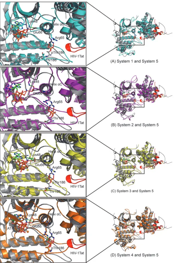
Fig 1. Architecture of CDK9/cyclin T1.(A) Ribbon representation of the overall crystal structure of pCDK9/ cyclin T1 bound to ATP and one Mg2+ion at site 2 (Mg
22+ion) complex (PDB code 3BLQ). (B) Ribbon
representation of the overall crystal structure of pCDK9/cyclin T1 bound to HIV-1 Tat and AMP-PNP and one Mg22+ion (PDB code 3MIA). CDK9 is purple blue, cyclin T1 is deep teal, and HIV-1 Tat is red. ATP,
AMP-PNP, and pThr186 are drawn as sticks, and Zn2+ions and Mg2+ions are drawn as yellow and green
spheres, respectively. The salt bridge network formed by pThr186 is shown as a red dotted line. (C) Structure of ATP with labeled oxygen and nitrogen atoms used in this article.
Force Field
The AMBER force field (ff99SB) [33] was applied to the CDK9 protein, cyclin T1 protein,
HIV-1 Tat protein, and Mg2+ions. Glu/Asp residues were deprotonated and Lys/Arg residues
were protonated at a simulated pH of 7. The protonated states for the His residues were as-signed according to the PROPKA calculation, with the exception of the involvement of those in the coordination with the zinc ion [His33 in the HIV-1 Tat coordinated to the zinc ion was
modeled as a negative charge state (−1 charge)]. We adopted the cationic dummy atom
ap-proach introduced by Pang et al. [34,35] to describe the zinc divalent cation. This approach
places four cationic dummy atoms in a tetrahedral arrangement in the zinc nucleus to mimic
the 4s4p3vacant orbitals of the zinc ion. Thus, the lone-pair electrons of the zinc coordinates
occupy the vacant orbitals and fulfill the orientation requirements for the tetrahedral coordina-tion geometry of zinc. The zinc nucleus was assigned with only van der Waals parameters (van
der Waalsr= 3.1 Å, van der Waals potential well depthε= 1E-6 kcal/mol, and chargeq= 0),
whereas the dummy atom was assigned only with charge (r= 0,ε= 0, andq= 0.5e). The force
field parameters for the -2 charged pThr186 and the -4 charged ATP were taken from the
AMBER parameter database [36,37].
MD Simulations
The hydrogen atoms and the missing atoms were added to the Leap module of AMBER 11
[32]. Each system was immersed in the truncated octahedron box of TIP3P [38] water
mole-cules with a 10 Å buffer in each direction. An appropriate number of Cl-counterions were then
added through the random substitution of solvent water molecules with Cl-ions at the most
fa-vorable electrostatic potential positions to maintain the electroneutrality of the five systems. Thus, the total number of atoms for Systems 1, 2, 3, 4, and 5 were 68110, 68297, 68308, 68309, and 93339, respectively. Energy minimizations and MD simulations were conducted using the SANDER module of AMBER 11 with periodic boundary conditions. Prior to the production run, each system was optimized by using three steps minimization. First, ATP, metal ions, and protein residues were fixed with harmonic force restraint, and only the positions of the water molecules were minimized. Second, ATP, metal ions, and protein residues from crystal struc-tures were constrained, whereas the added missing residues and water molecules were mini-mized. Finally, the whole system was allowed to fully relax. In each step, energy minimization was performed using the steepest descent method for the first 2500 steps and the conjugated gradient method for the next 2500 steps. Each system was then gradually raised from 0 K to 300 K in a 50 ps canonical ensemble (NVT) heating process. Finally, 50 ns production MD simulations were performed on the five systems in an isothermal isobaric ensemble (NPT) at a constant pressure (1 atm) and constant temperature (300 K) by applying the Langevin
algo-rithm [39]. A cutoff equivalent to 10 Å was set for short-range electrostatics and van der Waals
interactions. Long-range electrostatic interactions were processed using the particle mesh
Ewald method [40] with cubic fourth-order B-spline interpolation and a 10–5tolerance set for
Table 1. The compositions of the five simulations.
Complex System code Phosphorylaton state Composition
CDK9/cyclin T1/ATP/1Mg 1 Thr186 Dephosphorylated CDK9, cyclin T1, ATP, Mg22+
pCDK9/cyclin T1/ATP/1Mg 2 pThr186 Phosphorylated CDK9, cyclin T1, ATP, Mg22+
CDK9/cyclin T1/ATP/2Mg 3 Thr186 Dephosphorylated CDK9, cyclin T1, ATP, Mg12+, Mg22+
pCDK9/cyclin T1/ATP/2Mg 4 pThr186 Phosphorylated CDK9, cyclin T1, ATP, Mg12+, Mg22+
pCDK9/cyclin T1/ATP/2Mg/Tat 5 pThr186 Phosphorylated CDK9, cyclin T1, ATP, Mg12+, Mg22+, HIV-1 Tat
the direct sum tolerance. An integration step of 2 fs was set for the MD simulations. All cova-lent bonds involving hydrogen atoms were constrained at their equilibrium positions by the
SHAKE method [41] with a tolerance of 10–5Å.
All the MD trajectories were subsequently analyzed using PTRAJ module. The 1 ps interval saved coordinates were used to obtain the root-mean-square deviations (RMSDs), to calculate the change in distance between two atoms, and to analyze H bonds. The criteria for forming an H bond consists of an angle A-H-D larger than 120° and a distance between the acceptor atom and the donor atom smaller than 3.5 Å.
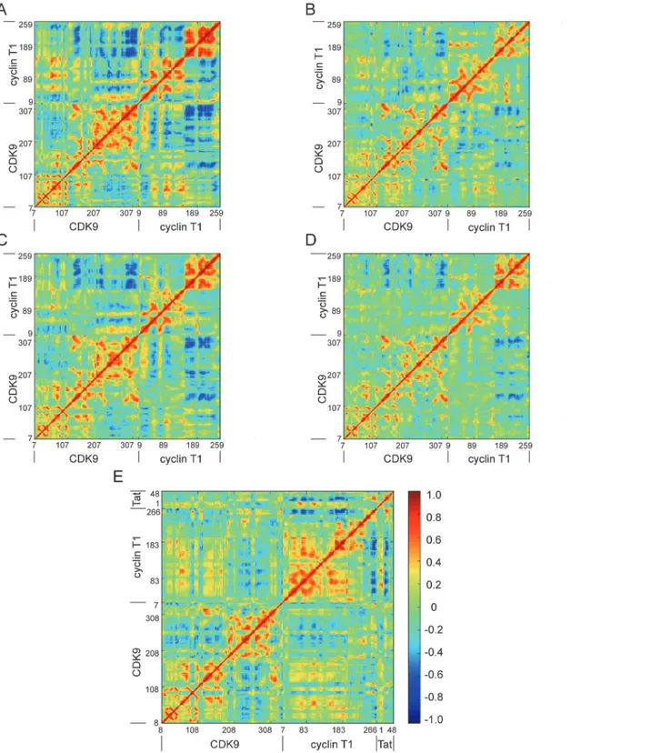
Dynamic Cross-correlation Matrices
The Cαdynamic cross-correlation matrices (DCCM) were computed to reveal the correlative
motions of proteins [42].C(i,j) was calculated as follows:
Cði;jÞ ¼ cði;jÞ cði;iÞ1=2
cðj;jÞ1=2
whereC(i,j) is the covariance matrix element of the proteinfluctuation between residuesiand
j.
The value ofC(i,j) ranges from -1 to 1. Positive values suggest positively correlated
move-ment (the same direction), whereas negative values suggest anticorrelated movemove-ment (the op-posite direction).
Results
System Stabilities during MD Simulations
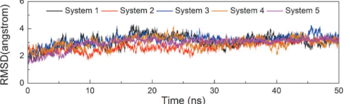
Conventional MD simulations of five systems, CDK9/cyclin T1/ATP/1MG complex (System 1), pCDK9/cyclin T1/ATP/1MG complex (System 2), CDK9/cyclin T1/ATP/2MG complex (System 3), pCDK9/cyclin T1/ATP/2MG complex (System 4), and pCDK9/cyclin T1/ATP/
2MG/Tat complex (System 5), were performed in explicit water for 50 ns. The Cαatom
RMSDs of the CDK9/cyclin T1 complexes in relation to the initial minimized structures as a
function of simulation time for five systems were monitored. As shown inFig 2, after
approxi-mately 10 ns of simulation, the RMSDs tended to converge in Systems 1, 2, 3, 4, and 5 with val-ues of 3.07 ± 0.37 Å, 2.71 ± 0.30 Å, 3.18 ± 0.25 Å, 2.95 ± 0.31 Å, and 2.94 ± 0.30 Å, respectively. These values, along with the time dependence of total energies (data not shown), indicate that the five systems achieved a state of equilibrium and were sufficient for exploring the dynamic behavior of the studied systems.
Fig 2. Time dependence of Cαatoms RMSDs of CDK9/cyclin T1 for five simulations in the 50 ns MD simulations.Systems 1, 2, 3, 4, and 5 are shown in black, red, blue, orange, and magenta, respectively. The same colors are maintained in the following Figs.
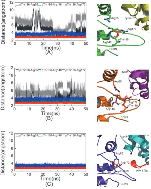
Salt Bridge Network in the pThr186 Binding Site
Thr186 at the active loop is phosphorylated in three constructed simulation systems (Systems
2, 4, and 5). A cluster of three arginine residues, including Arg65 at theαC helix, Arg148 at the
catalytic loop, and Arg172 at the activation loop, constitutes the pThr186 binding site. System 5 contains HIV-1 Tat and features two salt bridges that were formed by Arg65 and Ary148 and one salt bridge that was formed by Arg172. Negatively charged pThr186 served as the hub to organize the positively charged triad. All five salt bridges were very stable throughout the
simu-lation time (Fig 3C). By contrast, in Systems 2 and 4 without HIV-1 Tat binding, the resulting
salt bridge networks were less stable. For example, in System 4, only one salt bridge was formed Fig 3. Distances between phosphate group of pThr186 and guanidine group of arginine triad versus simulation time.The distance between pThr186 and Arg65 are shown in black and gray, the distance between pThr186 and Arg148 are shown in dark cyan and blue, and the distance between pThr186 and Arg172 are represented as red in System 2 (A), System 4 (B), and System 5 (C). The salt bridges in the pThr186 binding site at the 50 ns snapshot in three corresponding systems are shown in the right panel. Residues involved in salt bridge formation (red dotted line) are described by the stick with a red oxygen atom, blue nitrogen atom, and orange phosphorus atom.
by Arg65, and it was less stable than that in System 5 (Fig 3B). In the System 2, the salt bridge
formed between pThr186 and Arg65 was weakest (Fig 3A). Taken together, these data indicate
that both Mg12+ion and HIV-1 Tat contributed to the stability of the salt bridge network
formed by pThr186.
Hydrogen Bonds in the ATP Binding Pocket
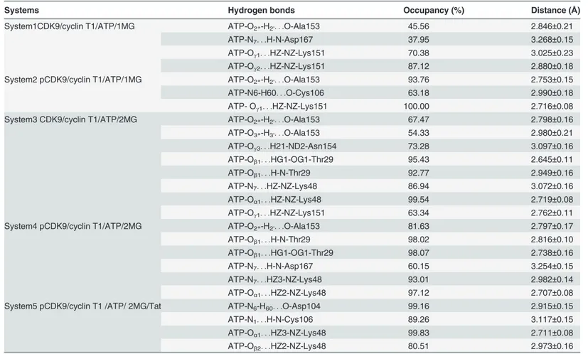
The hydrogen bonds formed by ATP with CDK9 are shown inFig 4and tabulated inTable 2.
Significant changes in ATP binding mode and ATP conformation were observed during simu-lation. System 5 showed that H bonds were formed between Asp104 backbone carbonyl O
atom and ATP N6atom, as well as between Cys106 backbone amide N atom and ATP N1
atom. As shown inFig 5, these two H bonds were very stable in System 5 throughout
simula-tion process. By contrast, the same H bonds were all broken in the other four systems. The breakage of these two H bonds resulted in the displacement of the adenine moiety of ATP from its original position. The occupancy values (%) of H bonds formed between the Asp104
car-bonyl O atom and the ATP N6atom were 2.01, 5.99, 3.19, and 5.30 in Systems 1, 2, 3, and 4,
re-spectively, whereas those between the Cys106 amide N atom and the ATP N1atom were 1.67,
5.40, 1.99, and 4.17, respectively. As shown inFig 5, the rupture of the two H bonds between
the adenine moiety of ATP and the hinge residues of CDK9 occurred at approximately 1 ns in nonphosphorylated Systems 1 and 3, and at approximately 2.7 ns in phosphorylated Systems 2 and 4. These observations suggest that the HIV-1 Tat is critical to the stabilization of H bonds between the adenine moiety of ATP and the hinge residues Asp104 and Cys106 of CDK9. In addition, another significant observation was related to the hydrogen bonding property of
Lys48. In System 5, the Lys48 side chain Nzatom was within the hydrogen bonding distance of
both the ATP Oα1and Oβ2atoms. However, in the systems occupied by one Mg2+(Systems 1
and 2), Lys48 was incapable of forming H bonds with ATP, whereas in the systems occupied
by two Mg2+ions (Systems 3 and 4), Lys48 formed one H bond with the ATP N7atom and one
H bond with the ATP Oα1atom. The pivotal H bond between ATP and invariant Lys48
(ATP-Oβ2. . .HZ-NZ-Lys48), which is crucial to the enzymatic catalysis reaction, was broken in
all no HIV-1 Tat binding simulations (Table 2). This critical H bond causes the triphosphate
moiety of ATP to assume the correct orientation during simulations. Although Lys48 was
inca-pable of positioning its side chain in its optimal orientation in the two Mg2+occupied systems
(Systems 3 and 4), the presence of Mg12+ion contributed to the arrangement of Lys48 to obtain
one correct H bond, that is, ATP-Oα1. . .HZ-NZ-Lys48 interaction.
ATP Conformation in the ATP Binding Pocket
Apart from the differences in the formation of H bonds between ATP and CDK9 in various
systems, the simulations also indicated that the binding of both the Mg12+ion and HIV-1 Tat
significantly reduced the magnitude of nanosecond timescale fluctuations in ATP phosphates. The heavy atom RMSDs of ATP triphosphate moiety in the five systems in relation to its con-formation in the initial minimized structure were monitored and calculated to be 0.89 ± 0.09 Å, 0.98 ± 0.04 Å, 0.72 ± 0.05 Å, 0.62 ± 0.04 Å, and 0.56 ± 0.06 Å for Systems 1, 2, 3, 4, and 5,
re-spectively (Fig 6A). The average RMSD values in the first two systems with one Mg2+ion
bind-ing were higher than those in the remainbind-ing three systems with the bindbind-ing of two Mg2+ions,
which is consistent with the observed reorientation of the triphosphate moiety in Systems
1 and 2. With the Mg12+ion at the binding pocket in Systems 3, 4, and 5, the additional
dihedral Oα3-Pβ-Oβ3-Pγand the distance between the Pαand Pγatoms in the five simulation
systems as a function of time (Fig 6B and 6C). The values of dihedral Oα3-Pβ-Oβ3-Pγin System
1 significantly fluctuated during simulation but were rather stable in the other four systems
(−98.56 ± 17.02°, 106.74 ± 7.86°, 91.84 ± 6.52°, and 97.21 ± 10.80° for Systems 2, 3, 4, and 5,
Fig 4. Hydrogen bonds and coordination bonds at the ATP binding pocket of CDK9 in five systems.
CDK9 is shown as a gray ribbon with a gray stick representing residues involved in hydrogen bond or coordination bond. ATP is depicted by a yellow stick. All oxygen atoms, nitrogen atoms, and phosphate atoms are depicted in red, blue, and orange, respectively. Mg12+and Mg22+ions are exhibited as green
spheres and water molecules are shown as red spheres. Red dotted lines indicate hydrogen bonds and blue dotted lines represent coordination bonds.
respectively). The distances between the Pαand Pγatoms were 3.78 ± 0.08 Å, 3.94 ± 0.07 Å,
3.99 ± 0.06 Å, 4.24 ± 0.06 Å, and 4.31 ± 0.06 Å in Systems 1, 2, 3, 4, and 5, respectively. In
Sys-tems 1 and 2, which lacked the Mg12+ion, the dihedral angle O
α3-Pβ-Oβ3-Pγand the distance
between the two phosphorus atoms (Pαand Pγ) were significantly different from those
ob-served in the binding systems with two Mg2+ions.
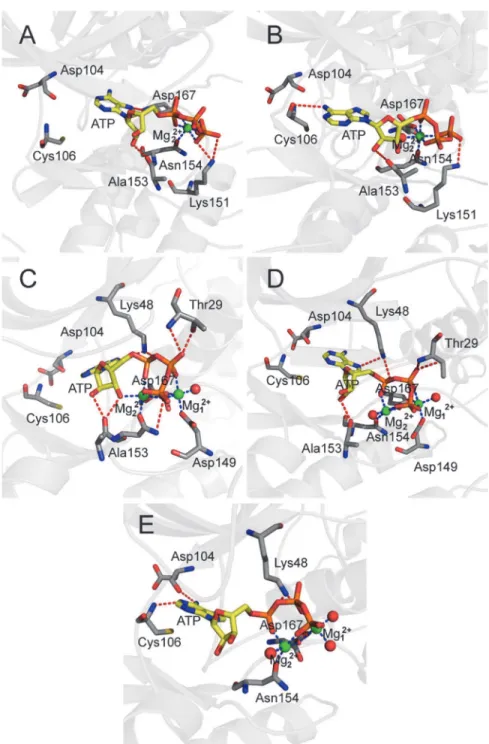
Magnesium
–
Ligand Coordination
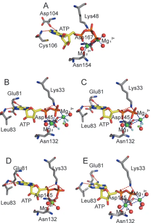
To analyze the magnesium–ligand coordination mode in our simulations, we compared our
simulation results with the X-ray crystal structures of CDK2. Yonget al. recently obtained
sev-eral ADP cocrystallized pCDK2/cyclin A structures [21,22]. Two of these structures comprise
pCDK2/cyclin A bound to ADP, substrate peptide, and trigonal-planar MgF3-ion, a mimic for
theγ-phosphate of ATP (PDB code 3QHR and 3QHW), which are very similar to the TS
com-plexes. Two crystal structures (Fig 7B and 7C) showed Mg12+ions that were coordinated by
one of the MgF3-fluorine atoms, an ADPβ-phosphate oxygen atom, two side chain carboxyl
oxygen atoms of Asp145 at the DFG motif, and oxygen atoms of two water molecules. Mg22+
ion also formed six coordination bonds with one of the MgF3-fluorine atoms, ADPα- andβ
-phosphate oxygen atoms, one carboxyl oxygen atom of Asp145, one side chain carbonyl
oxy-gen atom of Asn132, and one oxyoxy-gen atom of water molecule. Each Mg2+ion maintained a
hexa-coordinated octahedral geometry, which was also observed in the pCDK2/cyclin A
com-plex bound to ADP with one Mg2+ion (PDB code 4II5) or with two Mg2+ions (PDB code
Table 2. Summary of the average distances and occupancies of hydrogen bonds between ATP and CDK9 in five 50 ns simulated systems.
Systems Hydrogen bonds Occupancy (%) Distance (Å)
System1CDK9/cyclin T1/ATP/1MG ATP-O2*-H2'. . .O-Ala153 45.56 2.846±0.21
ATP-N7. . .H-N-Asp167 37.95 3.268±0.15
ATP-Oγ1. . .HZ-NZ-Lys151 70.38 3.025±0.23
ATP-Oγ2. . .HZ-NZ-Lys151 87.12 2.880±0.18
System2 pCDK9/cyclin T1/ATP/1MG ATP-O2*-H2'. . .O-Ala153 93.76 2.753±0.15
ATP-N6-H60. . .O-Cys106 63.18 2.990±0.18
ATP- Oγ1. . .HZ-NZ-Lys151 100.00 2.716±0.08
System3 CDK9/cyclin T1/ATP/2MG ATP-O2*-H2'. . .O-Ala153 67.47 2.798±0.16
ATP-O3*-H3'. . .O-Ala153 54.33 2.980±0.21
ATP-Oγ3. . .H21-ND2-Asn154 73.28 3.097±0.16
ATP-Oβ1. . .HG1-OG1-Thr29 95.43 2.645±0.11
ATP-Oβ1. . .H-N-Thr29 92.77 2.949±0.16
ATP-N7. . .HZ-NZ-Lys48 86.94 3.072±0.16
ATP-Oα1. . .HZ-NZ-Lys48 99.54 2.719±0.08
ATP-Oγ1. . .HZ-NZ-Lys151 63.34 2.762±0.11
System4 pCDK9/cyclin T1/ATP/2MG ATP-O2*-H2'. . .O-Ala153 81.63 2.797±0.17
ATP-Oβ1. . .H-N-Thr29 98.02 2.816±0.10
ATP-Oβ1. . .HG1-OG1-Thr29 98.07 2.738±0.16
ATP-N7. . .H-N-Asp167 60.15 3.254±0.15
ATP-N7. . .HZ3-NZ-Lys48 93.01 2.982±0.14
ATP-Oα1. . .HZ2-NZ-Lys48 97.12 2.707±0.08
System5 pCDK9/cyclin T1 /ATP/ 2MG/Tat ATP-N6-H60. . .O-Asp104 99.16 2.915±0.15
ATP-N1. . .H-N-Cys106 89.26 3.117±0.15
ATP-Oα1. . .HZ3-NZ-Lys48 99.83 2.711±0.08
ATP-Oβ2. . .HZ2-NZ-Lys48 80.51 2.973±0.16
4I3Z), as shown inFig 7D and 7E, respectively. The difference is that the coordinated MgF3 -fluorine atom in the 3QHR and 3QHW is replaced by the oxygen atom of water molecule in the 4II5 and 4I3Z.
In simulation System 5 (Fig 7A), the Mg12+ion also formed six coordination bonds with
ATPβ- andγ-phosphate oxygen atoms, two side chain carboxyl oxygen atoms of invariant
Asp167 (Asp145 in CDK2) of the DFG motif, and two oxygen atoms of water molecules.
Com-pared with Mg12+ion in the two CDK2 TS complex X-ray structures, five coordination bonds
were found to be similar, and only one coordination bond was different, that is, theγ
-phos-phate oxygen acted as a coordinating group in place of fluorine in MgF3-.
An Mg22+ion also exhibits hexa-coordinated octahedral geometry, but only five
coordina-tion bonds were observed during simulacoordina-tion. Mg22+was coordinated to ATPα- andγ
-phos-phate oxygen atoms, a carboxyl oxygen atom of Asp167 (Asp145 in CDK2), a side chain carbonyl oxygen of Asn154 (Asn132 in CDK2), and an oxygen atom in water. In this work, the
γ-phosphate oxygen, instead ofβ-phosphate oxygen, is a coordinating group, and an
unoccu-pied coordination position is observed. This position is occuunoccu-pied by fluorine ion in MgF3-in
the CDK2 TS complex.
All three solvent water molecules involved in the coordination at the ATP binding pocket rapidly reached their equilibrium positions at the beginning of the simulation in System 5 and remained at their equilibrium positions at the end of 50 ns simulation, but no water molecule Fig 5. Time dependence of the distances between adenine of ATP and Asp104/Cys106 in the five simulations.(A) The distance between Asp104 carbonyl O atom and ATP N6atom; (B) The distance between Cys106 amide N atom and ATP N1atom.
filled the unoccupied coordination position. This condition may be attributed to the fact that the unoccupied position is required for CDK9 to achieve its TS. A possible scenario is that the
β-phosphate oxygen atom will form a coordination bond with Mg22+, whereas the cleavedγ
-Fig 6. Time dependence of RMSDs of ATP in five systems versus simulation time in the five systems.(A) The triphophate moiety of ATP. (B) The dihedral Oα3-Pβ-Oβ3-Pγ. (C) The Pαand Pγatoms of triphosphate moiety of ATP.
phosphate oxygen moves into the unoccupied coordination position to form another coordination bond.
In binding simulation systems with two Mg2+ions (Systems 3, 4, and 5), the second Mg2+
ion (Mg12+) formed six coordination bonds, similar to that observed in the three CDK2 crystal
structures (Fig 7). Luet al. [43,44] showed that the hexa-coordinated octahedral geometry of
Mg2+ion at site 1 was important for GSK3βactivity, and dislodging this natural cofactor Mg2+
Fig 7. Schematic representation of the hydrogen bonds and coordination bonds in ATP active site in System 5 and in four CDK2 X-ray structures.(A) pCDK9/cyclin T1/ATP/2MG/Tat complex in System 5, (B) TS complex pCDK2/cyclinA/ADP/2MG/MgF3-/peptide (PDB code 3QHR) solved at pH 8.0, (C) TS complex
pCDK2/cyclinA/ADP/2MG/MgF3-/peptide (PDB code 3QHW) solved at pH 8.25, (D) pCDK2/cyclinA/ADP/
1MG complex (PDB code 4II5), (E) pCDK2/cyclinA/ADP/2MG complex (PDB code 4I3Z).
ion by a nonnative Ca2+ion, which preferred a hepta-coordinated geometry, eliminated enzymatic activity.
Critical Conformational Change
We superimposed the CDK9 subunit in pCDK9/cyclin T1/ATP/2Mg/Tat complex of System 5 with the CDK9 subunit in the other four systems to explore the critical conformation change of CDK9, which results in the nonproductive binding of ATP. Upon closer inspection, the
promi-nent helixαC, a key structural element, was shifted in non-HIV-1 Tat binding systems. As
shown inFig 8D, the unoccupied space above the activation loop in System 4 caused the
activa-tion loop to rotate upward, followed by the upward shift in the helixαC. As a consequence of
this conformational change, the position of two conserved hydrogen-bond interacting residues (Glu66 and Lys48) that are important for the correct localization of ATP triphosphate were re-located, which consequently caused Lys48 to form different H bonds with ATP. Systems 3 and
4 showed that Lys48 H bonds with Oα1and N7atoms instead of forming H bonds with Oα1
and Oβ2atoms, as is the case in System 5. In the one Mg2+binding systems, the location and
conformation of ATP were altered to a greater degree than those in the two Mg2+binding
sys-tems. The absence of Mg12+, ATPβ-phosphate, andγ-phosphate oxygen atoms in Systems 1
and 2 were rotated and displaced, which were also demonstrated by the significant differences
in RMSDs of the triphosphate moieties and the dihedral angles Oα3-Pβ-Oβ3-Pγ. In System 1,
the helixαC moved forward instead of exhibiting the upward shift observed in the other three
systems. The side chain of Arg65 was curled in the nonphosphorylated systems, possibly be-cause of charge repulsion.
Discussion
Phosphorylation of the activation loop is a prerequisite for kinase activation [45,46]. The ion
pairs between Arg65 and pThr186 in CDK9, between Arg50 and pThr160 in CDK2, and
be-tween His87 and pThr197 in catalytic subunit of PKA (PKAc) [47], which connect the
activa-tion loop of the C terminal lobe and helixαC of the N terminal lobe, are key structural
elements of active kinases. A more stable salt bridge network between pThr186 and the argi-nine triad was formed in the presence of the HIV-1 Tat binding system. Notably, cyclin T1 in
the first four systems was based on one crystal structure (PDB code 3BLQ) [8], which included
three point mutations at residues Q77R, E96G, and F241L. By contrast, cyclin T1 in the last
system is based on a different crystal structure (PDB code 3MIA) [9] and is a wild-type protein.
Among the three mutations, E96G is located at the CDK/cyclin interface, and Glu96T1was
found to form salt bridge interactions with Arg65CDK9in System 5. To investigate the function
of Glu96T1in the formation of Arg65–pThr186 ion pairs, we checked the twenty CDK9/cyclin
T1 crystal structures in the PDB database. Only seven of them contained wild-type Glu96 in
cy-clin T1. The crystal structures of seven CDK9/cycy-clin T1 complexes are listed inS1 Tableand
the salt bridge network formed by Glu96T1, Arg65CDK9and pThr186CDK9are shown inS1 Fig.
Two Arg65–pThr186 ion pairs were observed in the four crystal structures with Tat binding
(PDB code 3MIA [9], 3MI9 [9], 4OGR [17], and 4OR5 [18]). The CDK9/cyclin T1 complex
binding to AFF4 scaffold (PDB code 4IMY [11]) showed the existence of an Arg65–pThr186
ion pair. In other non-Tat binding crystal structures, only one Arg65–pThr186 ion pair was
found in the crystal structure (PDB code 3TNH [10]), and no ion pair was detected in the
other crystal structure (PDB code 3TNI [10]). These data from crystal structures are consistent
with our MD simulation results, which suggested that only one less stable Arg65–pThr186 salt
bridge was formed in the complex owing to the absence of Tat binding. Schulze-Gahmenet al.
the structure of CDK9 kinase subunit structure did not show significant change upon AFF4
binding to P-TEFb and further addition of AFF2–73did not stimulate the kinase activity of
Tat-P-TEFb complex. Taken together, these results demonstrate that the Glu96 in the cyclin T1 may contribute to the reorientation of the side chain of Arg65, but does not significantly affect the interaction between Arg65 and pThr186. Therefore, HIV-1 Tat binding contributed to the formation of a stable salt bridge network.
Although the conformation differences between the structures of pCDK9/cyclin T1/ATP/ 1MG complex (PDB code 3BLQ) and pCDK9/cyclin T1/AMP-PNP/1MG/Tat complex (PDB code 3MIA) were minor (RMSD = 1.220 Å), significant differences in dynamic behavior were observed in the last two systems. Both the wild-type and triple mutant complexes have the sameKMapp'ATPvalue determined by Baumliet al. [10], which indicated that ATP binding was unaffected by mutation. Therefore, the different dynamic behavior of Systems 4 and 5 can be
attributed to HIV-1 Tat binding. Repositioning of the helixαC is widely exploited to explore
the modulation of protein kinase activities. In this work, the shift of helixαC caused Glu66 and
its partner Lys48 to assume incorrect positions, thereby leading to the disruption of H bonds
between Lys48 Nzatom and ATP Oβ2atom.
The presence of Mg12+, along with regulator HIV-1 Tat binding, accounted for the
extreme-ly similar architecture in CDK9 as that observed in some other kinase crystal structures con-taining two metal ions and nonhydrolyzable ATP analog (or ADP). These structures include
pCDK2/cyclin A/ADP/2MG/MgF3-/peptide complex (PDB code 3QHR [21]), GSK3β/
AMP-PNP/2MG complex (PDB code 1PYX [30]), MST3/ADP/2MN complex (PDB code 3A7J
[48]), PKAc/AMP-PCP/2MG complex (PDB code 4IAC [49]), PKB/AMP-PNP/2MN complex
(PDB code 1O6L [50]) and p38γ/AMP-PNP/2MG complex(PDB code 1CM8 [51]) (S2 Table).
Schematic representation of the H bonds and coordination bonds in the ATP active site of
these kinase crystal structures are shown inS2 Fig. All share similar binding mode between
ki-nase and ADP or ATP analog as that found in System 5. Adenine forms two H bonds with two
residues in hinge region. The Lys interacts withα- andβ-phosphate oxygen atoms, and
resi-dues Asn and Asp coordinate to metal ions.
Many enzymes catalyze similar reactions that release theγ-phosphate from a nucleotide
tri-phosphate (NTP), but the number of catalytic metals is not always conserved. Myosin [52–53],
elongation factor Tu [54] and TIP49 AAA+ ATPase [55] have different ATP binding mode
and ATP hydrolysis mechanism is involved in a single Mg2+ion. Also, there are two metal ions
captured in several crystal structures of catalytic subunit of cAMP-dependent kinase (PKAc),
including the reactant, PKAc/ATP/2MG complex (PDB code 4IAC [49]), the product, PKAc/
ADP/2MG complex (PDB code 4IAD, 4IAF [49]), and the transition state analog, PKAc/ADP/
AlF3/substrate peptide/2MG complex (PDB code 1L3R [56]). In the transition state of PKAc
(S3A Fig), the arrangement of the side chain oxygen atom of Ser in the substrate peptide
(OγSer-P), Al3+ion and the oxygen atom ofβphosphate is in line. In PKAc, the transition
states of the phosphoryl-transfer reaction have been classified as associative or dissociative
transition states. In the associative mechanism, one of the oxygen atoms of theγphosphate
group to be transferred acts as a base, accepting the proton initially attached to OγSer-P [57].
In the dissociative mechanism, Asp166 residue serves as a catalytic base that accepts substrate
peptide proton during the phosphorylation process [58–60]. Although there are two disputed
phosphoryl-transfer mechanisms, OγSer-P has to attack ATPγphosphorus in SN2-like
reac-tion, producing the direct displacement of the ADP moiety, without the involvement of any solvent water. Alignment of the MD snapshot of System 5 with PKAc transition state analog is
shown inS3C Fig. With the engagement of both HIV-1 Tat and Mg12+, CDK9 in the System 5
System 5, CDK9 may share a similar mechanism of phosphoryl-transfer reaction with its ho-mologous protein PKAc.
The triphosphate moiety binding subpocket is extremely electronegative and includes a number of conserved negative charges, such as Glu66 (Glu51 in CDK2), Glu149 (Glu127 in
CDK2), and Asp167 (Asp145 in CDK2). The two Mg2+ions are utilized to accommodate the
phosphates into the active site environment. We modeled the second Mg2+ion (Mg12+ion at
site 1) in MD simulations to explore its functional role. Significant differences were found
be-tween one Mg2+ion binding simulations and binding simulations of two Mg2+ions in the
RMSDs of triphosphate moieties of ATP, the dihedral Oα3-Pβ-Oβ3-Pγ, and the distance
be-tween the Pαand Pγatoms. Owing to the loss of Mg12+mediated interaction with CDK9, the
triphosphate moiety was rotated and shifted in one Mg2+ion binding simulations. Szareket al.
[61] used the methodology of differential transition state stabilization to investigate the
phos-phoryl transfer reaction catalyzed by PKAc. They indicated that Mg2 (labeled Mg22+in our
MD simulation) and Mg1 (labeled Mg12+in our MD simulation) contributed -32.36 kcal/mol
and -15.15 kcal/mol to stabilize the transition state of PKAc, respectively. Zhaoet al. [21]
found that Mg2+concentration could represent an important regulator of CDK2 activity in
vivo. Furthermore, both of their data demonstrated that binding of the second Mg2+ion
ren-dered ATP in more orren-dered conformation and in additional interactions with the protein. The correct orientation and conformation of ATP triphosphate moiety is crucial to the
phosphoryl-transfer reaction between ATPγ-phosphate and the threonine hydroxyl group on the substrate,
which has been corroborated by experimental evidence and computational studies [62–68].
Our MD simulation study also provided convincing evidence that both Mg2+ions are needed
for CDK9 to recruit ATP and localize the triphosphate moiety of ATP in the correct position and conformation.
The DCCM for the five systems was further analyzed to determine the effect of the
phos-phorylated state of CDK9, Mg2+ions, and HIV-1 Tat on the conformational motions of
com-plexes. Inspection of the nonphosphorylated states of CDK9 (Fig 9A and 9C) and the
phosphorylated states of CDK9 (Fig 9B, 9D and 9E) revealed that the nonphosphorylated states
of CDK9 displayed stronger anticorrelated motions than the phosphorylated states of CDK9. These results indicate that phosphorylation of CDK9 has the potential to stabilize the
confor-mational plasticity of CDK9. A comparison between the one Mg2+ion binding (Fig 9A) and
the two Mg2+ion binding systems (Fig 9C) showed that the two Mg2+ion binding system
sig-nificantly reduced the conformational motions relative to the one Mg2+ion binding system.
However, this effect was not obviously observed in the phosphorylated states of CDK9 with
one Mg2+ion binding (Fig 9B) and two Mg2+ion binding systems (Fig 9D). In addition, when
compared with to the structure without HIV-1 Tat (Fig 9D), binding of HIV-1 Tat to the
CDK9/cyclin T1 complex reduced the conformational motions of complex (Fig 9E).
Cumula-tively, these data suggest that the phosphorylated state of CDK9, the second Mg2+ion, and
HIV-1 Tat binding are capable of stabilizing the conformational flexibility of complexes.
Conclusions
The present MD simulations provided valuable insight into the functional roles of regulator
HIV-1 Tat and a second Mg2+ion at site 1. HIV-1 Tat binding was is important to stabilize the
salt bridge network in the pThr186 binding site. HIV-1 Tat binding also occupied the space above the activation loop, hence blocking upward shift of the activation loop. The strong
Arg65–pThr186 ion pair and steric hindrance fixed the prominent helixαC in its appropriate
position, which consequently resulted in the correct location of the highly conserved glutamate
realize its function. The ion had a crucial role to fix the triphosphate moiety in its appropriate
position by establishing coordination bonds withβ- andγ-phosphate oxygen atoms. HIV-1
Tat binding, along with the appearance of two Mg2+ions, resulted in an optimized
Fig 9. DCCM for System 1 (A), System 2 (B), System 3 (C), System 4 (D), and System 5 (E).
magnesium-ligand coordination mode and the reproduction of the active site architectural characteristics in phosphorylated CDK9. The ATP binding mode in CDK9, which involves H bonds between the adenine moiety of ATP and hinge region residues, H bonds formed by the
conserved residue Lys48 with the Oα1and Oβ2atoms of ATP, and the hexa-coordinated
octa-hedral geometry of Mg2+with conserved residues Asp167 (Asp145) and Asn154 (Asn132), is
very similar to the conformation captured in crystal structures of the pCDK2/cyclin A TS com-plex. All these results provide significant insight into the CDK activation/regulation processes and might help with the design of P-TEFb inhibitors to target HIV-1 transcription.
Supporting Information
S1 Fig. The salt bridge network formed by Glu96T1, Arg65CDK9and pThr186CDK9in 7
crys-tal structures of CDK9/cyclin T1 complex.(A) pCDK9/cyclin T1/Tat/AMP-PNP complex (PDB code 3MIA) (B) pCDK9/cyclin T1/Tat complex (PDB ID 3MI9), (C) pCDK9/cyclin T1/ Tat/AFF4/adenosine complex (PDB code 4OGR), (D) pCDK9/cyclin T1/Tat/AFF4 complex (PDB code 4OR5), (E) pCDK9/cyclin T1/AFF4/AMP complex (PDB code 4IMY), (F) pCDK9/ cyclin T1/CAN508 complex(PDB code 3TNH) and (G) pCDK9/cyclin T1 complex (PDB code 3TNI). CDK9, cyclin T1 and HIV-1 Tat are shown in grey, magenta and red ribbon, respective-ly. The Arg65, pThr186 and Glu96 are drawn as sticks. The salt bridges are shown as red dotted lines.
(TIF)
S2 Fig. Schematic representation of the hydrogen bonds and coordination bonds in ATP active site in kinase crystal structures containing 2 metal ions and ATP analog.(A) pCDK2/
cyclin A/ADP/2MG/MgF3-/peptide complex (PDB code 3QHR), (B) GSK3β/AMP-PNP/2MG
complex (PDB code 1PYX), (C) MST3/ADP/2MN complex (PDB code 3A7J), (D) PKAc/ AMP-PCP/2MG complex (PDB code 4IAC), (E) PKB/AMP-PNP/2MN complex (PDB code
1O6L), (F) p38γ/AMP-PNP/2MG complex (PDB code 1CM8).
(TIF)
S3 Fig. Alignment of MD snapshot of System 5 with PKAc transition state analog.(A) PKAc/ADP/AlF3/substrate peptide/2MG complex (PDB code 1L3R), (B) MD snapshot of Sys-tem 5, (C) the alignment of MD snapshot of SysSys-tem 5 with PKAc transition state analog. (TIF)
S1 Table. The compositions of 7 crystal structures of CDK9/cyclin T1 complex. (DOCX)
S2 Table. Kinase crystal structures containing 2 metal ions and ATP analog. (DOCX)
Acknowledgments
This work was supported by Natural Science Foundation of Ningbo (2010A610025), National Natural Science Foundation of China (20903058, 31400683), Natural Science Foundation of Zhejiang Province (LQ14C050001), scientific research fund of Ningbo University (XYL11014) and K.C. Wong Magna Fund in Ningbo University.
Author Contributions
References
1. Peterlin BM, Price DH (2006) Controlling the elongation phase of transcription with P-TEFb. Mol cell 23: 297–305. PMID:16885020
2. Wang S, Fischer PM (2008) Cyclin-dependent kinase 9: a key transcriptional regulator and potential drug target in oncology, virology and cardiology. Trends Pharmacol Sci 29: 302–312. doi:10.1016/j. tips.2008.03.003PMID:18423896
3. Krystof V, Chamrád I, Jorda R, Kohoutek J (2010) Pharmacological targeting of CDK9 in cardiac hyper-trophy. Med Res Rev 30: 646–666. doi:10.1002/med.20172PMID:19757441
4. Zhu Y, Pe'ery T, Peng J, Ramanathan Y, Marshall N, Marshall T, et al. (1997) Transcription elongation factor P-TEFb is required for HIV-1 Tat transactivation in vitro. Genes Dev 11: 2622–2632. PMID: 9334325
5. Mancebo HS, Lee G, Flygare J, Tomassini J, Luu P, Zhu Y, et al. (1997) P-TEFb kinase is required for HIV Tat transcriptional activation in vivo and in vitro. Genes Dev 11: 2633–2644. PMID:9334326 6. Garber ME, Wei P, KewalRamani VN, Mayall TP, Herrmann CH, Rice AP, et al. (1998) The interaction
between HIV-1 Tat and human cyclin T1 requires zinc and a critical cysteine residue that is not con-served in the murine CycT1 protein. Genes Dev 12: 3512–3527. PMID:9832504
7. Zhou M, Halanski MA, Radonovich MF, Kashanchi F, Peng J, Price DH, et al. (2000) Tat modifies the activity of CDK9 to phosphorylate serine 5 of the RNA polymerase II carboxyl-terminal domain during human immunodeficiency virus type 1 transcription. Mol Cell Biol 20: 5077–5086. PMID:10866664 8. Baumli S, Lolli G, Lowe ED, Troiani S, Rusconi L, Bullock AN, et al. (2008) The structure of P-TEFb
(CDK9-cyclin T1), its complex with flavopiridol and regulation by phosphorylation. EMBO J 27: 1907– 1918. doi:10.1038/emboj.2008.121PMID:18566585
9. Tahirov TH, Babayeva ND, Varzavand K, Cooper JJ, Sedore SC, Price DH (2010) Crystal structure of HIV-1 Tat complexed with human P-TEFb. Nature 465: 747–751. doi:10.1038/nature09131PMID: 20535204
10. Baumli S, Hole AJ, Noble ME, Endicott JA (2012) The CDK9 C-helix exhibits conformational plasticity that may explain the selectivity of CAN508. ACS chem Biol 7: 811–816. doi:10.1021/cb2004516 PMID:22292676
11. Schulze-Gahmen U, Upton H, Birnberg A, Bao K, Chou S, Krogan NJ, et al. (2013) The AFF4 scaffold binds human P-TEFb adjacent to HIV Tat. Elife 2: e00327. doi:10.7554/eLife.00327PMID:23471103
12. Baumli S, Hole AJ, Wang LZ, Noble ME, Endicott JA (2012) The CDK9 tail determines the reaction pathway of positive transcription elongation factor b. Structure 20: 1788–1795. doi:10.1016/j.str.2012. 08.011PMID:22959624
13. Shao H, Shi S, Huang S, Hole AJ, Abbas AY, Baumli S, et al. (2013) Substituted 4-(thiazol-5-yl)-2-(phe-nylamino)pyrimidines are highly active CDK9 inhibitors: synthesis, X-ray crystal structures, structure-activity relationship, and anticancer activities. J Med Chem 56: 640–659. doi:10.1021/jm301475f PMID:23301767
14. Hole AJ, Baumli S, Shao H, Shi S, Huang S, Pepper C, et al. (2013) Comparative structural and func-tional studies of 4-(thiazol-5-yl)-2-(phenylamino)pyrimidine-5-carbonitrile CDK9 inhibitors suggest the basis for isotype selectivity. J Med Chem 56: 660–670. doi:10.1021/jm301495vPMID:23252711 15. Bettayeb K, Baunbæk D, Delehouze C, Loaëc N, Hole AJ, Baumli S, et al. (2010) CDK Inhibitors
Ros-covitine and CR8 Trigger Mcl-1 Down-Regulation and Apoptotic Cell Death in Neuroblastoma Cells. Genes cancer 1: 369–380. doi:10.1177/1947601910369817PMID:21779453
16. Baumli S, Endicott JA, Johnson LN (2010) Halogen bonds form the basis for selective P-TEFb inhibition by DRB. Chem Biol 17: 931–936. doi:10.1016/j.chembiol.2010.07.012PMID:20851342
17. Schulze-Gahmen U, Lu H, Zhou Q, Alber T (2014) AFF4 binding to Tat-P-TEFb indirectly stimulates TAR recognition of super elongation complexes at the HIV promoter. Elife 3:4.e02375 doi:10.7554/ eLife.02375PMID:24843025
18. Gu J, Babayeva ND, Suwa Y, Baranovskiy AG, Price DH, Tahirov TH (2014) Crystal structure of HIV-1 Tat complexed with human P-TEFb and AFF4. Cell Cycle 13:1788–1797. doi:10.4161/cc.28756 PMID:24727379
19. Mukherjee K, Sharma M, Urlaub H, Bourenkov GP, Jahn R, Südhof TC, et al. (2008) CASK Functions as a Mg2+-independent neurexin kinase. Cell 133: 328–339. doi:10.1016/j.cell.2008.02.036PMID: 18423203
21. Bao ZQ, Jacobsen DM, Young MA (2011) Briefly bound to activate: transient binding of a second cata-lytic magnesium activates the structure and dynamics of CDK2 kinase for catalysis. Structure 19: 675– 690. doi:10.1016/j.str.2011.02.016PMID:21565702
22. Jacobsen DM, Bao ZQ, O'Brien P, Brooks CL 3rd, Young MA (2012) Price to be paid for two-metal ca-talysis: magnesium ions that accelerate chemistry unavoidably limit product release from a protein ki-nase. J Am Chem Soc 134: 15357–15370. PMID:22891849
23. Jeffrey PD, Russo AA, Polyak K, Gibbs E, Hurwitz J, Massaqué J, et al. (1995) Mechanism of CDK acti-vation revealed by the structure of a cyclinA-CDK2 complex. Nature 376: 313–320. PMID:7630397 24. Brown NR, Noble ME, Endicott JA, Johnson LN (1999) The structural basis for specificity of substrate
and recruitment peptides for cyclin-dependent kinases. Nat Cell Biol 1: 438–443. PMID:10559988 25. Day PJ, Cleasby A, Tickle IJ, O'Reilly M, Coyle JE, Holding FP, et al. (2009) Crystal structure of human
CDK4 in complex with a D-type cyclin. Proc Natl Acad Sci U S A 106: 4166–4170. doi:10.1073/pnas. 0809645106PMID:19237565
26. Takaki T, Echalier A, Brown NR, Hunt T, Endicott JA, Noble ME (2009) The structure of CDK4/cyclin D3 has implications for models of CDK activation. Proc Natl Acad Sci U S A 106: 4171–4176. doi:10. 1073/pnas.0809674106PMID:19237555
27. Endicott JA, Noble ME (2013) Structural characterization of the cyclin-dependent protein kinase family. Biochem Soc Trans 41: 1008–1016. doi:10.1042/BST20130097PMID:23863171
28. Li Q, Price JP, Byers SA, Cheng D, Peng J, Price DH (2005) Analysis of the large inactive P-TEFb com-plex indicates that it contains one 7SK molecule, a dimer of HEXIM1 or HEXIM2, and two P-TEFb mole-cules containing Cdk9 phosphorylated at threonine 186. J Biol Chem 280: 28819–28826. PMID: 15965233
29. Huse M, Kuriyan J (2002) The conformational plasticity of protein kinases. Cell 109: 275–282. PMID: 12015977
30. Bertrand JA, Thieffine S, Vulpetti A, Cristiani C, Valsasina B, Knapp S, et al. (2003) Structural Charac-terization of the GSK-3βActive Site Using Selective and Non-selective ATP-mimetic Inhibitors. J Mol Biol 333: 393–407. PMID:14529625
31. Molecular Operation Enviroment, Montreal, Quebec, Canada,Chemical Computing Group Inc. 2012
32. Case DA, Darden TA CTE III, Simmerling CL, Wang J, Duke RE, Luo R, et al. (2010) AMBER 11, Uni-versity of California: San Francisco.
33. Hornak V, Abel R, Okur A, Strockbine B, Roitberg A, Simmerling C (2006) Comparison of multiple Amber force fields and development of improved protein backbone parameters. Proteins 65: 712–725. PMID:16981200
34. Pang YP (2001) Successful molecular dynamics simulation of two zinc complexes bridged by a hydrox-ide in phosphotriesterase using the cationic dummy atom method. Proteins 45: 183–189. PMID: 11599021
35. Tang J, Park JG, Millard CB, Schmidt JJ, Pang YP (2007) Computer-aided lead optimization: improved small-molecule inhibitor of the zinc endopeptidase of botulinum neurotoxin serotype A. PLOS One 2: e761. PMID:17712409
36. Homeyer N, Horn AH, Lanig H, Sticht H (2006) AMBER force-field parameters for phosphorylated amino acids in different protonation states: phosphoserine, phosphothreonine, phosphotyrosine, and phosphohistidine. J Mol Model 12: 281–289. PMID:16240095
37. Meagher KL, Redman LT, Carlson HA (2003) Development of polyphosphate parameters for use with the AMBER force field. J Comput Chem 24: 1016–1025. PMID:12759902
38. Jorgensen WL, Chandrasekhar J, Madura JD, Impey RW, Klein ML (1983) Comparison of simple po-tential functions for simulating liquid water. J Chem Phy 79: 926–935.
39. Wu X, Brooks BR (2003) Self-guided Langevin dynamics simulation method. Chem. Phys Letter 381: 512–518.
40. Darden T, York D, Pedersen L (1993) Particle mesh Ewald: An N log(N) method for Ewald sums in large systems. J Chem Phys 98: 10089–10092.
41. Ryckaert JP, Ciccotti G, Berendsen HJC (1977) Numerical integration of the cartesian equations of mo-tion of a system with constraints: molecular dynamics of n-alkanes. J Comput Phys 23: 327–341. 42. Li S, Zhang J, Lu S, Huang W, Geng L, Shen Q, et al. (2014) The mechanism of allosteric inhibition of
protein tyrosine phosphatase 1B. PLOS ONE 9: e97668. doi:10.1371/journal.pone.0097668PMID: 24831294
44. Lu SY, Jiang YJ, Zou JW, Wu TX (2011) Dissection of the differences between the group I metal ions in inhibiting GSK3β: a computational study. Phys Chem Chem Phys 13: 7014–7023. doi:10.1039/ c0cp02498hPMID:21409189
45. Lu S, Li S, Zhang J (2014) Harnessing allostery: a novel approach to drug discovery. Med Res Rev 34: 1242–1285. doi:10.1002/med.21317PMID:24827416
46. Lu S, Huang W, Wang Q, Shen Q, Li S, Nussinov R, et al. (2014) The structural basis of ATP as an allo-steric modulator. PLOS Comput Biol 10: e1003831. doi:10.1371/journal.pcbi.1003831PMID: 25211773
47. Zheng J, Trafny EA, Knighton DR, Xuong NH, Taylor SS, Ten Eyck LF, et al. (1993) 2.2Årefined crystal structure of the catalytic subunit of cAMP-dependent protein kinase complexed with MnATP and a pep-tide inhibitor. Acta Crystallogr D Biol Crystallogr 49: 362–365. PMID:15299527
48. Ko TP, Jeng WY, Liu CI, Lai MD, Wu CL, Chang WJ, et al. (2010) Structures of human MST3 kinase in complex with adenine, ADP and Mn2+. Acta Crystallogr D Biol Crystallogr. 66:145–154. doi:10.1107/ S0907444909047507PMID:20124694
49. Gerlits O, Waltman MJ, Taylor S, Langan P, Kovalevsky A (2013) Insights into the phosphoryl transfer catalyzed by cAMP-dependent protein kinase: an X-ray crystallographic study of complexes with vari-ous metals and peptide substrate SP20. Biochemistry 52: 3721–3727. doi:10.1021/bi400066aPMID: 23672593
50. Yang J, Cron P, Good VM, Thompson V, Hemmings BA, Barford D (2002) Crystal structure of an acti-vated Akt/protein kinase B ternary complex with GSK3-peptide and AMP-PNP. Nat Struct Biol 9:940– 944. PMID:12434148
51. Bellon S, Fitzqibbon MJ, Fox T, Hsiao HM, Wilson KP (1999) The structure of phosphorylated p38gamma is monomeric and reveals a conserved activation-loop conformation. Structure 7:1057– 1065 PMID:10508788
52. Grigorenko BL, Rogov AV, Topol IA, Burt SK, Martinez HM, Nemukhin AV (2007) Mechanism of the myosin catalyzed hydrolysis of ATP as rationalized by molecular modeling. Proc Natl Acad Sci U S A 104:7057–7061. PMID:17438284
53. Grigorenko BL, Kaliman IA, Nemukhin AV (2011) Minimum energy reaction profiles for ATP hydrolysis in myosin. J Mol Graph Model 31:1–4. doi:10.1016/j.jmgm.2011.07.005PMID:21839658
54. Grigorenko BL, Shadrina MS, Topol IA, Collins JR, Nemukhin AV (2008) Mechanism of the chemical step for the guanosine triphosphate (GTP) hydrolysis catalyzed by elongation factor Tu. Biochim Bio-phys Acta 1784:1908–1917 doi:10.1016/j.bbapap.2008.08.003PMID:18773979
55. Afanasyeva A, Hirtreiter A, Schreiber A, Grohmann D, Pobegalov G, McKay AR, et al. (2014) Lytic water dynamics reveal evolutionarily conserved mechanisms of ATP hydrolysis by TIP49 AAA+ ATPases. Structure 22:549–559. doi:10.1016/j.str.2014.02.002PMID:24613487
56. Madhusudan, Akamine P, Xuong NH, Taylor SS (2002) Crystal structure of a transition state mimic of the catalytic subunit of cAMP-dependent protein kinase. Nat Struct Biol 9: 273–277. PMID:11896404 57. Pérez-Gallegos A, Garcia-Viloca M, González-LafontÀ, Lluch JM (2014) A QM/MM study of the asso-ciative mechanism for the phosphorylation reaction catalyzed by protein kinase A and its D166A mu-tant. J Comput Aided Mol Des 28:1077–1091. doi:10.1007/s10822-014-9786-3PMID:25129483 58. Cheng Y, Zhang Y, McCammon JA (2005) How does the cAMP-dependent protein kinase catalyze the
phosphorylation reaction: an abinitio QM/MM study. J Am Chem Soc 127:1553–1562. PMID: 15686389
59. Valiev M, Kawai R, Adams JA, Weare JH (2003) The role of the putative catalytic base in the phospho-ryl transfer reaction in a protein kinase: first-principles calculations. J Am Chem Soc 125:9926–9927. PMID:12914447
60. Montenegro M, Garcia-Viloca M, Lluch JM, González-Lafont A (2011) A QM/MM study of the phospho-ryl transfer to the Kemptide substrate catalyzed by protein kinase A. The effect of the phosphophospho-rylation state of the protein on the mechanism. Phys Chem Chem Phys 13:530–539. doi:10.1039/c0cp01062f PMID:21052604
61. Szarek P, Dyquda-Kazimierowicz E, Tachibana A, Sokalski WA (2008) Physical nature of intermolecu-lar interactions within cAMP-dependent protein kinase active site: differential transition state stabiliza-tion in phosphoryl transfer reacstabiliza-tion. J Phys Chem B 112:11819–11826. doi:10.1021/jp8040633PMID: 18720966
62. Jin H, Wu T, Jiang Y, Zou J, Zhuang S, Mao X, et al. (2007) Role of phosphorylated Thr-197 in the cata-lytic subunit of cAMP-dependent protein kinase. J Mol Struc-Theochem 805: 9–15.
specificity for GSK3βsubstrates. J Chem Inf Model 51: 1025–1036. doi:10.1021/ci100493jPMID: 21495724
64. Lu S, Huang W, Li X, Huang Z, Liu X, Chen Y, et al. (2012) Insights into the role of magnesium triad in
myo-inositol monophosphatase: metal mechanism, substrate binding, and lithium therapy. J Chem Inf
Model 52: 2398–2409. PMID:22889135
65. Zhang B, Tan VBC, Lim KM, Tay TE (2007) The activation and inhibition of cyclin-dependent kinase-5 by phosphorylation. Biochemistry 46: 10841–10851. PMID:17713927
66. Bártová I, Otyepka M, Kríz Z, Koca J (2005) The mechanism of inhibition of the cyclin-dependent ki-nase-2 as revealed by the molecular dynamics study on the complex CDK2 with the peptide substrate HHASPRK. Protein Sci 14: 445–451. PMID:15632290
67. Wang PF, Flynn AJ, Naor MM, Jensen JH, Cui G, Merz KM Jr, et al. (2006) Exploring the role of the ac-tive site cysteine in human muscle creatine kinase. Biochemistry 45: 11464–11472 PMID:16981706 68. Sun H, Li Y, Tian S, Wang J, Hou T (2014) P-loop conformation governed crizotinib resistance in