Biophysicochemical characterization of Pyocin SA189 produced by
Pseudomonas aeruginosa
SA189
Sehar Afshan Naz
1, Nusrat Jabeen
1, Muhammad Sohail
2, Sheikh Ajaz Rasool
2 1Department of Microbiology, Federal Urdu University of Arts, Science and Technology, Karachi, Pakistan.
2
University of Karachi, Karachi, Pakistan.
Submitted: August 29, 2014; Approved: May 2, 2015.
Abstract
Pseudomonas aeruginosa, in spite of being a ubiquitous organism (as it is found in soil, water, and humans), is also an opportunistic pathogen. In order to maintain its diversity in the community, it pro-duces various toxic proteins, known as, bacteriocins. In the present study, pyocin SA189, which is a bacteriocin produced byP. aeruginosaSA189 (isolated from a clinical sample) was characterized.P. aeruginosaSA189, as identified by the conventional and 16S rRNA gene amplification, produced pyocin SA189 of molecular weight of 66 k Da. The pyocin showed antimicrobial activity against sev-eral clinically relevant Gram-positive and Gram-negative bacteria and was substantially stable for wide ranges of temperature and pH. Furthermore, the pyocin also retained its biological activity upon treatment with metal ions, organic solvents, and various proteolytic and lipolytic enzymes. The data from the growth kinetics indicated that the maximum bacteriocin production occurred in the late log phase. Overall, our results signify the potential of pyocin SA189 as a bio-control agent.
Key words:bacteriocin, pyocin,P. aeruginosa.
Introduction
Pseudomonas aeruginosa, during the process of es-tablishment in any habitat-soil, water, plants or human and animal systems synthesizes different antimicrobial weap-ons in order to dominate over the other competing organ-isms. Pyocin, among others, is a well-known weapon, which facilitates the bacterium to not only invade, but also to defend its ecological niche (Kerret al., 2002; Rileyet al., 2003). In the first report of its kind, Jacob (1954) described a protease resistant pyocin that was obtained from UV irra-diatedP. aeruginosa.Further investigations elaborated that it adhered to the cell surface of sensitive bacteria that led to their ultimate killing (Michel-Briand and Baysse, 2002).
Afterwards, it was revealed that pyocin is a high mo-lecular weight bacteriocin, which is produced by certain strains ofP. aeruginosaand aids in destruction of otherP. aeruginosastrains (Ritchieet al., 2011; Dingemans, 2014). However, it has been argued that the antimicrobial spec-trum of pyocin exceeds such definition. Activities against
other Pseudomonas species, such as, P. fluorescens, P. putida,Burkholderia cepaciacomplex as well as strains of Neisseriasuch asN. meningitidis, N. gonorrhoeae, Haemo-philus ducreyiandCampylobacterspp. have also been re-ported (Williamset al., 2008; Bakkalet al., 2010).
Based on the structure and the mode of action, pyo-cins may be classified as R, F or S-types. A single strain of P. aeruginosamay produce more than one pyocin type at a particular time. Among 1400 strains ofP. aeruginosa, iso-lated from different sources, more than 90% produced one or more types of pyocins (Riley, 1998). For instance,P. aeruginosastrain PA01 produces three types of pyocins, i.e., R2, F2 and S2 (Parret and De Mot, 2002). Riley (1998) observed that the pyocin types, R and F, are produced by more than 90% of the clinical isolates, and the S-type is produced by more than 70% of the strains.
R-type pyocins are high molecular weight proteins, which are resistant to proteases and nucleases. Eight sub-types of R-type pyocin have been described, which are R-1, C-9, R-2, R-3, R-4, R-5, 21 and 430C (Michel-Briand and
Send correspondence to S.A. Naz. Department of Microbiology, Federal Urdu University of Arts, Science and Technology, Gulshan Iqbal, Karachi, Paki-stan. E-mail: [email protected], [email protected].
Baysse, 2002). These subtypes differ in specificities for their hosts, however, are similar in their morphological fea-tures, antigenic properties, and composition of proteins (Leeet al., 1999). Based on the morphological features, as elucidated by transmission electron microscopy (TEM), and the genetic background, the R-type pyocins tend to bear a close resemblance to the T-even phages (Kageyamaet al., 1964). However, the physical and chemical stabilities of R-type pyocins, such as, resistance to acids and proteases, make them different from the phages (Scholl and Martin, 2009). The R-type pyocins destroy the target cells without lysing them, this is in contrast to the mode of killing of bac-teriophages, where the phage infection results in lysis of the cells (Strauch et al., 2001). The bactericidal activity of R-type pyocins is characterized by its rapidity and specific-ity, where every individual pyocin molecule is capable of killing a sensitive cell (Schollet al., 2009).
F-type pyocins are high molecular weight protease-resistant proteins, which were first described by Takeyaet al.(1967). The electron microscopic examination revealed that they have a distinct morphological structure, however, are similar to the phage tails. S-type pyocins are soluble, and sensitive to proteases and heat, these features differen-tiate them from the R and the F- types. The S-type pyocins are composed of two protein sub-units which remain intact throughout the purification process, but they may be sepa-rated by gel-filtration in the presence of urea. The larger subunit is the effector component of the S-type pyocin and possesses the killing ability including a DNase activity. On the other hand, the smaller subunit of the pyocin is the im-munity component, which provides imim-munity to the pyocin-producing strains from the destructive effects of the pyocin produced by themselves (Duportet al., 1995; Ling et al., 2010). The present study was designed to produce, purify and study bio-physicochemical characterization of a pyocin produced by an indigenously isolated clinical strain ofP. aeruginosa, SA189, which also bears antagonistic ac-tivity against the bacterial pathogens.
Materials and Methods
Selection and identification of bacteriocin producing strain
P. aeruginosaSA 189 was originally isolated from a pus sample, which was obtained from a hospitalized pa-tient. The strain has already been reported for its antibacte-rial activity (Naz and Rasool, 2013). The selected strain, for the current study, was identified by 16S rDNA gene ampli-fication (Spilkeret al., 2004) using forward and reverse primers, PA-SS-F “GGGGGATCTTCGGACCTCA” and PA-SS-R “TCCTTAGAGTGCCCACCCG”, respectively.
Inhibitory spectrum of Pyocin SA189
For determination of the inhibitory spectrum of pyo-cin SA189, several clinical bacterial strains were tested by
agar well diffusion assay (Jabeen et al., 2009; Naz and Rasool, 2013). Briefly in this method, the cell free super-natant (CFS) from a 24 hour culture of P. aeruginosa SA189 was filter sterilized using a membrane filter of 0.45mm pore size. A volume of 100mL of the CFS was
poured onto a 7-mm diameter well in nutrient agar plates that were previously seeded with the indicator bacteria (ap-proximately 1 x 10⁶cfu/mL). The plates were incubated overnight at 37 °C. A clear zone of inhibition surrounding the well of test organism was considered to be positive for the test (Table 1).
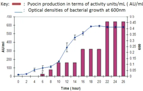
Optimization of culture conditions and their kinetics
In order to achieve the maximum production of bac-teriocin fromP.aeruginosa SA189, the culture was inocu-lated in different growth media (i.e., nutrient broth, trypti-case soya broth, brain heart infusion broth (BHI), lactose broth, Luria-basal broth and pseudo agar base) with varied incubation periods (10, 15, 20, 24, 36, 48 and 72 h) and temperatures (29, 37 and 40 °C for 24 h). The bioactivity of the cell free supernatants ofP. aeruginosa SA189was mea-sured by agar well diffusion assay (AWDA) using Staphy-lococcus aureusSA 84 as the indicator strain. The inhibi-tory strength (bacteriocin titer) was expressed as arbitrary units (AU/mL) or activity units/mL (Rajaramet al., 2010). The kinetics of bacteriocin production byP. aeruginosa SA189 was studied by growing the producer strain under optimum conditions for at least 24 h. The samples were
col-Table 1- Inhibitory spectrum of Pyocin SA189.
Indicator strains No of strains tested
Average Zone of inhibition (mm)
Acinetobacter lwoffi 6 0
Bacillus subtilis 5 12
B. cereus 7 10
Corynebacterium xerosis 4 11
Enterobacter aerogenes 8 0
Enterococcus faecalis 6 17
Escherichia coli 10 14
Klebsiella pneumoniae 5 14
Listeria monocytogenes 1 17
Proteus mirabilis 6 0
Pseudomonas aeruginosa 7 9
Salmonella typhi 8 0
Serratia marcescens 2 0
Shigella dysenteriae 8 0
Staphylococcus aureus 16 26
S. epidermidis 6 20
Streptococcus pyogenes 6 12
S. pneumoniae 2 10
lected after each hour to record the optical density at 600 nm. Thereafter, the samples were centrifuged and the cell free supernatant ofP. aeruginosaSA189 was used to determine the activity in units/mL (Jabeenet al., 2009)
Preparation and purification of Pyocin SA189
After optimization of culture conditions, P. aeruginosaSA189 was grown in BHI broth and was incu-bated at 37 °C for 24 hours. The following day, the culture broth was subjected to centrifugation at 10,000 x g for 30 min at 4 °C for separation of bacterial cells. The super-natant was filter sterilized by passing through 0.45mm pore
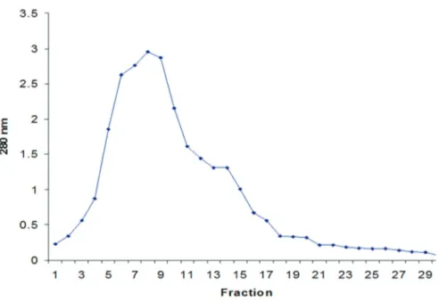
sized filter membrane (Millipore, MA, USA) and was con-centrated to 3 to 5-fold using a pre-chilled (4 °C) rotary evaporator (Buschi, Germany). This cell free supernatant (CFS) was referred to as the `crude bacteriocin prepara-tion’. This CFS was further partially purified by ammo-nium sulphate precipitation (Harris, 1989). In order to achieve the maximum saturation of pyocin SA189, differ-ent concdiffer-entrations (50%, 60%, 70% and 80%) of ammo-nium sulphate were added by constant agitation at 4 °C and precipitates were recovered by centrifugation (10,000 xg for 30 min at 4 °C). The resulting pellets were re-suspended in 50 mM sodium phosphate buffer (pH 7.0) and were des-ignated as `partially purified pyocin’ (Jabeenet al., 2009). For removal of the salt from the partially purified bac-teriocin, ultra-filtration was done through a pre-treated di-alysis tubing of 12 kDa cutoff size (Harris, 1989). The dialyzed bacteriocin was further subjected to gel chroma-tography on a Sephadex G-75 column of dimensions, 30 x 1.5 cm (Amersham Pharmacia Biotech, USA). The column was pre-equilibrated and the suspension was eluted with 50 mM Sodium phosphate buffer of pH 7.0. The flow rate was maintained at 0.2 mL/min and the eluates were sub-jected to absorbance measurement at 280 nm. The active fractions, thus obtained, were collected and pooled for as-sessment of inhibitory activity (DeCourcy, 2004). The bio-activity of pyocin SA189 was analyzed at each point of purification by agar well diffusion assay (AWDA), in terms of AU/mL. After every step of purification, protein concen-tration was measured (Lowryet al., 1951).
Molecular weight estimation of Pyocin SA189 by SDS-PAGE and related antibacterial assay
The ammonium sulphate precipitate and their active fractions that were obtained after gel filtration chromatog-raphy were subjected to SDS-PAGE (10% polyacrylamide gel) analysis using the standard protein marker (range 14.5 kDa to 200 kDa (Sigma)) loaded on a vertical slab gel (BioRad, USA). After the complete run, the gel was cut into two halves. One half of the gel that contained protein sam-ple and standard molecular weight marker was visualized by Coomassie blue staining to visualize protein bands (Jabeenet al., 2007), while the other half of the gel, con-taining only the sample protein, was treated with a solution
of 20% isopropanol (v/v) and 10% acetic acid (v/v) for 2 h followed by washing with distilled water for 4 hours. The gel was placed on a nutrient agar plate and overlaid with soft agar (0.6%) containing the indicator strain. The plate was incubated overnight at 37 °C and next day observed for the zone of inhibition (Bhuniaet al., 1988).
Physico-chemical characterization of Pyocin SA189
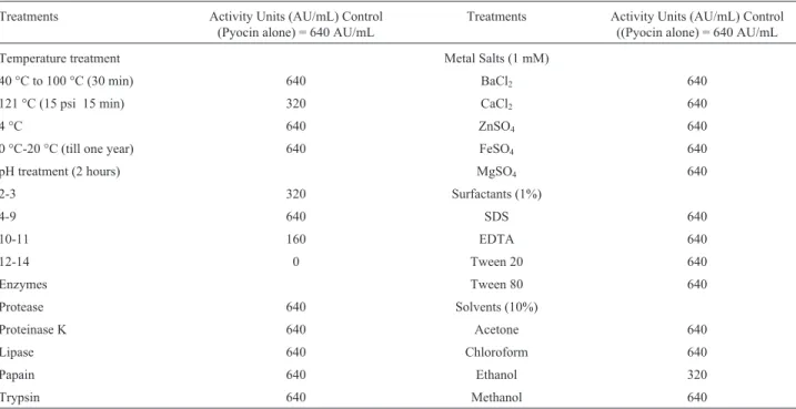
Thermostability of pyocin SA189 was determined by exposing the preparations to elevated temperatures, i.e., from 40 °C to 80 °C for 30 min, and to 100 °C and 121 °C for 15 min. After treatment, the residual activity was deter-mined by AWDA (Jabeenet al., 2009; Benreguieget al., 2013). To determine the temperature and duration of the bacteriocin stability, the preparation was stored at tempera-tures, 0 °C, 4 °C or -20 °C, and their bioactivity was moni-tored till one year using AWDA. To assess the effect of different pH levels on its bioactivity, bacteriocin prepara-tion was adjusted to different pH values, ranging between 1-14, with 1N NaOH (Merck) or 1 N HCl (Merck). The samples were incubated at 37 °C for 2 hours, followed by re-adjustment to neutral (7.0) pH and assessment of the bioactivity by AWDA (Vamanu and Vamanu, 2010). Bio-logical stability of pyocin SA189 was performed after giv-ing a treatment with different enzymes includgiv-ing proteases, proteinase K, pepsin, papain and lipase (Sigma, USA) at a final concentration of 1 mg /mL (Vamanu and Vamanu, 2010). Similarly, equal volumes of the pyocin SA189 were mixed separately with pre-chilled (at 4 °C) 10% concen-trated preparations of various organic solvents, acetone, ethanol, methanol and chloroform, with 1 mM solutions of metal ions (CaCl2, FeSO4,ZnSO4,MgSO4,BaCl2) and with different surfactants or detergents (at a final concentration of 1%) including sodium dodecyl sulphate (SDS) and eth-ylene diamine tetra acetic acid (EDTA), Tween 20, and Tween 80 (Sigma, USA). The mixtures were stirred and in-cubated at 37 °C for 2 hours and further analyzed by AWDA. The sets of respective positive and negative con-trols were also processed simultaneously in the same way (Rajaramet al., 2010).
Results and Discussions
The present study basically focused on the bacterio-cin, which is produced by a clinical strainP. aeruginosa SA189. This producer strain as well as the indicator strain, S. aureus SA84, were previously isolated and reported (Naz and Rasool, 2013). The identity of the producer strain (P. aeruginosaSA189) was confirmed by 16S rDNA gene amplification (conventional PCR, product size 956 bp).
Antimicrobial spectrum
fungi (Reaet al., 2011). The broad spectrum activity of bacteriocins fromPseudomonas spp. has widely been re-ported (Parret and De Mot, 2000). Pyocin SA189 also ex-hibited bioactivity against a number of sensitive organisms, notably the Gram-positive bacteria includingS. aureus, S. pyogenes andListeria monocytogenes. However, Esche-richia coli,Klebsiella pneumoniae, Shigella dysenteriae,P. aeruginosa andProteus mirabilis were also found to be susceptible to a certain extent (Table 1). These finding in-spired another study where an isolated pyocin, JU-Ch, ex-hibited significant antagonistic activity against Gram-positive and Gram-negative bacteria along with different fungi (Grewalet al., 2014). Such bio-active potential might be attributed to the presence of high number of bacteriocin adsorption receptors in the peptidoglycan-based cell wall of Gram-positive bacteria (Padillaet al., 2002).
Optimization of conditions for bacteriocin production and their kinetics
Like other microbial products, bacteriocin production is a genetically regulated phenomenon and is influenced by a number of environmental and nutritional factors such, as the composition of medium, incubation time, temperature, and pH (Lucaset al., 2006, Rajaramet al., 2010). In order to obtain maximum production of the pyocin (bacteriocin) fromP. aeruginosaSA189, the mentioned parameters were optimized in our current study. The results indicated that BHI broth (an enriched medium) enhanced the production of pyocin (data not shown). Enriched medium not only fa-vors the growth ofP. aeruginosabut also helps the bacte-rium to yield high titers of bacteriocins (MacKinnon, 2011). The maximum yield of pyocin SA189 was obtained when the organism was cultivated at 37 °C and pH 7.0. Our finding is in concordance with the previous study con-ducted by Scholl and Martin (2008). We recorded the
maxi-mum titer of pyocin SA189 as 640 AU/mL (data not shown).
The analysis of the growth and production kinetics re-vealed that the pyocin SA189 production varies in different phases of the growth cycle. The protein production is initi-ated in the early logarithmic phase, reaches the maximum level after 18 to 22 hours (late log phase) and remained con-stant till the late stationary phase (Figure 1).
Purification of Pyocin SA189
Production of pyocin is regulated in such a manner that only a few cells in a population actively produce pyocin. Further, the bacteriocin production can be en-hanced by induction with DNA damage treatments, such as ultraviolet irradiation (Higerdet al., 1967), mitomycin C (Kageyama, 1964) or by DNA gyrase inhibiting antibiotics (Brazas and Hancock, 2005). However, in the present study, the pyocin SA189 was harvested by propagation of the respective strain as per the optimum growth require-ments followed by partial purification by ammonium sul-phate precipitation, of the cell free supernatant (CFS). The salts, in fact, make the proteins resistant to denaturation, proteolysis or bacterial contamination (Harris, 1989). The maximum antagonistic activity, in case of pyocin SA189, was observed to be 1,280 AU/mL in the resolved precipi-tate with 70% saturation, which is in concordance with the previously reported findings of Scholl and Martin (2008). After ultra-filtration of partially purified pyocin SA189, it was noted that the pyocin almost retained their activity; however, a little loss might be attributed to the adsorption of the bacteriocin on the dialysis membrane. In the follow-ing step of conventional gel permeation chromatography, on Sephadex G-75 column, pyocin SA189 depicted the ac-tivity to reside in fraction 6, 7 and 8 and the chromatogram also showed a single peak of protein (Figure 2). These
ings correlate with the study, where pyocin was eluted from the CM Sephadex A-50 (pH 7.2) column as a single protein peak (Al-Shibibet al., 1985). Furthermore, Duportet al. (1995) determined pyocin S3 as a single protein peak upon Sephacryl S-200 gel filtration (Mr of 90,000). In contrast to these results, a protease sensitive S-type pyocin AP41 was observed as a complex of two proteins that were observed as two separate peaks when subjected to gel filtration Sephacryl S 200 column in the presence of 6 M urea (Sano and Kageyama, 1981).
In the protein purification profile of pyocin SA189, the specific activity in CFS was found to be 220.7 AU/mg, which increased up to 752.9 AU/mg after ammonium sul-fate precipitation and 1,828.6 AU/mg after gel filtration (Table 2). The final purification that was achieved after gel filtration chromatography was 8.2 folds with 2% recovery. These findings are in accordance with another study which also reported the purification of pyocin with an increase in the specific activity as well as purification up to 434-fold (Al-Shibibet al., 1985). In another study, the specific activ-ity of the pyocin after every step of purification was in-creased and final recovery after chromatography using Sephacryl S 200 was observed to be 13% (Sano and Kageyama, 1981). Increase in the specific activity was also demonstrated in each purification step for the pyocin 42A (Abdi-Ali etal.,2004).
SDS-PAGE and related bioactivity
The purity of pyocin protein SA189 was further checked by electrophoresis and confirmed by gel overlay technique. Polyacrylamide gel electrophoresis (PAGE) in the presence of sodium dodecyl sulphate (SDS), an anionic detergent, is one of the important procedures, used
success-fully to characterize proteins with respect to the molecular sizes of their constituent polypeptides. Moreover, in bac-teriocin studies, it has been a widely used technique, which is sensitive enough to detect the activity of the bacteriocin directly on the gel. This technique was employed to check the purity of pyocin SA189. Ammonium sulphate precipi-tates and the active fractions from gel filtration were subjected to 10% acrylamide denaturing gel. Electrophero-gram of pyocin SA189 revealed a single band, which corre-sponded to the molecular mass of approximately 66 kDa. This band also showed bioactivity by gel overlay method (data not shown). The high molecular weight pyocins, par-ticularly the R-type pyocins are well-documented in litera-ture (Ritchieet al., 2011). A study in this relation demon-strated the isolation of five pyocins from clinical isolates, which were resistant to trypsin treatment and had molecular masses within the 54.9-282 Kg/mol. range (Al-Rubieeet al., 1988). Similar protease-resistant pyocins with 20-135 kg/mol. molecular masses have been reported ear-lier (Al-Shibib et al., 1985). In another study, R-type pyocin, resistant to all tested proteases, depicted a major band of about 30 kDa on SDS-PAGE (Fontoura et al., 2009). On the other hand, a study on protease-sensitive pyocin showed a single band corresponding to molecular weight 62.4 kDa on the SDS-PAGE (Linget al., 2010).
Physicochemical characterization of pyocin SA189
Different physical and chemical factors may have sig-nificant impact on the bioactivity of bacteriocins. The de-termination of such impacts bears significance for possible application(s) of bacteriocins in food preservation, as pro-biotics or as chemotherapeutic agents. Interestingly, in the present study, pyocin SA189 was found to be resistant to
the action of almost all the physical and chemical factors tested (Table 3). Thus, as per our analysis, pyocin SA189 could be stated as thermotolerant in nature. Our observa-tions suggested that the pyocin retained its bioactivity even at the autoclaving temperature. Such high thermostability is in contrast to a previous report where R-type pyocin lost ac-tivity at a temperature above 60 °C (Al-Rubieeet al., 1988). However, Padillaet al.(2002) found a pyocin to be resis-tant to proteolytic enzymes as well as high temperatures. Moreover, an antimicrobial peptide produced by Pseudo-monasspp. also showed resistance to high ranges of tem-perature (Fontouraet al., 2009). Regarding the influence of pH on the biological activity, pyocin SA189 remained stable within the pH range 2 to 11. Retention of bioactivity of the pyocins at various pH values has been reported ear-lier (Sano and Kageyema, 1981; Padilla et al., 2002; Saleemet al., 2009). However, in contrast to our findings, an R-type pyocin demonstrated stability only in a pH range of 6 to 8 (Al-Rubieeet al., 1988).
Besides the physical agents, certain chemicals may modulate the bioactivity of pyocins. The most important finding was the resistance of pyocins SA189 to different proteolytic enzymes (Table 3). Although, this pyocin mani-fested an apparent proteinaceous nature in various
experi-ments, such as ammonium sulphate precipitation, SDS-PAGE analysis and gel filtration, but when treated with proteolytic enzymes, like proteinase K, proteases, trypsin and papain, it survived digestion and retained its bioa-ctivity. In addition, lipase also had no effect on the activity of this pyocin. These results were not surprising when com-pared with previous findings on pyocins, where protease re-sistant pyocins were obtained from the strains isolated from well water (Padillaet al., 2002), pond water (Fontouraet al., 2009) and clinical materials (Al-Rubieeet al., 1988). Similarly, pyocins that are sensitive to proteases were also reported from different environments (Sano and Kageya-ma, 1981). The resistance to proteases might be due to the cyclic peptide nature of some of the bacteriocins, which may have unusual amino acids rendering them resistant to hydrolysis by proteolytic enzymes (Bizani and Brandelli, 2002). Exposure of pyocin SA189 to surfactants also re-sulted in complete retention of their activity. Incubation of pyocin SA189 with various concentrations of organic sol-vents and metal salts had no effect on its bioactivity. These findings are in accordance with a previously reported study where a pyocin was observed to be resistant to action of all the organic solvents (Saleemet al., 2009).
Table 3- Physicochemical characteristics of Pyocin SA189.
Treatments Activity Units (AU/mL) Control (Pyocin alone) = 640 AU/mL
Treatments Activity Units (AU/mL) Control ((Pyocin alone) = 640 AU/mL
Temperature treatment Metal Salts (1 mM)
40 °C to 100 °C (30 min) 640 BaCl2 640
121 °C (15 psi 15 min) 320 CaCl2 640
4 °C 640 ZnSO4 640
0 °C-20 °C (till one year) 640 FeSO4 640
pH treatment (2 hours) MgSO4 640
2-3 320 Surfactants (1%)
4-9 640 SDS 640
10-11 160 EDTA 640
12-14 0 Tween 20 640
Enzymes Tween 80 640
Protease 640 Solvents (10%)
Proteinase K 640 Acetone 640
Lipase 640 Chloroform 640
Papain 640 Ethanol 320
Trypsin 640 Methanol 640
Table 2- Purification Profile of Pyocin SA189.
Sample/steps Vol./mL AU/mL Total AU Total Protein Specific activity % recovery Purification fold
Cell free Supernatant/ Crude extract 300 640 192 000 870 220.7 100 1
Ammonium Sulfate Precipitate (70%) 30 1 280 38 400 51 752.9 20 3.4
References
Abdi-Ali A, Worobec EA, Deezagi Aet al.(2004) Cytotoxic ef-fects of pyocin S2 produced byPseudomonas aeruginosaon the growth of three human cell lines. Can J Microbiol 50:375-381.
Al-Rubiee R, Al-Mudhaffar S, Hassan Fet al.(1988) Purification and characterization of pyocins R fromPseudomonas aeru-ginosa. Folia Microbiol 33:520-524.
Al-Shibib A, Al-Mudhaffar S, Al-Ani Fet al.(1985). Purification and characterization of pyocins from Pseudomonas aeru-ginosa. Folia Microbiol 30:25-29.
Bakkal S, Robinson SM, Ordonez CL et al. (2010) Role of bacteriocins in mediating interactions of bacterial isolates taken from cystic fibrosis patients. Microbiol 156:2058-2067.
Benreguieg M, Dalache F, Gacemi B (2013) Characterization of antibacterial activity and potential as probiotic of lactic acid bacteria isolated from goat’s milk in Algeria. J Life Sci 7:802-813.
Bhunia AK, Johnson MC, Ray B (1988) Purification, character-ization and antimicrobial spectrum of a bacteriocin pro-duced by Pediococcus acidilactici. J Appl Bacteriol 65:261-268.
Bizani D, Brandelli A (2002) Characterization of a bacteriocin produced by a newly isolatedBacillus sp.strain 8A. J Appl Microbiol 93:512-519.
Brazas MD, Hancock RE (2005) Ciprofloxacin induction of a sus-ceptibility determinant in Pseudomonas aeru-ginosa.Antimicrob. Agents Chemother 49:3222-3227. DeCourcy K (2004) Column chromatography information
man-ual. Fralin Biotechnology Center. Virginia Technology pp. 5-17.
Dingemans J, Ye L, Hildebrand Fet al.(2014) The deletion of TonB-dependent receptor genes is part of the genome reduc-tion process that occurs during adaptareduc-tion ofPseudomonas aeruginosato the cystic fibrosis lung. Pathog Dis 71:26-38. Duport C, Baysse C, Michel-Briand Y (1995) Molecular
charac-terization of pyocin S3, a novel S-type pyocin from Pseudo-monas aeruginosa. J Biol Chem 270:8920-8927.
Fontoura R, Spada JC, Silveira STet al.(2009) Purification and characterization of an antimicrobial peptide produced by
Pseudomonassp. Strain 4B. World J Microbiol Biotechnol 25:205-213.
Grewal S, Bhagat M, VakhluJ (2014) Antimicrobial protein pro-duced bypseudomonas aeruginosaJU-Ch 1, with a broad spectrum of antimicrobial activity. Biocatal Agri Biotech 8:1290-1305.
Harris ELV (1989) Concentration of the extract: Protein Purifica-tion Methods. In: A Practical Approach. E.L.V. Harris and S. Angal (eds) IRL Press, Oxford, pp. 125-172.
Higerd TB, Baechler CA, Berk RS (1967)In vitroandin vivo
characterization of pyocin. J Bacteriol 93:1976-1986. Jabeen N (2007) Bacteriocins for the control of indigenous
phyto-pathogenic bacteria. Ph.D. Thesis. Department of Microbi-ology, University of Karachi, pp. 49-52.
Jabeen N, Gul H, Subhan SAet al.(2009) Biophysicochemical characterization of bacteriocin(s) from indigenously iso-latedAgrobacterium radiobacterNA 6. Pak J Bot 41:3227-3237.
Jacob F (1954) Induced biosynthesis and mode of action of a pyocine, antibiotic produced byPseudomonas aeruginosa. Ann Inst Pasteur 86:149-160.
Kageyama M, Ikeda K, Egami F (1964) Studies of a pyocin. III. Biological properties of the pyocin. J Biochem 55:59-64. Kerr B, Riley MA, Feldman MWet al.(2002) Local dispersal
pro-motes biodiversity in a real-life game of rock-paper-scis-sors. Nature 418:171-174.
Lee FK, Dudas KC, Hanson JAet al.(1999) The R-type pyocin of
Pseudomonas aeruginosaC is a bacteriophage tail-like parti-cle that contains a single-stranded DNA. Infect Immun 67:717-725.
Ling H, Saeidi N, Rasouliha BHet al.(2010) A predicted S-type pyocin shows a bactericidal activity against clinical
Pseudo-monas aeruginosa isolates through membrane damage.
FEBS Lett 584:3354-3358.
Lowry OH, Rosebrough NJ, Farr ALet al.(1951) Protein mea-surement with the Folin phenol reagent. J Biol Chem 193:265-275.
Lucas R, Grande MA, Abriouel Het al.(2006) Application of the broad-spectrum bacteriocinenterocin AS-48 to inhibit B. coagulansin canned fruit and vegetable foods. Food Chem Toxicol 44:1774-1781.
MacKinnon EM (2011) Characterizing the pyocin activity of di-verse Pseudomonas aeruginosaisolates, M.Sc. thesis, De-partment of Molecular Genetics, University of Toronto, Canada.
Michel-Briand AY, Baysse C (2002) The pyocins of Pseudomo-nas aeruginosa.Biochemie 84:499-510.
Naz SA, Rasool SA (2013) Isolation, production and characteriza-tion of bacteriocins produced by strains from indigenous en-vironments. Pak J Bot 45:261-267.
Padilla C, Lobos O, Brevis Pet al.(2002) Effects of the bacterio-cin PsVP-10 produced by Pseudomonas sp. on sensitive bacterial strains. De Microbiologia 44:19-23.
Parret AHA, De Mot R (2002) Bacteria killing their own kind: novel bacteriocins of Pseudomonas and otherg -proteobac-teria. Trends Microbiol 10:107-112.
Rajaram G, Manivasagan P, Thilagavathi Bet al.(2010) Purifica-tion and CharacterizaPurifica-tion of a bacteriocin produced by
Lactobacillus lactisisolated from marine environment. Adv J Food Sci Technol 2:138-144.
Rea MC, Ross RP, Cotter PD et al. (2011) Classification of bacteriocins from gram positive bacteria. In: Prokaryotic Antimicrobial Peptides. From Genes to Application. Drider, D. and Rebuffat, S. (eds). Springer, Berlin, pp. 29-54. Riley MA (1998) Molecular mechanisms of bacteriocin
evolu-tion. Annu Rev Genet 32:255-278.
Riley MA, Goldstone CM, Wertz JEet al.(2003) A phylogenetic approach to assessing the targets of microbial warfare. J Evol Biol 16:690-697.
Ritchie JM, Greenwich JL, Davis BMet al.(2011) An Esche-richia coliO157-specific engineered pyocin prevents and ameliorates infection byE. coliO157:H7 in an animal model of diarrheal disease. Antimicrob Agents Chemother 55:5469-5474.
Saleem F, Ahmed S, Yaqoob Zet al.(2009) Comparative study of two bacteriocins produced by representative indigenous soil bacteria. Pak J Pharma Sci 22:252-258.
Scholl D, Martin DW (2008) Antibacterial efficacy of R-type pyocins towardsPseudomonas aeruginosain a murine peri-tonitis model. Antimicrob Agents Chemother 52:1647-1652
Scholl D, Cooley M, Williams SRet al.(2009) An Engineered R-Type Pyocin is a highly specific and sensitive bactericidal agent for the food-borne pathogen Escherichia coli
O157:H7. Antimicrob Agents Chemother 53:3074-3080.
Spilker T, Coenye T, Vandamme Pet al.(2004) PCR-based assay for differentiation of Pseudomonas aeruginosafrom other
Pseudomonas species recovered from cystic fibrosis pa-tients. J Clin Microbiol 42:2074-2079.
Strauch E, Kaspar H, Schaudin Cet al.(2001) Characterization of enterocoliticin, a phage tail-like bacteriocin, and its effect on
pathogenic Yersinia enterocoliticastrains. Appl Environ Microbiol 67:5634-5642.
Takeya K, Minamishima Y, Amako K et al. (1967) A small rod-shaped pyocin. Virol 31:166-168.
Vamanu E, Vamanu A (2010) The influence of prebiotics on bacteriocin synthesis using the strain Lactobacillus paracaseiCMGB16. Afr J Microbiol Res 4:534-537. Williams S, Gebhart D, Martin D et al. (2008) Retargeting
R-typing pyocins to generate
novel bactericidal protein complexes. Appl Environ Microbiol 74:3868-3876.
Associate Editor: Elizabeth de Andrade Marques