online | memorias.ioc.fiocruz.br
Malaria remains a major public health problem, which affects approximately 225 million people worldwide and causes around 781,000 deaths, mostly in children under five years old (WHO 2010). In areas where malaria is highly endemic, the parasite rate and density decline with increasing age and severe disease and mortality are usu-ally restricted to early childhood (McGregor 1974, 1987, Cattani et al. 1986, Greenwood et al. 1987). Adolescents and adults are usually free of clinical symptoms of malar-ia, although they may maintain low parasitemias through-out the transmission season (Marsh & Snow 1999). It has been proposed that these changes reflect the acquisition of clinical immunity against malaria infection. The results from in vivo studies suggest that one of the mechanisms underlying clinical immunity against malaria is the con-tainment of parasite multiplication by antibodies (Cohen
et al. 1961, McGregor & Carrington 1963, Sabchareon 1991). Antibodies that inhibit blood stage replication of Plasmodium falciparum are believed to be important in mediating both naturally acquired and artificially in-duced immunity generated by blood-stage vaccine can-didates (Cohen et al. 1969, Good et al. 2004). Therefore, research has pursued a vaccine capable of inducing the formation of specific antibodies in sufficient quantities and mainly functionally able to participate in protect-ing against the parasite. In this context, several proteins have been identified and selected as candidate molecules for use in the composition of a malaria vaccine. Among these, the P. falciparum glutamate-rich protein (GLURP) appears to be a promising candidate (Hogh et al. 1992, 1993, Dziegiel et al. 1993, Theisen et al. 1998, Soe et al. 2004, Pratt-Riccio et al. 2005).
The GLURP protein is an exoantigen expressed at all stages of development in the parasite life cycle in human hosts, including on the surface of newly released mero-zoites (Borre et al. 1991). It is highly antigenic and the gene encoding GLURP exhibits low polymorphism in geographically different P. falciparum isolates (Theisen et al. 1995, Stricker et al. 2000). GLURP contains an N-terminal non-repeat region (R0), a central non-repeat re-gion (R1) and a C-terminal repeat rere-gion (R2). Immuno-epidemiological studies performed in high transmission areas have demonstrated a high prevalence of antibodies Financial support: FIOCRUZ, FAPERJ, CNPq
+ Corresponding author: riccio@ioc.fiocruz.br
Present address (VE-V): Laboratório de Biologia Molecular e Doen-ças Endêmicas, IOC-Fiocruz
Received 15 April 2011 Accepted 1 June 2011
Antibodies against the Plasmodium falciparum glutamate-rich
protein from naturally exposed individuals living in a Brazilian
malaria-endemic area can inhibit in vitro parasite growth
Lilian Rose Pratt-Riccio1/+, Cesare Bianco-Junior1, Paulo Renato Rivas Totino1,
Daiana De Souza Perce-Da-Silva1, Luciene Aquino Silva1, Evelyn Kety Pratt Riccio1,
Vítor Ennes-Vidal1, Ana Gisele Costa Neves-Ferreira2, Jonas Perales2,
Surza Lucia Gonçalves Da Rocha2, Fabrício Dias-Da-Silva1, Maria de Fátima Ferreira-da-Cruz1,
Cláudio Tadeu Daniel-Ribeiro1, Joseli De Oliveira-Ferreira3, Michael Theisen4,
Leonardo José De Moura Carvalho1, Dalma Maria Banic1
1Laboratório de Pesquisas em Malária 2Laboratório de Toxinologia 3Laboratório de Imunoparasitologia,
Instituto Oswaldo Cruz-Fiocruz, Av. Brasil 4365, 21040-900 Rio de Janeiro, RJ, Brasil 4Statens Seruminstitut, Copenhagen, Denmark
The glutamate-rich protein (GLURP) is an exoantigen expressed in all stages of the Plasmodium falciparum life cycle in humans. Anti-GLURP antibodies can inhibit parasite growth in the presence of monocytes via antibody-dependent cellular inhibition (ADCI), and a major parasite-inhibitory region has been found in the N-terminal R0 region of the protein. Herein, we describe the antiplasmodial activity of anti-GLURP antibodies present in the sera from individuals naturally exposed to malaria in a Brazilian malaria-endemic area. The anti-R0 antibodies showed a potent inhibitory effect on the growth of P. falciparum in vitro, both in the presence (ADCI) and absence (GI) of monocytes. The inhibitory effect on parasite growth was comparable to the effect of IgGs purified from pooled sera from hyperimmune African
individuals. Interestingly, in the ADCI test, higher levels of tumour necrosis factor alpha (TNF-α) were observed in the
supernatant from cultures with higher parasitemias. Our data suggest that the antibody response induced by GLURP-R0 in naturally exposed individuals may have an important role in controlling parasitemia because these antibodies are able to inhibit the in vitro growth of P. falciparum with or without the cooperation from monocytes. Our results also
indicate that TNF-α may not be relevant for the inhibitory effect on P. falciparum in vitro growth.
against GLURP in immune adults (Boudin et al. 1993, Dziegiel et al. 1993) and have shown that high levels of GLURP-specific antibodies are significantly associ-ated with low parasite densities (Hogh et al. 1992, 1993) and protection against clinical malaria (Dziegiel et al. 1993, Dodoo et al. 2000, Oeuvray et al. 2000). Moreover, GLURP is a target antigen for antibodies involved in an-tibody-dependent cellular inhibition (ADCI) (Theisen et al. 1998), which is believed to be involved in acquired protective immunity against malaria (Khusmith & Druilhe 1983, Lunel & Druilhe 1989, Bouharoun-Tay-oun et al. 1990, BouharBouharoun-Tay-oun-TayBouharoun-Tay-oun & Druilhe 1992). In the ADCI mechanism, cytophilic antibodies (IgG1 and IgG3 but not IgG2 and IgG4) act in conjunction with blood monocytes to contain parasite multiplication. In vitro studies have indicated that affinity-purified human IgG against the non-repeat R0 and R2 repeat regions can inhibit parasite growth in the presence of monocytes, al-though anti-R0 antibodies exerted a greater ADCI effect than anti-R2 antibodies (Theisen et al. 1998).
In Brazil, malaria is hypo to meso-endemic, is pres-ent throughout the year with clear seasonal fluctuations and is frequently associated with the migratory move-ments of non-immune individuals to areas where malaria is endemic (Oliveira-Ferreira et al. 2010). The population exposed to malaria in these areas is vulnerable and infec-tions tend to be followed by clinical symptoms (Marques 1987, Castilla & Sawyer 1993). For a long time, asymp-tomatic cases have been considered rare in Brazil (Prata et al. 1998). However, asymptomatic infection by P. fal-ciparum and Plasmodium vivax have been detected in the states of Rondônia (RO) and Amazonas, suggesting that subjects exposed to malaria in Brazil can also de-velop acquired resistance to clinical malaria despite the different epidemiological profile from the one observed in Africa (Camargo et al. 1999, Alves et al. 2002, Coura et al. 2006). Because the great majority of growth-inhib-itory antibody studies have been conducted in African countries, we aimed to verify the antiplasmodial activity of anti-GLURP antibodies present in the sera from indi-viduals living in a Brazilian malaria-endemic area with a low level of transmission.
SUBJECTS, MATERIALS AND METHODS
Study site and volunteers - Written informed consent was obtained from all donors before blood samples were collected. Donors giving informed consent answered a standard questionnaire to evaluate the possible degree of malaria exposure. Venous peripheral blood was col-lected into heparinised tubes. Plasma was obtained from the blood samples by centrifugation and was aliquoted and stored at -20ºC.
The plasma donors comprised nine adult individu-als living in rural villages situated near Porto Velho, the capital of RO, in the Brazilian Amazon malaria endemic region. In this region, malaria transmission is unstable with an increase in the number of cases between April-September (Rodrigues et al. 2008). The population of these villages is composed of natives and Brazilian mi-grants that have inhabited this area for variable periods of time since the 1970s. The age range of the studied
in-dividuals was 18-74 years old (41 ± 22). The subjects had spent all or most of their lives in these locations (32 ± 24) and they referred to repeated malaria attacks (7 ± 4). All donors presented parasite-negative thin and thick Giem-sa-stained blood smears at the time of blood collection.
For the inhibition assays, non-endemic control se-rum (NCS) samples from two individuals from the ma-laria laboratory staff [Rio de Janeiro (RJ), Brazil], who had neither a history of malaria nor contact with ma-laria transmission areas, were included in our study as negative controls. Positive control IgG (PIAG, kindly provided by Dr Pierre Druilhe, Institut Pasteur, Paris) was purified from a pool of sera obtained from 30 Af-rican adults living permanently in Garitenga, Burkina Faso, where malaria is holo-endemic. Sera donors were free of clinical symptoms and of heavy parasitemia and thus were regarded as immune individuals (Bouharoun-Tayoun et al. 1990, Lundquist et al. 2006). PIAG has pre-viously been found to confer passive protection against malaria and to cause a high ADCI effect (Sabchareon et al. 1991, Galamo et al. 2009).
For the enzyme linked immunosorbent assay (ELI-SA) against the R0 recombinant protein, sera from25 RJ controls who had neither a history of malaria nor contact with malaria transmission areas were used to establish the normalrange of the assay.
The study was reviewed and approved by the Oswal-do Cruz Foundation Ethical Committee (258/04).
Recombinant GLURP-R0 protein - The recombinant protein R094-489 corresponding to the non-repeat amino-terminal region was expressed in Escherichia coli and purified, as described elsewhere (Theisen et al. 1995).
de-termined as themean optical density (OD) plus three stan- dard deviations fromRJ controls (cut-off values: IgG = 0.122, IgG1 = 0.132, IgG2 = 0.156, IgG3 = 0.143, IgG4 = 0.151). To standardise the OD data obtained in different experiments, OD index was calculated for each immu-noglobulindetermination as the ratio of the observed OD to the cut-offvalues. A sample with an OD index > 1.0 was considered positive.
IgG purification - IgG was purified from individual plasma samples and from NCS and PIAG by affinity chromatography using a 1-mL Hi-Trap Protein G column (GE Healthcare Life Sciences), according to the manu-facturer’s instructions. After centrifugation (14,000 rpm, 5 min), the supernatant from each plasma sample was filtered through a 0.45 µm filter (Millipore) and loaded onto a column equilibrated with 0.02 M sodium phos-phate buffer, pH 7.0, at a flow rate of 1.0 mL/min. The IgG fraction was eluted with 0.1 M glycine/HCl buffer, pH 2.7, at the same flow rate. The eluate was immedi-ately neutralised with 1 M Tris-HCl, pH 9.0, pooled and dialysed against PBS. The IgG purity was estimated by sodium dodecyl sulphate-polyacrylamide gel electropho-resis upon staining the gels with Coomassie blue dye.
GLURP-specific IgG antibody purification - The GLURP-specific IgG antibody was purified from total IgG by affinity chromatography using a 1-mL Hi-Trap NHS-activated affinity column (GE Healthcare Life Sciences) containing GLURP94-489 (R0) immobilised ac-cording to the manufacturer’s instructions. The total IgG in column buffer (0.02 M sodium phosphate, pH 7.0) was applied to the R0 column at a flow rate of 1.0 mL/min. The column was washed extensively with column buf-fer. The bound GLURP-specific IgG was eluted with 0.1 glycine/HCl, pH 2.7 and collected over 1 M Tris-HCl, pH 9.0, to neutralise the pH at the same flow rate. Soon after, the eluate was dialysed against PBS and then Ro-swell Park Memorial Institute (RPMI) 1640 medium and concentrated using an Ultrafree Cl centrifugal filter de-vice (Millipore). The GLURP-specific IgG fraction was filtered through a 0.22 µm Millex filter (Millipore) and the levels were quantified using the Bicinchoninic Acid Protein Assay kit (BCA, Sigma) with bovine serum al-bumin (BSA) as a standard.
Indirect immunofluorescence assays (IFAs) - IFAs were performed to verify if the purified R0 anti-bodies recognised the native protein. IFA slides were prepared with synchronous cultures of P. falciparum schizonts (strain PSS1) using the method described by Trager and Jensen (1976). IFAs were performed at 37ºC in moist chambers after a 10-min fixation in cold ac-etone (-20ºC). After incubation with purified ies (2-factor dilution from 1:40-1:5120), bound antibod-ies were detected by reaction with appropriate fluorescein isothiocyanate-conjugated antisera (Sigma).
Parasite culture - The P. falciparum line PSS1 (Bra-zil) was cultured according to a modification of the meth-od described by Trager and Jensen (1976) with serum-free medium. Briefly, parasites were cultured in vitro with freshly prepared blood bank-derived O+ erythrocytes in
RPMI 1640 (Sigma) supplemented with 25 mM HEPES buffer, 23 mM NaHCO3 (Sigma), 40 μg/L gentamicin, 4 g/L glycose (Sigma), 0.18 mM hypoxanthine (Sigma) and 10% albumax (Gibco), in an atmosphere containing 5% O2,5% CO2 and 90% N2 (White Martins, RJ, Brazil). The parasites were synchronised by repeated sorbitol treatments and schizonts were enriched by flotation on plasmagel, as described elsewhere (Lambros & Vander-berg 1979, Reese et al. 1979).
Monocyte preparation - Monocytes were obtained from freshly drawn heparinised peripheral blood from a single Brazilian donor without previous exposure to malaria. Peripheral blood mononuclear cells (PBMC) were isolated by density gradient centrifugation (Ficoll-Hypaque). The cells obtained were washed three times in serum-free RPMI 1640 medium and resuspended in RPMI 1640 with 10% albumax (Gibco) at 2 x 10⁷ mL.
The PBMC were distributed in 96-well flat-bottomed microdilution plates at 2 x 10⁶ cells/well and cultured
at 37ºC in 5% CO2 for 2 h and washed with RPMI 1640 (Sigma). This method allows the recovery of about 2 x 10⁵ cells, which are mostly monocytes (Theisen et al.
2004). For assays in which activated monocytes were used, cells were pretreated for 24 h with recombinant
human interferon gamma (IFN-γ) (BD Bioscience) at a
final concentration of 100 ng/mL in RPMI 1640 contain-ing 10% albumax. Monocytes were then washed with serum-free RPMI 1640 before use in the ADCI assay.
Growth inhibition (GI) and ADCI assays - GI and ADCI assays, in the absence or presence of monocytes, respectively, were performed in parallel in triplicate wells of 96-well flat-bottom culture plates (Falcon). For the GI assay, 50 µL volumes of anti-R0 purified antibodies, at the final concentrations of 50, 150 and 450 µg/mL in RMPI containing 10% albumax, were added to 50 µL of syn-chronised P. falciparum cultures at the schizont stage at an initial parasitemia of 0.5% and with 5% hematocrit.
In the ADCI assay, 50 μL of P. falciparum synchronised cultures at the schizont stage at an initial parasitemia of 0.5% and with 5% hematocrit and 50 µL of anti-R0 pu-rified antibodies at final concentrations of 50, 150 and 450 µg/mL in RMPI with 10% albumax were added to wells containing the adhered monocytes.
The experimental controls comprised the following: (i) P. falciparum synchronised cultures at the schizont stage alone, (ii) P. falciparum synchronised cultures at the schi-zont stage with monocytes, (iii) P. falciparum synchro- nised cultures at the schizont stage with IgG purified from non-endemic control serum, (iv) P. falciparum synchro-nised cultures at the schizont stage with IgG purified from NCS and monocytes, (v) P. falciparum synchronised cul-tures at the schizont stage with IgG purified from pooled hyperimmune serum and (vi) P. falciparum synchro-nised cultures at the schizont stage with IgG purified from pooled hyperimmune serum and monocytes.
The plates were maintained at 37ºC in a 5% CO2 for
72 h and, at intervals of 24 h, 50 μL volumes of RPMI
the 72 h, the cell culture supernatants from each well were collected individually and stored at -70ºC for later
deter-mination of tumour necrosis factor alpha (TNF-α). The
mean of parasitemia determined in each series of triplicate wells was calculated in both GI and ADCI assays by mi-croscopic examination and flow cytometry. The specific growth inhibitory index (SGI), which takes into account the possible inhibition induced by antibodies alone (GI) or by antibodies and monocytes (ADCI) was independently calculated as follow: SGI = 100 x [1 - (% parasitemia in the test sample/% parasitemia in the control sample)].
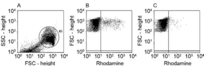
Parasitemia - The parasitemia in each triplicate well was evaluated by flow cytometry using rhodamine 123, a cationic, lipophilic fluorochrome that is incorporated into the mitochondria of viable parasites (Totino et al. 2008). After 72 h of culture, the parasitized erythro- cytes were washed in RPMI medium (RPMI-1640, 25 mM HEPES, 0.2% glucose, 23 mM sodium bicar-bonate), centrifuged at 350 g for 5 min and resuspended
and incubated at 37ºC for 5 min in 50 volumes of 1 μg/
mL rhodamine in RPMI. The rhodamine solution was removed by centrifugation (350 g, 5 min) and the parasit-ized erythrocytes were washed in RPMI and incubated at 37ºC for 30 min in 100 volumes of complete medium. After incubation, the parasitized erythrocytes were re-suspended in fresh complete medium and analysed in a flow cytometer (FACSCalibur, Becton Dickinson). To discriminate parasitized (viable parasites) from non-par-asitized erythrocytes (no parasite or dead parasites) in the culture samples, non-infected erythrocytes stained with rhodamine 123 were used as negative controls in the flow cytometry analysis (Fig. 1).
ELISA for determination of TNF-α levels in culture
supernatants - TNF-α was measured using commercial ELISA kits (BD Biosciences). Briefly, enzyme-linked immunosorbent assay 96-well plates (Maxisorp, NUNC, Denmark) were coated overnight at 4ºC with 2 µg/mL of capture antibody in a volume of 100 µL/well in 0.1 M carbonate-bicarbonate buffer, pH 8.2. After washing with PBS-T20, pH 7.4, uncoated sites were blocked for 2 h
at RT with 200 μL/well of 0.1% (wt/vol) BSA (Sigma)
in PBS with 0.05% sodium azide (Sigma) (PBS/BSA/ NaN3).After washing with PBS, the plates were incu-bated with 100 µL/well of cell culture supernatant or
100 µL/well of recombinant cytokines diluted succes- sively to determine the standard curve. After washing with PBS-T20, the plates were incubated for 1 h at RT with
100 μL/well of 200 ng/mL biotinylated detection
anti-body. After washing with PBS-T20, 100 µL of a solution of streptavidin-peroxidase (Sigma) diluted 1:500 in PBS/ BSA/NaN3 was added to each well and incubated for 30 min at RT in the dark. The reaction was visualised with 100 mL/well of 2,2’-azino-bis(3-ethylbenzthiazoline-6-sulphonic acid) (Sigma) containing 10 µL of 30% H2O2 for 30 min and 50 µL/well of 20% sodium dodecyl sul-fate (Sigma) was used to stop the reaction. Plates were read at 405 nm in a spectrophotometer (Spectramax 250,
Molecular Devices). The TNF-α OD values were con -verted to concentration values (ng/mL) using sigmoidal curve-fit equations derived from the standard curve
gen-erated using recombinant TNF-α.
Statistical analysis - The data were stored in the Fox-plus® (Borland International, Inc Perrysburg, OH) data
bank software. The Statistica (Microsoft, Inc Redmond, WA) and Epi-Info 6 (Centres for Disease Control and Pre-vention, Atlanta, GA) statistical software programs were used for data analysis. The Student’s t-test was used to analyse the differences in mean values; the Chi-square test was used to analyse the difference in prevalence of the positive responses and the Spearman rank coefficient test was used to analyse the correlations between variables.
RESULTS
Natural infections with P. falciparum induced high levels of IgG antibodies against the GLURP-R0 region in most naturally exposed individuals from RO analysed in this study (Table I). The levels of anti-GLURP-R0 IgG an-tibodies were positively correlated with age (p = 0.03, r = 0.7167) and with the time of residence in malaria-endemic areas (p = 0.04, r = 0.7000). No association between the anti-R0 IgG antibody response and the reported number of previous malaria episodes was observed. There was a predominance of IgG1 R0-specific antibodies over IgG2, IgG3 and IgG4 (p = 0.01, IgG1 vs. IgG2; p = 0.02, IgG1 vs. IgG3; p = 0.002, IgG1 vs. IgG4). No associations be-tween age, time of residence in malaria endemic areas or the number of previous malaria infections and the levels of anti-R0 IgG subclasses could be detected (Table I).
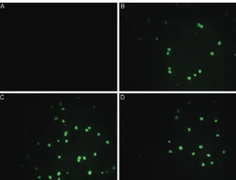
IgG antibodies were affinity-purified against GLURP-R0 from the nine Brazilian individuals and from a pool of serum from hyperimmune African adults (PIAG). All 10 of the anti-R0 IgG preparations recognised the natu-ral parasite protein by IFAs (Fig. 2). As expected, IgG purified from two blood donors that were never exposed to malaria (NCS) did not react with the parasite. The anti-R0 IgG antibodies purified from Brazilian donors strongly inhibited the growth of P. falciparum in vitro independently of the presence of monocytes. Indeed, eight of nine (89%) samples exhibited a parasite GI of more than 50% at a concentration of 50 µg/mL. Inter-estingly, this inhibitory activity tended to decrease with increasing antibody concentration. The samples R006, R007, R008 and R009 inhibited parasite growth to the same extent as the anti-R0 IgG antibodies purified from Fig. 1: flow cytometry analysis of in vitro inhibition of Plasmodium
PIAG, especially in the absence of monocytes (Table II). A higher level of GI was observed as compared to ADCI at a concentration of 50 µg/mL IgG (p = 0.03) (Fig. 3).
In the presence of monocytes, there was an inverse relationship between the ADCI activity and the concen-tration of anti-R0 affinity-purified IgG in the assay. The mean parasitemia at the concentrations of 50 µg/mL and 150 µg/mL was lower than the mean at the 450 µg/mL concentration (p = 0.007; 50 µg/mL vs. 450 µg/mL; p = 0.008; 150 µg/mL vs. 450 µg/mL) (Table II).
Interest-ingly, higher levels of TNF-α in the supernatant were
observed in the ADCI test performed in the presence of monocytes at a concentration of 450 µg/mL of purified anti-R0 (p = 0.01; 50 µg/mL vs. 450 µg/mL; p = 0.003; 150 µg/mL vs. 450 µg/mL) (Fig. 4). In parallel, we per-formed P. falciparum in vitro culture in the presence of
10, 5, 2.5 or 1.25 ng/mL of recombinant TNF-α. Inde
-pendently of the TNF-α concentration, no difference in
the mean parasitemia was observed (mean of 3.3 ± 0.3
without TNF-α, 3.4 ± 0.2 at 10 ng/mL of TNF-α 3.3 ± 0.1 at 5 ng/mL of TNF-α 3.5 ± 0.2 at 2.5 ng/mL of TNF-α and 3.4 ± 0.5 at 1.25 ng/mL of TNF-α, p > 0.05).
DISCUSSION
Several studies have shown that antibodies, espe-cially cytophilic antibodies, may have an important role in the development of antimalarial immunity. Malaria-associated clinical and anti-parasite immunity, known as premunition, is not sterilising, is strain-independent, re-quires many years of continuous challenges and is short-lived (Bouharoun-Tayoun & Druilhe 1992, Luty et al. 1994, Bouharoun-Tayoun et al. 1995, Aribot et al. 1996, Shi et al. 1996, Sarthou et al. 1997, Taylor et al. 1998, Oeuvray et al. 2000, Ndungu et al. 2002). Although anti-bodies can act directly by inhibiting merozoite invasion and preventing the schizont rupture (Bolad & Berzins 2000), cytophilic antibodies can also act in cooperation with monocytes to contain the proliferation of parasites, using a mechanism called ADCI.
Based on the considerations that (i) GLURP is an anti-gen target for antibodies involved in ADCI (Theisen et al. 1998, 2000, Oeuvray et al. 2000, Hermsen et al. 2007) and (ii) that anti-R0 antibodies showed a more potent effect than anti-R2 antibodies (Theisen et al. 1998), we evaluat-ed the role of anti-R0 antibodies purifievaluat-ed from individuals living in RO in inhibiting, both directly and in cooperation with monocytes, the in vitro growth of P. falciparum.
The potential importance of GLURP in immunity to malaria has been demonstrated in the present study by the fact that antibodies against the R0 region were able to promote a potent inhibition of parasite growth in vitro. This finding suggests that whatever the native conformation and the schizont/merozoite distribution of GLURP, the R0 region is accessible to antibodies at some critical point during parasite development.
TABLE I
Demographical, epidemiological and serological data of individuals naturally exposed to malaria living in Porto Velho, state of Rondônia
Age (years)
Time of residence in malaria endemic area
(years)
Previous infection (n)
Optical density index Relation cytophilic/
non-cytophilic antibodies
Relation IgG1/IgG3 antibodies
IgG IgG1 IgG2 IgG3 IgG4
R001 74 74 2 15.5 15.0 18.4 5.0 1.3 1.0 3.0
R002 34 34 11 14.4 24.0 6.0 5.2 0.8 4.3 4.6
R003 17 17 11 7.9 21.6 13.4 0.8 0.9 1.6 27
R004 49 49 2 15.6 27.5 1.8 12.0 1.6 11.6 2.3
R005 51 51 2 24.5 37.6 1.9 21.3 1.1 19.6 1.8
R006 70 70 5 20.5 19.8 1.1 4.7 1.2 10.7 4.2
R007 46 46 9 1.2 1.1 1.1 7.0 0.6 4.8 0.2
R008 12 6 10 7.0 1.9 4.0 1.2 0.6 0.7 1.6
R009 21 9 11 9.7 15.4 2.7 6.7 0.7 6.5 2.3
R0: N-terminal non-repeat region.
Fig. 2: immunofluorescence on asexual Plasmodium falciparum
para-sites (PSS1 strain). Reactivity of 450 μg/mL of IgG purified from non-endemic control serum (A), 450 μg/mL of IgG purified from positive control IgG (B) or 450 μg/mL of anti-N-terminal non-repeat region
Our results showed that anti-R0 IgG antibodies pu-rified from individuals living in RO, a Brazilian en-demic area with low levels of transmission, were able to promote a strong inhibition of P. falciparum growth at 50 µg/mL and that, curiously, there was a tendency towards reduced frequency of GI with increasing concentration of antibodies. These results were independent of the presence of monocytes. Our findings are different from those reported by Theisen et al. (1998), who observed that anti-R0 antibodies purified from eight adults im-mune to malaria living in Liberia had no direct effect, but promoted a strong inhibition of P. falciparum growth in cooperation with monocytes in ADCI. Furthermore, studies have also reported that antibodies directed to other P. falciparum antigens, such as P126 and MSP-3, have little or no direct effect on the growth of P. falci-parum in vitro and can only inhibit the parasite growth in cooperation with monocytes (Bouharoun-Tayoun et al. 1990, 1995, Oeuvray et al. 1994).
A limitation in our ADCI assay was the use of a sin-gle monocyte donor for the experiments. Both the source of the monocytes and the existence of different subpopu-lations can affect the outcome of ADCI assays. Shi et al. (1999) have shown that monocytes from different donors or from the same donor collected at different times, ex-hibited wide variation in inhibiting parasite growth in ADCI assays. More recently, Chimma et al. (2009) have shown that monocytes from healthy individuals differed in their ADCI effect depending on whether the donor was exposed or not to malarial infection. In addition, monocyte subsets defined by the expression of CD14 and CD16 presented different inhibitory activities against P. falciparum, with CD14hiCD16+ monocytes showing
greater activity than the classical CD14hiCD16-
pheno-type (Chimma et al. 2009). Therefore, the use of a single donor in our study does not allow us to take into account the variations in the intrinsic ability of monocytes to in-hibit parasite growth.
TABLE II
Evaluation of Plasmodium falciparum in vitro growth inhibition by anti-glutamate-rich protein (GLURP)-N-terminal
non-repeat region (R0) antibodies in presence and absence of monocytes
Presence of monocyte Absence of monocyte
50 µg/mL mean ± SD
(%)
150 µg/mL mean ± SD
(%)
450 µg/mL mean ± SD
(%)
50 µg/mL mean ± SD
(%)
150 µg/mL mean ± SD
(%)
450 µg/mL mean ± SD
(%)
NCS01 2.92 ± 0.19 2.98 ± 0.10 3.56 ± 0.91 3.95 ± 0.04 3.78 ± 0.30 3.67 ± 0.24
NCS02 2.96 ± 0.47 2.99 ± 0.41 2.94 ± 0.08 3.71 ± 0.35 3.66 ± 0.16 3.57 ± 0.38
PIAG 0.48 ± 0.12
(84)
0.43 ± 0.14 (86)
1.45 ± 0.23 (51)
0.50 ± 0.03 (87)
0.58 ± 0.04 (85)
1.85 ± 0.15 (49)
R001 0.81 ± 0.13
(73)
0.94 ± 0.15 (69)
2.37 ± 0.11 (34)
1.56 ± 0.21 (61)
0.70 ± 0.12 (82)
2.78 ± 1.20 (25)
R002 0.88 ± 0.15
(70)
1.04 ± 0.10 (66)
2.49 ± 0.16 (30)
0.46 ± 0.04 (89)
0.51 ± 0.03 (87)
0.59 ± 0.02 (84)
R003 0.96 ± 71.33
(67)
1.25 ± 0.33 (58)
0.78 ± 0.17 (69)
1.01 ± 0.44 (75)
0.54 ± 0.08 (86)
0.44 ± 0.03 (88)
R004 1.46 ± 0.19
(50)
2.86 ± 0.13 (4)
3.18 ± 0.51 (10)
0.86 ± 0.09 (78)
3.26 ± 0.10 (14)
4.51 ± 0.23 (0)
R005 2.53 ± 0.70
(14)
2.60 ± 0.17 (13)
3.99 ± 0.19 (0)
2.91 ± 0.69 (26)
2.91 ± 0.38 (23)
4.81 ± 0.63 (0)
R006 0.6 ± 0.16
(80)
1.14 ± 0.15 (62)
1.71 ± 0.24 (42)
0.44 ± 0.06 (89)
1.59 ± 0.37 (57)
2.52 ± 0.25 (30)
R007 0.64 ± 0.11
(79)
1.79 ± 0.17 (41)
2.39 ± 0.09 (19)
0.40 ± 0.04 (90)
1.55 ± 0.05 (58)
1.48 ± 0.15 (59)
R008 0.77 ± 0.15
(74)
0.71 ± 0.29 (77)
2.54 ± 0.19 (14)
0.54 ± 0.04 (86)
0.59 ± 0.06 (84)
2.84 ± 0.11 (21)
R009 0.76 ± 0.25
(75)
1.40 ± 0.12 (54)
1.28 ± 0.16 (57)
0.47 ± 0.03 (88)
2.19 ± 0.21 (41)
1.84 ± 0.16 (49)
R001-09 1.01 ± 0.63a
(65)
1.52 ± 0.74 (49)
2.30 ± 0.96 (30)
0.96 ± 0.82b (76)
1.53 ± 1.05 (59)
2.42 ± 1.53 (39)
a: p = 0.005, 50 µg/mL vs. 450 µg/mL; b: p = 0.02, 50 µg/mL vs.450 µg/mL;NCS: non-endemic control serum; PIAG: positive
Our data suggest that anti-R0 antibodies purified from individuals living in RO are able to directly inhibit the in vitro growth of P. falciparum. We could argue that the direct effect of GLURP antibodies on inhibit-ing parasite growth could be due to the neutralisation of free merozoites and, consequently, inhibiting the mero-zoite invasion of erythrocytes (Perkins 1991). One could also hypothesise that these antibodies could access the parasite inside the red blood cells just before merozo-ite release, thus interfering with dispersion, because the erythrocyte membrane undergoes extensive morpholog-ical, structural and functional changes during Plasmodi-um infection, leading to changes in permeability (Lyon et al. 1989). Moreover, it has been demonstrated that in-tracellular P. falciparum parasites can capture fluores-cent macromolecules through the parasitophorous duct, which would be permeable to IgG molecules (Pouvelle et al. 1991, Pouvelle & Gysin 1997). In fact, Jensen et al. (1982) have demonstrated that incubation of P. fal-ciparum infected-erythrocytes with sera from immune humans caused the appearance of “crisis forms”, i.e., intracellular degeneration of the parasite. Thus, the anti-bodies could also act by inhibiting intracellular schizog-ony. The observation in this study of a direct inhibitory effect (in the absence of monocytes) does not exclude the possibility that the ADCI phenomenon exists and may effectively operate in vivo.
There is a limited knowledge about how growth-inhibitory antibodies are acquired, and it is unclear whether these antibodies can be acquired quickly after a limited exposure or if repeated exposure over an ex-tended period is required (McCallum et al. 2008). In this study, we demonstrate that inhibitory antibodies can be quickly acquired after a limited number of previous ma-laria infections because four out of the nine studied indi-viduals reported up to five episodes of malaria.
Our data show a significant decrease in the frequency of GI with increasing antibody concentration. Previous studies using anti-GLURP and anti-MSP3 antibodies have shown a dose-related ADCI effect up to a certain IgG concentration, followed by a decline in the ADCI effect at increasing IgG concentrations (Theisen et al. 2001). This observation may be due the fact that in the ADCI assay, bridging of a monocyte and a merozoite by a specific antibody recognising a surface structure on the merozoite leads to secretion of soluble factors by the monocyte that mediate parasite killing in vitro. At low an-tibody concentrations, the ADCI effect is weak due to the low number of activated monocytes. At increasing IgG concentrations, there will be an increasing ADCI-effect, which persists up to an optimal ratio between the antigen and antibody. At very high IgG concentrations, competi-tion can occur between specific IgG antibodies bound to
the Fcγ receptors on the monocytes and soluble specific
IgG antibodies for the binding to surface epitopes on the merozoite; this would lead to a relatively lower number of activated monocytes and, consequently, a lower ADCI effect. These data suggest that the optimal anti-parasite activity will occur within a limited range of antibody concentration and therefore either a lack or excess of
an-Fig. 3: specific growth inhibitory index (SGI) induced independently by antibodies alone [growth inhibition (GI)] and by antibodies and monocytes [antibody-dependent cellular inhibition (ADCI)]. Asterisk
means p = 0.03, GI 50 μg/mL vs. ADCI 50 μg/mL. Bar represents
median. R0: N-terminal non-repeat region antibodies.
tibody could decrease the effectiveness of the response. If this effect observed in vitro is also true in vivo, it may raise additional concerns for vaccine development.
Since the experiments performed by Clark et al. (1990), demonstrating the participation of TNF-α in the death of intracellular parasites, many researchers have attempted to evaluate the possible antiplasmodial role of this cytokine. Studies performed by Stevenson et al. (1990, 1995) have demonstrated that protection against infection by Plasmodium chabaudi depends on the
pro-duction of IFN-γ and TNF-α. Another study showed that repeated injections of TNF-α in mice infected with
Plasmodium yoelii reduced parasitemia and prolonged the survival of mice infected with a lethal strain of the parasite (Taverne et al. 1987). Bouharoun-Tayoun et al. (1995) have demonstrated that one of the consequences of monocyte activation in ADCI is the release of
parasi-tostatic factors, including TNF-α. Considering this, we
verified whether the levels of this cytokine produced by monocytes in the presence of anti-R0 antibodies could be related to the degree of inhibition of in vitro growth of P. falciparum in ADCI. Interestingly, the highest levels of
TNF-α were observed in wells containing 450 µg/mL of
anti-R0 purified, which were exhibited lower inhibition
of parasite growth, suggesting that TNF-α had no effect
or even stimulated, parasite growth. To test this
hypoth-esis, we added recombinant TNF-α to the P. falciparum cultures at concentrations of 10, 5, 2.5, and 1.25 ng/mL and compared the results with the results obtained with
cultures without recombinant TNF-α. We did not ob -serve an inhibition of parasite growth at any of the
con-centrations of TNF-α. These results corroborate previous
studies showing that incubation of erythrocytes parasit-ized with P. yoelii with high concentrations of TNF-α does not affect infectivity (Taverne et al. 1987) and that
the addition of TNF-α to co-culture of monocytes and
erythrocytes parasitized by P. falciparum has no effect on parasite growth (Hviid et al. 1988, Muniz-Junqueira et al. 2001). However, if lymphocytes are added to this co-culture, a decrease in the P. falciparum growth has
been reported, suggesting that TNF-α has a pleiotropic effect and that the protective effect of TNF-α depends on
the interaction between different factors, such as mono-cytes, lymphomono-cytes, antibodies and other cells and mol-ecules (Muniz-Junqueira et al. 2001).
In conclusion, our data suggest that the antibody re-sponse induced by the R0 region of the GLURP protein in naturally exposed individuals in the Brazilian popu-lation may have an important role in controlling para-sitemia because these antibodies were able to inhibit the in vitro growth of P. falciparum, regardless the pres-ence of monocytes. Furthermore, our results indicate
that TNF-α has no apparent direct effect on the in vitro
growth of P. falciparum.
REFERENCES
Alves FP, Durlacher RR, Menezes MJ, Krieger H, Silva LH, Camargo EP 2002. High prevalence of asymptomatic Plasmodium vivax
and Plasmodium falciparum infections in native Amazonian populations. Am J Trop Med Hyg 66: 641-648.
Aribot G, Rogier C, Sarthou JL, Trape JF, Balde AT, Druilhe P, Rous-silhon C 1996. Pattern of immunoglobulin isotype response to
Plasmodium falciparum blood-stage antigens in individuals liv-ing in a holoendemic area of Senegal (Dielmo, west Africa). Am J Trop Med Hyg 54: 449-457.
Bolad A, Berzins K 2000. Antigenic diversity of Plasmodium falci-parum and antibody-mediated parasite neutralization. Scand J Immunol 52: 233-239.
Borre MB, Dziegiel M, Hogh B, Petersen E, Rieneck K, Riley E, Meis JF, Aikawa M, Nakamura K, Harada M, Wind A, Jacobsen PH, Cowland J, Jepsen S, Axelsen NH, Vuust J 1991. Primary struc-ture and localization of a conserved immunogenic Plasmodium falciparum glutamate rich protein (GLURP) expressed in both the pre-erythrocytic and erythrocytic stages of the vertebrate life cycle. Mol Biochem Parasitol49: 119-131.
Boudin C, Chumpitazi B, Dziegiel M, Peyron F, Picot S, Hogh B, Am-broise-Thomas P 1993. Possible role of specific immunoglobulin M antibodies to Plasmodium falciparum antigens in immunopro-tection of humans living in a hyperendemic area, Burkina Faso.
J Clin Microbiol31: 636-641.
Bouharoun-Tayoun H, Attanath P, Sabchareon A, Chongsuphajaisid-dhi T, Druilhe P 1990. Antibodies that protect humans against
Plasmodium falciparum blood stages do not, on their own, inhibit parasite growth and invasion in vitro, but act in cooperation with monocytes. J Exp Med172: 1633-1641.
Bouharoun-Tayoun H, Druilhe P 1992. Plasmodium falciparum
malaria: evidence for an isotype imbalance which may be re-sponsible for delayed acquisition of protective immunity. Infect Immun60: 1473-1481.
Bouharoun-Tayoun H, Oeuvray C, Lunel F, Druilhe P 1995. Mecha-nisms underlying the monocyte-mediated antibody-dependent killing of Plasmodium falciparum asexual blood stages. J Exp Med 182: 409-418.
Camargo EP, Alves F, Pereira-da-Silva LH 1999. Symptomless Plas-modium vivax infections in native Amazonians. Lancet 353: 1415-1416.
Castilla RE, Sawyer DO 1993. Malaria rates and fate: a socioeconom-ic study of malaria in Brazil. Soc Sci Med37: 1137-1145.
Cattani JA, Tulloch JL, Vrbova H, Jolley D, Gibson FD, Moir JS, Hey-wood PF, Alpers MP, Stevenson A, Clancy R 1986. The epide-miology of malaria in a population surrounding Madang, Papua New Guinea. Am J Trop Med Hyg 35: 3-15.
Chimma P, Roussilhon C, Sratongno P, Ruangveerayuth R, Pattana-panyasat K, Perignon JL, Roberts DJ, Druilhe P 2009. A distinct peripheral blood monocyte phenotype is associated with parasite inhibitory activity in acute uncomplicated Plasmodium falcipar-um malaria. PLoS Pathog 5: e1000631.
Clark IA, Cowden WB, Butcher GA 1990. TNF and inhibition of growth of Plasmodium falciparum. Immunol Lett 25: 175-178.
Cohen S, Butcher GA, Crandall RB 1969. Action of malarial antibody
in vitro.Nature223: 368-371.
Cohen S, McGregor A, Carrington S 1961. Gamma-globulin and ac-quired immunity to human malaria. Nature192: 733-737.
Coura JR, Suárez-Mutis M, Ladeia-Andrade S 2006. A new challenge for malaria control in Brazil: asymptomatic Plasmodium infec-tion - A review. Mem Inst Oswaldo Cruz101: 229-237.
Dodoo D, Theisen M, Kurtzhals JA, Akanmori BD, Koram KA, Jepsen S, Nkrumah FK, Theander TG, Hviid L 2000. Naturally acquired antibodies to the glutamate-rich protein are associated with protection against Plasmodium falciparum malaria. J Infect Dis181: 1202-1205.
responses to Plasmodium falciparum glutamate-rich protein: correlation with clinical immunity in Gambian children. Infect Immun61: 103-108.
Galamo CD, Jafarshad A, Blanc C, Druilhe P 2009. Anti-MSP1 block 2 antibodies are effective at parasite killing in an allele specific manner by monocyte-mediated antibody-dependent cellular in-hibition. J Infect Dis199: 1151-1154.
Good MF, Stanisic D, Xu H, Elliott S, Wykes M 2004. The immuno-logical challenge to developing a vaccine to the blood stages of malaria parasites. Immunol Rev 201: 254-267.
Greenwood BM, Bradley AK, Greenwood AM, Byass P, Jammeh K, Marsh K, Tulloch S, Oldfield FS, Hayes R 1987. Mortality and morbidity from malaria among children in a rural area of The Gambia, West Africa. Trans R Soc Trop Med Hyg81: 478-486.
Hermsen CC, Verhage DF, Telgt DS, Teelen K, Bousema JT, Roesten-berg M, Bolad A, Berzins K, Corradin G, Leroy O, Theisen M, Sauerwein RW 2007. Glutamate-rich protein (GLURP) induces antibodies that inhibit in vitro growth of Plasmodium falciparum
in a phase 1 malaria vaccine trial. Vaccine25: 2930-2940.
Hogh B, Marbiah NT, Petersen E, Dolopaye E, Willcox M, Björkman A, Hanson AP, Gottschau A 1993. Classification of clinical falci-parum malaria and its use for the evaluation of chemosuppression in children under six years of age in Liberia, west Africa. Acta Trop54: 105-115.
Hogh B, Petersen E, Dziegiel M, David K, Hanson A, Borre M, Holm A, Vuust J, Jepsen S 1992. Antibodies to a recombinant gluta-mate-rich Plasmodium falciparum protein: evidence for protec-tion of individuals living in a holoendemic area of Liberia. Am J Trop Med Hyg 46: 307-313.
Hviid L, Reimert CM, Theander TG, Jepsen S, Bendtzen K 1988. Re-combinant human tumour necrosis factor is not inhibitory to Plas-modium falciparumin vitro. Trans R Soc Trop Med Hyg82: 48-49.
Jensen JB, Boland MT, Akood M 1982. Induction of crisis forms in cultured Plasmodium falciparum with human immune serum from Sudan. Science 216: 1230-1233.
Khusmith S, Druilhe P 1983. Cooperation between antibodies and monocytes that inhibit in vitro proliferation of Plasmodium falci-parum. Infect Immun41: 219-223.
Lambros C, Vanderberg JP 1979. Syncronization of Plasmodium fal-ciparum erythrocytic stages in culture. J Parasitol65: 418-420.
Lundquist R, Nielsen LK, Jafarshad A, SoeSoe D, Christensen LH, Druilhe P, Dziegiel MH 2006. Human recombinant antibod-ies against Plasmodium falciparum merozoite surface protein 3 cloned from peripheral blood leukocytes of individuals with immunity to malaria demonstrate antiparasitic properties. Infect Immun 74: 3222-3231.
Lunel F, Druilhe P 1989. Effector cells involved in nonspecific and antibody-dependent mechanisms directed against Plasmodium falciparum blood stages in vitro. Infect Immun57: 2043-2049.
Luty AJ, Mayombo J, Lekoulou F, Mshana R 1994. Immunologic responses to soluble exoantigens of Plasmodium falciparum in Gabonese children exposed to continuous intense infection. Am J Trop Med Hyg51: 720-729.
Lyon JA, Thomas AW, Hall T, Chulay JD 1989. Specificities of an-tibodies that inhibit merozoite dispersal from malaria-infected erythrocytes. Mol Biochem Parasitol36: 77-85.
Marques AC 1987. Human migration and the spread of malaria in Bra-zil. Parasitol Today3: 166-170.
Marsh K, Snow RW 1999. Malaria transmission and morbidity. Paras-sitologia41: 241-246.
McCallum FJ, Persson KE, Mugyenyi CK, Fowkes FJ, Simpson JA, Richards JS, Williams TN, Marsh K, Beeson JG 2008.
Acquisi-tion of growth-inhibitory antibodies against blood-stage Plasmo-dium falciparum. PLoS ONE 3: e3571.
McGregor A, Carrington SP 1963. Treatment of east African P. falci-parum malaria with west African human gamma-globulin. Trans R Soc Trop Med Hyg 57: 170-175.
McGregor IA 1974. Mechanisms of acquired immunity and epide-miological patterns of antibody responses in malaria in man. Bull World Health Organ 50: 259-266.
McGregor IA 1987. Malarial immunity: current trends and prospects.
Ann Trop Med Parasitol81: 647-656.
Muniz-Junqueira MI, dos Santos-Neto LL, Tosta CE 2001. Influence of tumor necrosis factor-alpha on the ability of monocytes and lymphocytes to destroy intraerythrocytic Plasmodium falcipar-umin vitro. Cell Immunol208: 73-79.
Ndungu FM, Bull PC, Ross A, Lowe BS, Kabiru E, Marsh K 2002. Naturally acquired immunoglobulin (Ig)G subclass antibodies to crude asexual Plasmodium falciparum lysates: evidence for as-sociation with protection for IgG1 and disease for IgG2. Parasite Immunol24: 77-82.
Oeuvray C, Bouharoun-Tayoun H, Gras-Masse H, Bottius E, Kaidoh T, Aikawa M, Filgueira MC, Tartar A, Druilhe P 1994. Merozoite surface protein-3: a malaria protein inducing antibodies that pro-mote Plasmodium falciparum killing by cooperation with blood monocytes. Blood84: 1594-1602.
Oeuvray C, Theisen M, Rogier C, Trape JF, Jepsen S, Druilhe P 2000. Cytophilic immunoglobulin responses to Plasmodium falcipar-um glutamate-rich protein are correlated with protection against clinical malaria in Dielmo, Senegal. Infect Immun 68: 2617-2620.
Oliveira-Ferreira J, Lacerda MV, Brasil P, Ladislau JL, Tauil PL, Daniel-Ribeiro CT 2010. Malaria in Brazil: an overview. Malar J9: 115.
Perkins ME 1991. Approaches to study merozoite invasion of erythro-cytes. Res Immunol142: 662-665.
Pouvelle B, Gysin J 1997. Presence of the parasitophorous duct in
Plasmodium falciparum and P. vivax parasitized Saimiri monkey red blood cells. Parasitol Today13: 357-361.
Pouvelle B, Spiegel R, Hsiao L, Howard RJ, Morris RL, Thomas AP, Taraschi TF 1991. Direct access to serum macromolecules by in-traerythrocytic malaria parasites. Nature 353: 73-75.
Prata A, Urdaneta M, McGreevy PB, Tada MS 1998. Infrequency of asymptomatic malaria in an endemic area in Amazonas Brazil.
Rev Inst Med Trop Sao Paulo 21: 51-54.
Pratt-Riccio LR, Lima-Junior JC, Carvalho LJ, Theisen M, Espíndola-Mendes EC, Santos F, Oliveira-Ferreira J, Goldberg AC, Daniel-Ribeiro CT, Banic DM 2005. Antibody response profiles induced by Plasmodium falciparum glutamate-rich protein in naturally exposed individuals from a Brazilian area endemic for malaria.
Am J Trop Med Hyg 73: 1096-1103.
Reese RT, Langreth SG, Trager W 1979. Isolation of stages of the human parasite Plasmodium falciparum from culture and from animal blood. Bull World Health Organ57 (Suppl. 1): 53-61.
Rodrigues AF, Escobar AL, Souza-Santos R 2008. Spatial analy-sis and determination of malaria control areas in the state of Rondônia. Rev Soc Bras Med Trop41: 55-64.
Sabchareon A, Burnouf T, Ouattara D, Attanath P, Bouharoun-Tayoun H, Chantavanich P, Foucault C, Chongsuphajaisiddhi T, Druilhe P 1991. Parasitologic and clinical human response to immuno-globulin administration in falciparum malaria. Am J Trop Med Hyg45: 297-308.
anti-Plasmodium falciparum-specific immunoglobulin G3, cytokines, and their soluble receptors in West African patients with severe malaria. Infect Immun 65: 3271-3276.
Shi YP, Sayed U, Qari SH, Roberts JM, Udhayakumar V, Oloo AJ, Haw-ley WA, Kaslow DC, Nahlen BL, Lal AA 1996. Natural immune re-sponse to the C-terminal 19-kilodalton domain of Plasmodium fal-ciparum merozoite surface protein 1. Infect Immun64: 2716-2723.
Shi YP, Udhayakumar V, Oloo AJ, Nahlen BL, Lal AA 1999. Dif-ferential effect and interaction of monocytes, hyperimmune sera, and immunoglobulin G on the growth of asexual stage Plasmo-dium falciparum parasites. Am J Trop Med Hyg 60: 135-141.
Soe S, Theisen M, Roussilhon C, Aye KS, Druilhe P 2004. Associa-tion between protecAssocia-tion against clinical malaria and antibodies to merozoite surface antigens in an area of hyperendemicity in Myanmar: complementarity between responses to merozoite surface protein 3 and the 220-kilodalton glutamate-rich protein.
Infect Immun72: 247-252.
Stevenson MM, Tam MF, Nowotarski M 1990. Role of interferon-gamma and tumor necrosis factor in host resistance to Plasmo-dium chabaudi AS. Immunol Lett25: 115-121.
Stevenson MM, Tam MF, Wolf SF, Sher A 1995. IL-12-induced pro-tection against blood-stage Plasmodium chabaudi AS requires IFN-gamma and TNF-alpha and occurs via a nitric oxide-depen-dent mechanism. J Immunol 155: 2545-2556.
Stricker K, Vuust J, Jepsen S, Oeuvray C, Theisen M 2000. Conser-vation and heterogeneity of the glutamate-rich protein (GLURP) among field isolates and laboratory lines of Plasmodium falci-parum. Mol Biochem Parasitol111: 123-130.
Taverne J, Tavernier J, Fiers W, Playfair JH 1987. Recombinant tu-mour necrosis factor inhibits malaria parasites in vivo but not in vitro. Clin Exp Immunol67: 1-4.
Taylor RR, Allen SJ, Greenwood BM, Riley EM 1998. IgG3 antibodies to Plasmodium falciparum merozoite surface protein 2 (MSP2):
increasing prevalence with age and association with clinical im-munity to malaria. Am J Trop Med Hyg 58: 406-413.
Theisen M, Dodoo D, Toure-Balde A, Soe S, Corradin G, Koram KK, Kurtzhals JA, Hviid L, Theander T, Akanmori B, Ndiaye M, Druilhe P 2001. Selection of glutamate-rich protein long synthetic peptides for vaccine development: antigenicity and relationship with clinical protection and immunogenicity. Infect Immun 69: 5223-5229.
Theisen M, Soe S, Brunstedt K, Follmann F, Bredmose L, Israels-en H, MadsIsraels-en SM, Druilhe P 2004. A Plasmodium falciparum
GLURP-MSP3 chimeric protein; expression in Lactococcus lac-tis, immunogenicity and induction of biologically active antibod-ies. Vaccine 22: 1188-1198.
Theisen M, Soe S, Jessing SG, Okkels LM, Danielsen S, Oeuvray C, Druilhe P, Jepsen S 2000. Identification of a major B-cell epitope of the Plasmodium falciparum glutamate-rich protein (GLURP), targeted by human antibodies mediating parasite kill-ing. Vaccine 19: 204-212.
Theisen M, Soe S, Oeuvray C, Thomas AW, Vuust J, Danielsen S, Jepsen S, Druilhe P 1998. The glutamate-rich protein (GLURP) of Plasmodium falciparum is a target for antibody-dependent monocyte-mediated inhibition of parasite growth in vitro. Infect Immun66: 11-17.
Theisen M, Vuust J, Gottschau A, Jepsen S, Hogh B 1995. Antigenic-ity and immunogenicAntigenic-ity of recombinant glutamate-rich protein of Plasmodium falciparum expressed in Escherichia coli. Clin Diagn Lab Immunol 2: 30-34.
Totino PR, Daniel-Ribeiro CT, Corte-Real S, Ferreira-da-Cruz MF 2008.
Plasmodium falciparum: erythrocytic stages die by autophagic-like cell death under drug pressure. Exp Parasitol118: 478-486.
Trager W, Jensen JB 1976. Human malaria parasites in continuous culture. Science193: 673-675.
![Fig. 3: specific growth inhibitory index (SGI) induced independently by antibodies alone [growth inhibition (GI)] and by antibodies and monocytes [antibody-dependent cellular inhibition (ADCI)]](https://thumb-eu.123doks.com/thumbv2/123dok_br/15785064.644896/7.892.450.785.106.603/inhibitory-independently-antibodies-inhibition-antibodies-monocytes-dependent-inhibition.webp)
